. 2018 Jan 9;63(2):025011.
doi: 10.1088/1361-6560/aa9a2f.

The role of trapped bubbles in kidney stone detection with the color Doppler ultrasound twinkling artifact

Affiliations

The role of trapped bubbles in kidney stone detection with the color Doppler ultrasound twinkling artifact

Julianna C Simon et al. Phys Med Biol. .

Abstract

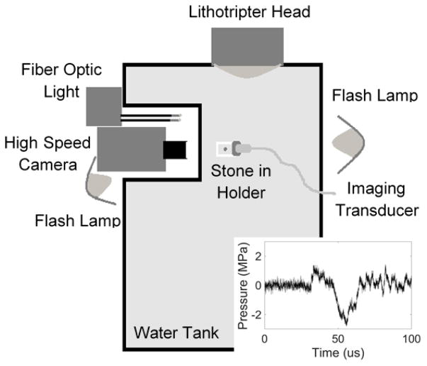
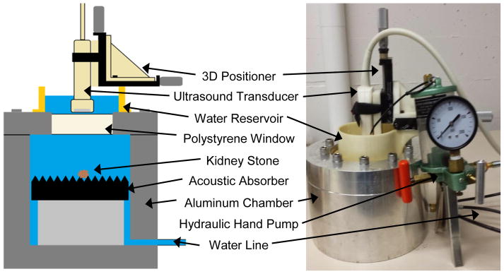
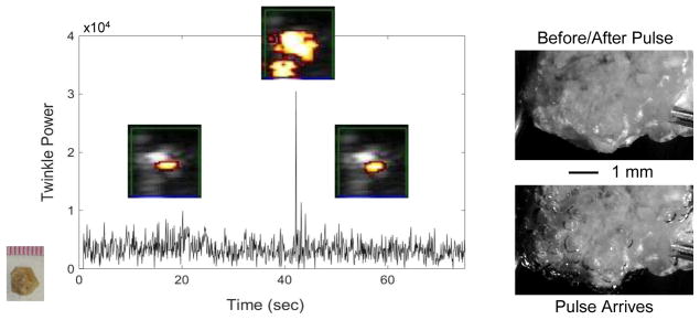
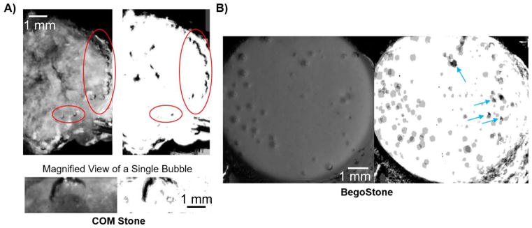
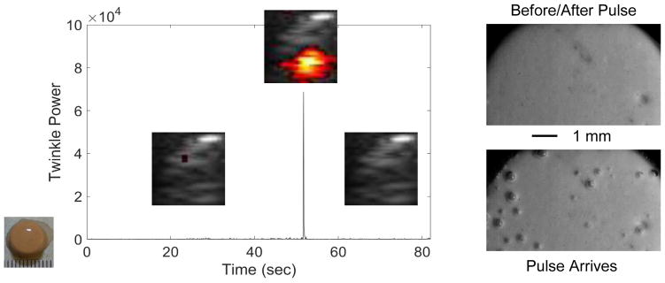
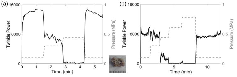
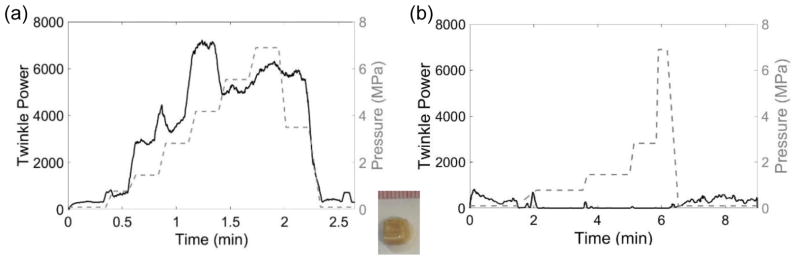
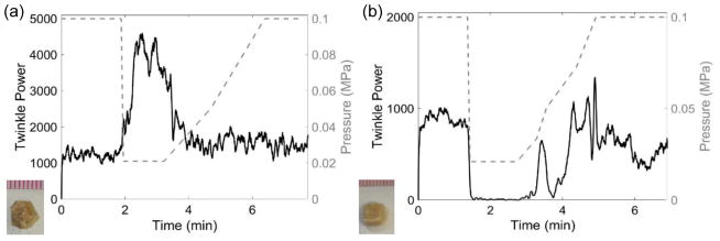
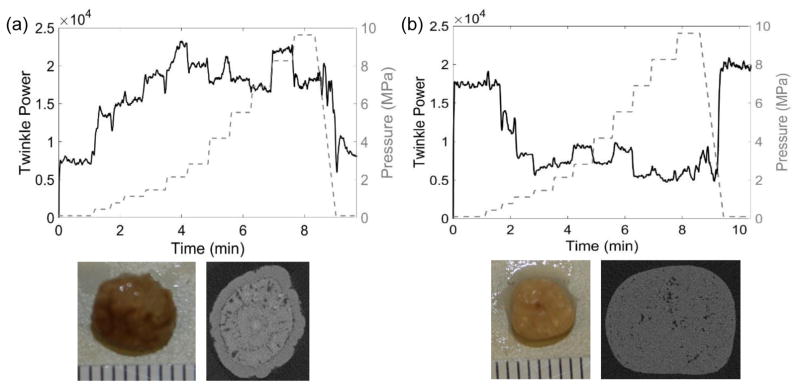
The color Doppler ultrasound twinkling artifact, which highlights kidney stones with rapidly changing color, has the potential to improve stone detection; however, its inconsistent appearance has limited its clinical utility. Recently, it was proposed stable crevice bubbles on the kidney stone surface cause twinkling; however, the hypothesis is not fully accepted because the bubbles have not been directly observed. In this paper, the micron or submicron-sized bubbles predicted by the crevice bubble hypothesis are enlarged in kidney stones of five primary compositions by exposure to acoustic rarefaction pulses or hypobaric static pressures in order to simultaneously capture their appearance by high-speed photography and ultrasound imaging. On filming stones that twinkle, consecutive rarefaction pulses from a lithotripter caused some bubbles to reproducibly grow from specific locations on the stone surface, suggesting the presence of pre-existing crevice bubbles. Hyperbaric and hypobaric static pressures were found to modify the twinkling artifact; however, the simple expectation that hyperbaric exposures reduce and hypobaric pressures increase twinkling by shrinking and enlarging bubbles, respectively, largely held for rough-surfaced stones but was inadequate for smoother stones. Twinkling was found to increase or decrease in response to elevated static pressure on smooth stones, perhaps because of the compression of internal voids. These results support the crevice bubble hypothesis of twinkling and suggest the kidney stone crevices that give rise to the twinkling phenomenon may be internal as well as external.


Figures

Figure 1
Figure 1
Experimental arrangement to visualize crevice bubbles on the kidney stone surface by expanding them with a lithotripter pulse. Stones were placed pre-focal and off axis and were visualized with a high-speed camera and ultrasound transducer when the lithotripter pulse (inset) arrived.
Figure 2
Figure 2
Schematic (left) and photograph (right) of the aluminum-walled hyperbaric chamber. The diagram shows the internal arrangement of the tank for the hyperbaric and hypobaric experiments.
Figure 3
Figure 3
Plot of twinkle power versus time before, during, and after a pre-focal, off axis lithotripter pulse arrives at a COM stone. Overlaid on the plot are selected ROI Doppler images (image scale: 1-cm width) showing the stone (grey) and twinkling (color) on the stone. When the lithotripter pulse arrives at about 45 sec, bubbles are excited on the stone surface, as observed with high-magnification, high-speed photography (right), and twinkle power increases transiently (for one Doppler imaging frame) by more than six times. After the lithotripter pulse and cessation of bubble oscillation, twinkling returns to approximately initial levels. These data were collected with the P4-2 transducer.
Figure 4
Figure 4
Each set of images shows (left) a high-speed photograph of bubbles on the stone surface from a single lithotripter pulse and (right) the average of four binary images from repeated lithotripter pulses for A) a COM stone and B) a BegoStone. In A), a chain of bubbles arose on the right side of the stone with every pulse, as indicated by the black bubble outline in the binary image. While not every bubble arose with each lithotripter pulse, bubbles repeatedly arose from certain locations on the stone surface. The magnified image shows the dark outline of a bubble that arose in one particular location in all four lithotripter pulses. In B), the bubble distribution was variable with successive lithotripter pulses, as evidenced by the grey as opposed to black scattered across the stone surface in the binary image. Blue arrows indicate the four locations on the BegoStone surface with noticeable imperfections that could not be filtered out of the binary image.
Figure 5
Figure 5
Plot of twinkle power versus time before, during, and after a pre-focal, off axis lithotripter pulse arrives at a cylindrical, artificial BegoStone. Overlaid on the plot are selected ROI Doppler images (image scale: 1-cm width) showing the stone (grey) and twinkling in color (if present). Twinkling is virtually nonexistent until the lithotripter pulse arrived at about 52 sec. When the pulse arrives, bubbles are excited on the BegoStone surface as observed with high-magnification, high-speed photography (right), and twinkling increases significantly for the duration of the pulse plus time for bubble oscillations. After the lithotripter pulse, twinkling returned to initial levels of little to no twinkling. These data were collected with the P4-2 transducer.
Figure 6
Figure 6
Plots of twinkle power versus time and absolute static pressure in MPa (dashed grey line, right axis) showing the response of the same COM stone (shown in inset) to hyperbaric pressures with (a) taken 24 hours before (b). Both plots show similar trends; however, the hyperbaric threshold to eliminate twinkling is >2 times higher in (b) compared to (a). These data were collected with the L7-4 transducer.
Figure 7
Figure 7
Plots of twinkle power versus time and absolute static pressure in MPa (dashed grey line, right axis) showing the effect of hyperbaric pressures for the same brushite stone (shown in inset) with the plot in (a) taken 4 days before the plot in (b). Twinkling is found to increase with hyperbaric pressures in (a), whereas twinkling decreases with the increasing pressure in (b). In both cases, twinkle powers are similar at both the beginning and the ends of the plots. These data were collected with the P4-2 transducer.
Figure 8
Figure 8
Plots of twinkle power versus time and absolute static pressure in MPa (dashed, right y-axis) showing the effect of hypobaric conditions on (a) COM and (b) brushite stones. (a) When pressure was reduced, the twinkle power on the macroscopically rough COM stone increased before returning to initial levels when the pressure was returned to atmosphere. (b) Twinkling on the macroscopically smooth brushite stone decreased when the pressure was reduced before returning to approximately the initial levels when the pressure was returned to atmosphere. These data were collected with the P4-2 transducer.
Figure 9
Figure 9
Plots of twinkle power versus time and absolute static pressure (MPa; right y-axis) for (a) a calcium oxalate dihydrate (COD) stone and (b) a cystine stone. (a) The μCT cross section of this COD stone shows a slightly rough stone surface with a ringed structure and some internal micro-crevices comprising 8.6% of the center slice area. The twinkle power was initially of moderate amplitude and generally increased with pressure. (b) The μCT cross section of this cystine stone shows a macroscopically smooth surface with a scattering of micro-crevices throughout the stone comprising 3.7% of the center slice void. Twinkling was initially quite strong and decreased with elevated pressure and then stayed at a constant, non-zero level. In both cases, twinkling returned to its initial amplitude when pressure was returned to ambient levels. These data were collected with the P4-2 transducer.

References

    1. Alan C, Koçoğlu H, Kosar S, Karatag O, Ersay AR, Erhan A. Role of twinkling artifact in characterization of urinary calculi. Actas Urológicas Españolas (English Edition) 2011;35(7):396–402. doi: 10.1016/j.acuroe.2011.02.006. - DOI - PubMed
    1. Apfel RE. Role of impurities in cavitation-threshold determination. J Acoust Soc Am. 1970;48(5):1179–1186.
    1. Aytaç SK, Özcan H. Effect of color Doppler system on the twinkling sign associated with urinary tract calculi. J Clinical Ultrasound. 1999;27(8):433–439. doi: 10.1002/(SICI)1097-0096(199910)27:8<433::AID-JCU4>3.0.CO;2-1. - DOI - PubMed
    1. Chelfouh N, Grenier N, Higueret D, Trillaud H, Levantal O, Pariente J-L, Ballanger P. Characterization of Urinary Calculi: In Vitro Study of “Twinkling Artifact” Revealed byColor-Flow Sonography. AJR. 1998;171:1055–1060. - PubMed
    1. Crum LA. Tensile strength of water. Nature. 1979;278:148–149.

Publication types

MeSH terms