. 2017 Nov 13;12(11):e0187516.
doi: 10.1371/journal.pone.0187516. eCollection 2017.

Expanded repertoire of kinetoplast associated proteins and unique mitochondrial DNA arrangement of symbiont-bearing trypanosomatids

Affiliations

Expanded repertoire of kinetoplast associated proteins and unique mitochondrial DNA arrangement of symbiont-bearing trypanosomatids

Silvana Sant Anna de Souza et al. PLoS One. .

Abstract

In trypanosomatids, the kinetoplast is the portion of the single mitochondrion that is connected to the basal body and contains the kDNA, a network composed by circular and interlocked DNA. The kDNA packing is conducted by Kinetoplast Associated Proteins (KAPs), which are similar to eukaryotic histone H1. In symbiont-harboring trypanosomatids (SHTs) such as Angomonas deanei and Strigomonas culicis, a ß-proteobacterium co-evolves with the host in a mutualistic relationship. The prokaryote confers nutritional benefits to the host and affects its cell structure. Atomic force microscopy showed that the topology of isolated kDNA networks is quite similar in the two SHT species. Ultrastructural analysis using high-resolution microscopy techniques revealed that the DNA fibrils are more compact in the kinetoplast region that faces the basal body and that the presence of the symbiotic bacterium does not interfere with kDNA topology. However, RT-PCR data revealed differences in the expression of KAPs in wild-type protozoa as compared to aposymbiotic cells. Immunolocalization showed that different KAPs present distinct distributions that are coincident in symbiont-bearing and in symbiont-free cells. Although KAP4 and KAP7 are shared by all trypanosomatid species, the expanded repertoire of KAPs in SHTs can be used as phylogenetic markers to distinguish different genera.


Conflict of interest statement

Competing Interests: The authors have declared that no competing interests exist.

Figures

Fig 1
Fig 1. Phylogenetic relationships among KAPs as determined by Bayesian inference.
Branches are colored according to the putative KAP type identified. Numbers on nodes represent the Bayesian posterior probability for the corresponding clade (clades without numbers are those presenting 100% posterior probability). Black circles next to taxon names indicate sequences used by Cavalcanti et al. 2009 [14]; red circles indicate sequences used by Motta et al. 2013 [30].
Fig 2
Fig 2. Quantification of KAP transcripts for KAP4, aKAP23 and stKAPy by RT-qPCR.
The relative amount of KAPs was estimated by comparison with the GAPDH expression of WT trypanosomatids. (A) A. deanei KAPs, (B) S. culicis KAPs. APO, aposymbiotic strain; WT, wild type strain. AdKAP4—p > 0.05, aKAP23—p < 0.05 (*), stKAPy—p > 0.05, ScKAP4—p < 0.01 (**).
Fig 3
Fig 3. Immunolocalization of KAPs on WT and APO strains of S. culicis and A. deanei.
DAPI was used to label the nucleus and the kinetoplast, and the anti-porin antibody to identify the symbiotic bacterium in WT cells. (A-B): The anti-KAP4 antibody labeled the kinetoplast region that faces the anterior end of A. deanei in WT (A) and APO (B) cells. Note that this labeling in part coincides with DAPI staining. (C-D): For S. culicis, KAP4 labeling is dispersed through the kinetoplast of both strains and overlaps those for DAPI. (E-F): The anti-aKAP23 antibody generated a similar and specific labeling for WT (E) and APO (F) cells of A. deanei. In this case, an overlap with DAPI was not observed indicating that aKAP23 does not co-localize with the kDNA and is probably located at the KFZ. (G-H) Regarding stKAPy, this protein also faces the anterior part of the cell body in both strains, however the labeling is more disperse when compared to that obtained for aKAP23. Arrowheads indicate the kinetoplast. Scale bars equal to 1 μm.
Fig 4
Fig 4. Kinetoplast ultrastructure of WT and APO strains of A. deanei and S. culicis.
A-D: Kinetoplast ultrastructure observed by transmission electron microscopy (TEM). A-B: Both strains of A. deanei present a similar kinetoplast ultrastructure that presents a trapezoid shape and a looser arrangement of the central area in relation to the region that faces the basal body (bb) where kDNA is more condensed (black arrows). C-D: In WT and APO strains of S. culicis, the kinetoplast displays an arch shape and the more condensed layer close to the basal body (black arrows). E-H: Kinetoplast ultrastructure observed by electron tomography (ET). E-F: This high-resolution technique confirmed that both A. deanei strains have a similar kDNA network topology presenting the densely packed kDNA region facing the basal body (brackets). (E, F) top, (E’, F’) middle and (E”, F”) bottom regions of the kinetoplast as visualized by electron tomography. G-H: In both strains of S. culicis, the kinetoplast is flatter when compared to that found in A. deanei, and the compact DNA fibers occupy approximately half of the kDNA network (dashed rectangle). (G, H) top, (G’, H’) middle and (G”, H”) bottom regions of the kinetoplast as visualized by electron tomography. In G’ and H’ arrows indicate the condensed kDNA fibers.
Fig 5
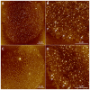
Fig 5. AFM analysis of isolated kDNA networks of A. deanei (A-B) and S. culicis (C-D).
Both species present an arrangement composed of circles uniformly distributed throughout a massive network of kDNA molecules. Clusters of DNA forming rosette-like structures (B and D, arrows) were seen along the network and correspond to regions where the DNA molecules crossover themselves.

Similar articles

Cited by

References

    1. Schmid MB. More than just "histone-like" proteins. Cell. 1990; 63: 451–453. - PubMed
    1. Luijsterburg MS, Noom MC, Wuite GJ, Dame RT. The architectural role of nucleoid-associated proteins in the organization of bacterial chromatin: a molecular perspective. J Struct Biol. 2006; 156: 262–272. doi: 10.1016/j.jsb.2006.05.006 - DOI - PubMed
    1. Kucej M, Butow RA. Evolutionary tinkering with mitochondrial nucleoids. Trends Cell Biol. 2007; 17:586–592. doi: 10.1016/j.tcb.2007.08.007 - DOI - PubMed
    1. Timmis J.N., Ayliffe MA, Huang CY, Martin W. Endosymbiotic gene transfer: organelle genomes forge eukaryotic chromosomes. Nature reviews: Genetics. 2004; 5: 123–135. doi: 10.1038/nrg1271 - DOI - PubMed
    1. Holt IJ, He J, Mao CC, Boyd-Kirkup JD, Martinsson P, Sembongi H, et al. Mammalian mitochondrial nucleoids: organizing an independently minded genome. Mitochondrion. 2007; 7: 311–321. doi: 10.1016/j.mito.2007.06.004 - DOI - PubMed

LinkOut - more resources