. 2018 Apr;23(2):154-163.
doi: 10.1177/2472630317742071. Epub 2017 Nov 13.

Embedded Multimaterial Extrusion Bioprinting

Affiliations

Embedded Multimaterial Extrusion Bioprinting

Marco Rocca et al. SLAS Technol. 2018 Apr.

Abstract

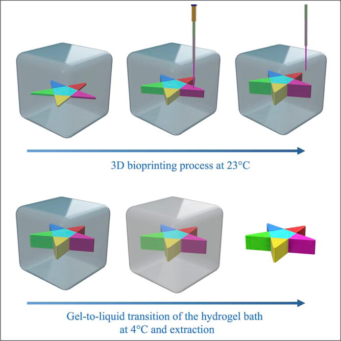
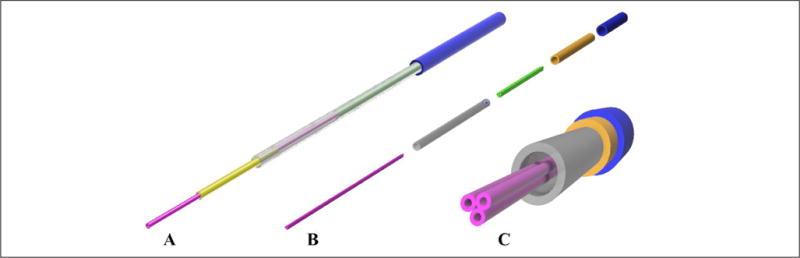
Embedded extrusion bioprinting allows for the generation of complex structures that otherwise cannot be achieved with conventional layer-by-layer deposition from the bottom, by overcoming the limits imposed by gravitational force. By taking advantage of a hydrogel bath, serving as a sacrificial printing environment, it is feasible to extrude a bioink in freeform until the entire structure is deposited and crosslinked. The bioprinted structure can be subsequently released from the supporting hydrogel and used for further applications. Combining this advanced three-dimensional (3D) bioprinting technique with a multimaterial extrusion printhead setup enables the fabrication of complex volumetric structures built from multiple bioinks. The work described in this paper focuses on the optimization of the experimental setup and proposes a workflow to automate the bioprinting process, resulting in a fast and efficient conversion of a virtual 3D model into a physical, extruded structure in freeform using the multimaterial embedded bioprinting system. It is anticipated that further development of this technology will likely lead to widespread applications in areas such as tissue engineering, pharmaceutical testing, and organs-on-chips.

Keywords: bioprinting; embedded; extrusion; freeform; multimaterial.


Conflict of interest statement

Declaration of Conflicting Interests

The authors declared no potential conflicts of interest with respect to the research, authorship, and/or publication of this article.

Figures

Figure 1
Figure 1
Concept of the embedded multimaterial 3D bioprinting. Top: A custom-designed multinozzle printhead is used to deposit different bioinks into the supporting hydrogel bath, PF-127. The bioprinting process occurs at room temperature (23°C), after the PF-127 bath has formed a stable hydrogel bath. Bottom: To release the bioprinted structure, the supporting PF-127 hydrogel bath is cooled down to 4°C, hence inducing its gel-to-liquid transition and subsequent retrieval of the structure.
Figure 2
Figure 2
(A) Design of the single-nozzle printhead. A thin tip (30G) with an inner diameter of 159 μm was encased in increasingly larger metal tubes to maintain a small nozzle size at the tip while at the same time keeping a stiff and rigid overall structure. (B) Exploded view of the design of a multinozzle printhead. Three thin tips (30G) were fixed together to form the extrusion nozzle. This allowed for different materials to be extruded simultaneously or sequentially, while avoiding intermixing of the hydrogels prior to extrusion. Similar to the single-nozzle approach, a sequence of three metal tubes with increasing diameters were used to make the structure stiffer and avoid its bending. (C) Front view of the multinozzle printhead.
Figure 3
Figure 3
Plots showing (A) gelation time and (B) liquefaction time vs. PF-127 concentration. (A) Gelation of PF-127 solutions at different concentrations of 21%–30%. The liquid solutions initially at 4°C were placed at 37°C until gelation was observed. (B) Liquefaction of PF-127 hydrogels at different concentrations of 10%–30%. The gelled samples were maintained at room temperature (23°C) for 12 h to observe if they could reach the liquid phase. All samples at concentrations of >20% did not show any liquefaction and hence were not included in the graph.
Figure 4
Figure 4
Characterization of the filament diameter vs. nozzle speed and pressure. (A) Patterns consisting of 10 straight lines each with a different nozzle moving speed ranging from 1 to 30 mm/s were bioprinted inside the PF-127 supporting hydrogel bath. The same patterns were bioprinted again for different pressures, from 10 to 70 psi. (B) Top view of the alginate lines bioprinted inside the PF-127 bath at a speed of 1 up to 30 mm/s at 50 psi. (C) Fluorescence micrograph showing a line bioprinted at 70 psi and 5 mm/s.
Figure 5
Figure 5
Young’s moduli of a series of bioprinted cylindrical structures at different interline spacing, equally varied along the x, y, and z directions.
Figure 6
Figure 6
Embedded bioprinting of single-material constructs. (A–C) Photographs showing (A) a cube, (B) a heart cartoon, and (C) MIT letters, indicating that the structures were sufficiently stiff to be extracted from the supporting bath and were able to self-support their weights. (D–I) Models and photographs showing (D–F) a Mobius strip and (G–I) a Klein bottle, proving that complex shapes that would conventionally require supporting pillars in normal extrusion 3D bioprinting methods could be readily achieved using embedded bioprinting. (J–L) Photographs showing (J,K) a heart-like structure and (L) a kidney-like structure, demonstrating that more complex macroscopic structures mimicking human organs could be bioprinted and have their shapes maintained after extraction from the sacrificial hydrogel bath.
Figure 7
Figure 7
Embedded bioprinting of multimaterial constructs. (A–C) Photographs showing the switching test for the multinozzle printhead. Alginate doped with three different dyes was used to visualize the switch between the different extrusion nozzles in a single printhead. (D) Photograph showing a structure with three alternating materials bioprinted on a glass slide. (E–H) Photographs showing embedded bioprinting of complex patterns containing multiple materials, including (E) a star, (F) MIT letters, (G) a Klein bottle, and (H) a Mobius strip.

References

    1. Murphy SV, Atala A. 3D Bioprinting of Tissues and Organs. Nat Biotechnol. 2014;32(8):773–785. - PubMed
    1. Dababneh AB, Ozbolat IT. Bioprinting Technology: A Current State-of-the-Art Review. J Manuf Sci Eng. 2014;136(6):061016.
    1. Groll J, Boland T, Blunk T, et al. Biofabrication: Reappraising the Definition of an Evolving Field. Biofabrication. 2016;8(1):013001. - PubMed
    1. Hyungseok L, Dong-Woo C. One-Step Fabrication of an Organ-on-a-Chip with Spatial Heterogeneity Using a 3D Bioprinting Technology. Lab Chip. 2016;16:2618–2625. - PubMed
    1. Huh D, Matthews BD, Mammoto A, et al. Reconstituting Organ-Level Lung Functions on a Chip. Science. 2010;328:1662–1668. - PMC - PubMed

Publication types

MeSH terms

Substances

LinkOut - more resources