. 2017 Nov 13;17(1):759.
doi: 10.1186/s12885-017-3729-z.

Gene expression signatures of neuroendocrine prostate cancer and primary small cell prostatic carcinoma

Affiliations

Gene expression signatures of neuroendocrine prostate cancer and primary small cell prostatic carcinoma

Harrison K Tsai et al. BMC Cancer. .

Abstract

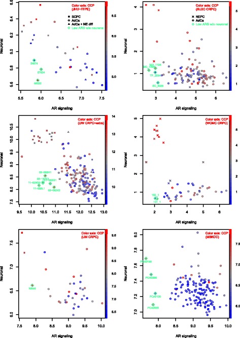
Background: Neuroendocrine prostate cancer (NEPC) may be rising in prevalence as patients with advanced prostate cancer potentially develop resistance to contemporary anti-androgen treatment through a neuroendocrine phenotype. While prior studies comparing NEPC and prostatic adenocarcinoma have identified important candidates for targeted therapy, most have relied on few NEPC patients due to disease rarity, resulting in thousands of differentially expressed genes collectively and offering an opportunity for meta-analysis. Moreover, past studies have focused on prototypical NEPC samples with classic immunohistochemistry profiles, whereas there is increasing recognition of atypical phenotypes. In the primary setting, small cell prostatic carcinoma (SCPC) is frequently admixed with adenocarcinomas that may be clonally related, and a minority of SCPCs express markers typical of prostatic adenocarcinoma while rare cases do not express neuroendocrine markers. We derived a meta-signature of prototypical high-grade NEPC, then applied it to develop a classifier of primary SCPC incorporating disease heterogeneity.

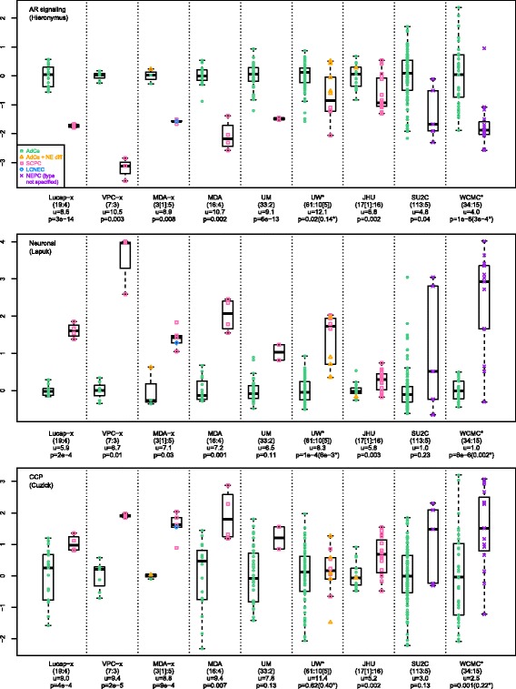
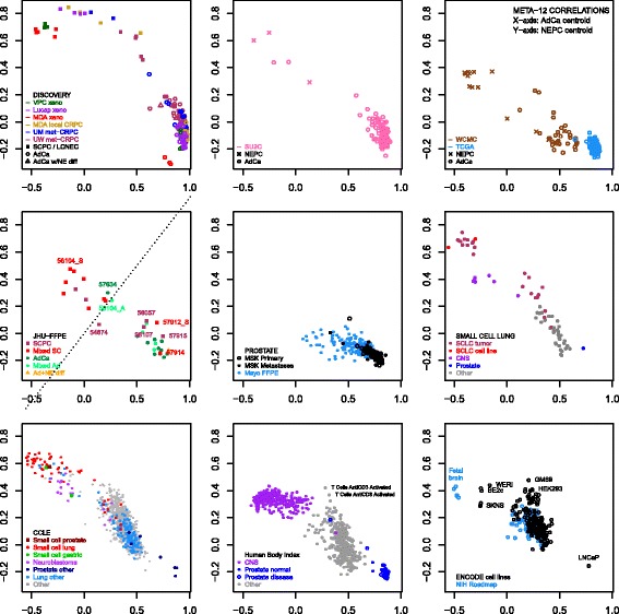
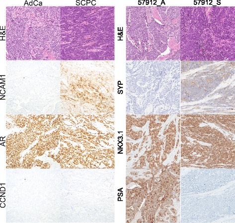
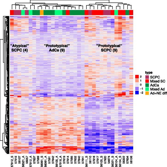
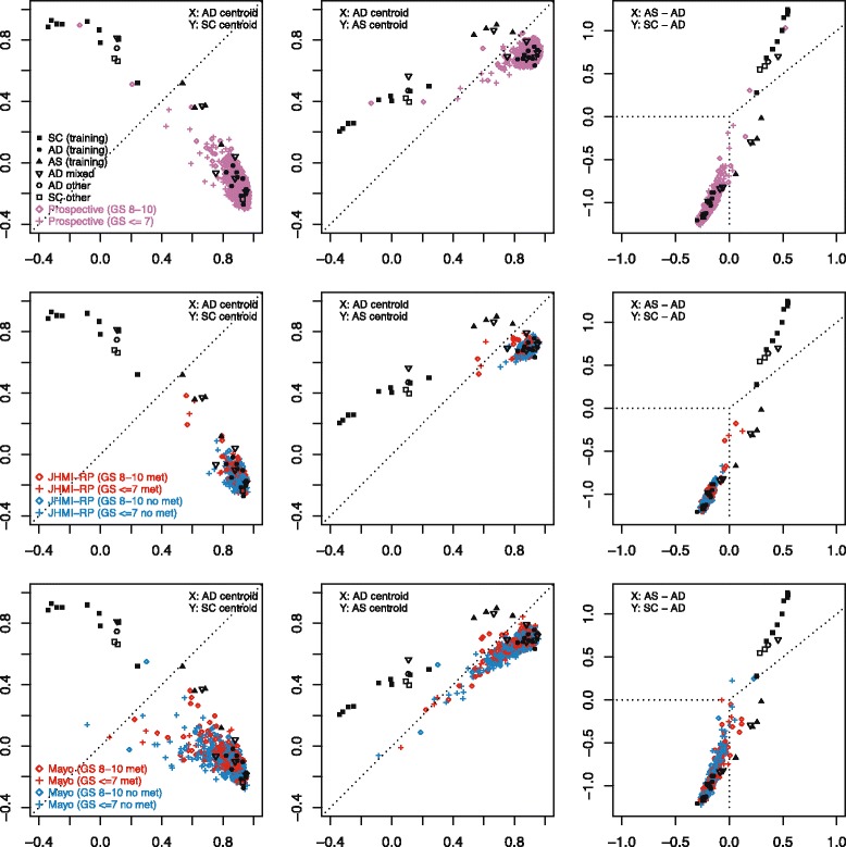
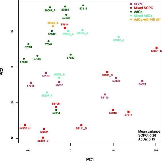
Methods: Prototypical NEPC samples from 15 patients across 6 frozen tissue microarray datasets were assessed for genes with consistent outlier expression relative to adenocarcinomas. Resulting genes were used to determine subgroups of primary SCPCs (N=16) and high-grade adenocarcinomas (N=16) profiled by exon arrays using formalin-fixed paraffin-embedded (FFPE) material from our institutional archives. A subgroup classifier was developed using differential expression for feature selection, and applied to radical prostatectomy cohorts.

Results: Sixty nine and 375 genes demonstrated consistent outlier expression in at least 80% and 60% of NEPC patients, with close resemblance in expression between NEPC and small cell lung cancer. Clustering by these genes generated 3 subgroups among primary samples from our institution. Nearest centroid classification based on the predominant phenotype from each subgroup (9 prototypical SCPCs, 9 prototypical adenocarcinomas, and 4 atypical SCPCs) achieved a 4.5% error rate by leave-one-out cross-validation. The classifier identified SCPC-like expression in 40% (2/5) of mixed adenocarcinomas and 0.3-0.6% of adenocarcinomas from prospective (4/2293) and retrospective (2/355) radical prostatectomy cohorts, where both SCPC-like retrospective cases subsequently developed metastases.

Conclusions: Meta-analysis generates a robust signature of prototypical high-grade NEPC, and may facilitate development of a primary SCPC classifier based on FFPE material with potential prognostic implications.

Keywords: FFPE; Gene signature; Meta-analysis; Mixed prostatic adenocarcinoma; Nearest centroid classifier; Neuroendocrine prostate cancer; Small cell carcinoma.


Conflict of interest statement

Ethics approval and consent to participate

Informed consent to use the tissue samples in this study was waived by the John Hopkins School of Medicine Institutional Review Board.

Consent for publication

Not applicable.

Competing interests

JL, MA, NE and ED are employees of GenomeDx Biosciences; TLL has received research funding from GenomeDx. HKT declares no competing interests.

Publisher’s Note

Springer Nature remains neutral with regard to jurisdictional claims in published maps and institutional affiliations.

Figures

Fig. 1
Fig. 1
Gene signature scores across datasets profiling NEPC and adenocarcinomas. (A[A’]:B[B’]) denotes cohort sizes of A adenocarcinomas and B NEPCs including A’ or B’ adenocarcinomas with NE differentiation, u denotes mean score of the adenocarcinoma cohort, p denotes p-value under t-test comparison of NEPCs versus adenocarcinomas, and (*) signifies p-values after averaging over multiple samples from the same patient. ARS and neuronal phenotype scores completely separated cohorts (AUC 100%) in xenograft and frozen tissue primary datasets (Lucap-x, VPC-x, MDA-x, MDA), and ARS demonstrated significant cohort differences (p<0.05) across all datasets. CCP was highly correlated to an RB loss signature (mean r=0.96 across datasets; not shown), in agreement with reports showing correlation of CCP and E2F1 targets [7]. In UW, NEPCs annotated as adenocarcinomas with NE differentiation mostly demonstrated ARS and CCP scores similar to adenocarcinomas. In WCMC and SU2C, NEPCs also sometimes demonstrated gene signature scores similar to adenocarcinomas, and may have corresponded to adenocarcinomas with NE differentiation, however NEPC subtypes were not specified in annotations provided. In JHU-FFPE, 5 SCPCs exhibited ARS scores similar to adenocarcinomas (fold-change > -0.5 and z-score > -1), and are investigated further in the JHU-FPE results section. JHU-FFPE scores also demonstrated the least dynamic range across gene signatures, likely related to RNA degradation in FFPE. Gene signature scores were formed by average expression of genes. Among single-sample scoring methods, SVD-based PLAGE has been recognized as a top performer and is equivalent to (signed) average expression for perfectly correlated (and anti-correlated) genes. Indeed, PLAGE and average expression were highly correlated across the NEPC datasets (correlations for CCP > 0.99, Neuronal > 0.96, ARS > 0.95)
Fig. 2
Fig. 2
Correlation profiles relative to meta-12 adenocarcinoma and NEPC centroids across datasets. Nearest centroid classification of NEPC datasets demonstrated NEPC sensitivities and specificities of 91% and 100% on training samples, 60% and 98% in SU2C, 80% and 100% in WCMC, and 63% and 94% in JHU-FFPE. Centroid correlation profiles were also evaluated for prostatic adenocarcinoma datasets (TCGA, MSK, Mayo-FFPE) and various human tissue or cell line datasets including SCLC (GSE43346), CCLE (cBioPortal), Human Body Index (GSD7307), ENCODE (GSE19090), and NIH Roadmap (GSE18927). Correlations were generally weaker in FFPE datasets (JHU-FFPE, Mayo-FFPE) and in WCMC derived primarily from biopsies. Rare outlier adenocarcinomas were present across datasets, usually related to low ARS. SCLCs generally had the most similar centroid profile to NEPC followed by small cell gastric carcinoma and CNS-related samples. In JHU-FFPE, 5 SCPCs appeared to cluster with adenocarcinomas, demonstrated ARS scores similar to adenocarcinomas (Fig. 1, Additional file 3: Figure S3), and are discussed further in the JHU-FFPE results section
Fig. 3
Fig. 3
Hybrid immunohistochemistry of an unusual mixed tumor. A hybrid IHC profile was observed in an unusual mixed case from JHU-FFPE with concurrent small cell (57912_S) and Gleason 5+4 adenocarcinoma (57912_A) components. The SCPC component appeared to uniformly co-express androgen-related markers (Nkx3.1, AR) and neuroendocrine markers (synaptophysin and CD56/NCAM1 but not chromogranin) by IHC. Unusually, IHC was negative for PSA despite moderate expression of the underlying gene KLK3 (Additional file 3: Figure S11). By contrast, the adenocarcinoma component was IHC positive for PSA and negative for synaptophysin and CD56. Both components were IHC negative for cyclin D1, a proposed marker of SCPC [21]
Fig. 4
Fig. 4
Hierarchical clustering of JHU-FFPE relative to meta-9 genes. There were 3 main groups, which we labeled “prototypical” adenocarcinomas, “prototypical” SCPCs, and “atypical” SCPCs, and which generally corresponded to pure adenocarcinomas, SCPCs with reduced ARS, and SCPCs with retained ARS respectively. The only exceptions were one SCPC outlier with retained ARS (57914) that clustered with prototypical adenocarcinomas, one pure adenocarcinoma outlier (57634) described previously in a case report that clustered with prototypical SCPCs, and heterogeneous behavior of mixed adenocarcinomas. The adenocarcinoma 57634 clustered near 56322, an SCPC with negative IHC for all 3 neuroendocrine markers synaptophysin, chromogranin, and CD56. The oldest SCPC’s (54674 and 56321_S) had low CCP and also clustered together. Meta-9 clustering was consistent with subsequent nearest centroid classification based on 9 prototypical SCPC, 9 prototypical adenocarcinoma, and 4 atypical SCPC with LIMMA-based feature selection (Fig. 6)
Fig. 5
Fig. 5
Low ARS without elevated neuronal phenotype samples across clinical datasets. Twenty samples with low ARS and low/average neuronal phenotype scores were identified based on outlier-style cut-offs relative to adenocarcinomas (fold-change < -1, z-score < -2 for ARS; fold-change < 0.5, z-score < 1 for neuronal phenotype), including known unusual cases such as the case report adenocarcinoma 57634 (JHU-FFPE), and also samples from pure adenocarcinoma datasets (MSKCC). Differential expression analysis was notable for down-expression of RAB3B in this group relative to the remaining adenocarcinomas or NEPCs (Additional file 3: Figure S4). These samples also demonstrated a wide range of CCP scores (color axis), where low CCP possibly reflected response to treatment (Additional file 3: Figure S5)
Fig. 6
Fig. 6
Nearest 3-centroid classification of GRID® RP adenocarcinoma cohorts. We assessed performance of nearest centroid classification in GRID® RP adenocarcinoma cohorts Prospective (N=2993), JHU-RP (N=355), and Mayo (N=780) relative to centroids AD (prototypical adenocarcinoma), SC (prototypical SCPC), and AS (atypical SCPC). Two Prospective (0.09%) and no JHU-RP (0%) samples were classified as prototypical SCPC while 4 Prospective (0.17%) and 2 JHU-RP (0.6%) samples were classified as atypical SCPC. A greater proportion of Mayo samples (1.6%) were classified as SCPC but likely included false positives with low correlations to all 3 centroids
Fig. 7
Fig. 7
Principal components analysis of JHU-FFPE. SCPCs and adenocarcinomas were generally separated by principal components analysis. Mixed adenocarcinomas exhibited roughly intermediate behavior, although we questioned whether 56104_A contained an accidental admixture with its neighboring small cell component 56104_S. One SCPC (57915) and one pure adenocarcinoma (57634) clustered with opposite phenotypes, similar to meta-9 clustering. Among SCPCs with known AR or PSA-positivity, two clustered side by side in a relatively intermediate territory (57915, 56107) while the third was loosely in the vicinity (57912_S). Of all principal components, PC2 separated SCPCs from adenocarcinomas best (AUC 86%) and was highly correlated to the difference between ARS and CCP (r=0.93). By contrast in frozen tissue primary and xenograft NEPC datasets, respective PC1's separated SCPCs from adenocarcinomas best (AUC 100%) and was highly correlated to the difference between CCP and ARS (r=0.75-0.98) (Supp Figure 9). Thus in JHU-FFPE, PC1 represented a different source of greatest variability. Examination of its top coefficients by magnitude revealed that PC1 was highly anti-correlated to the average expression of various ribosomal subunits (r = -0.93) including RPL19, known to be an effective reference gene. Two SCPCs (56321_S, autopsy 54674) had the largest PC1 magnitudes and were archived 14-16 years (versus 0-6 years for other SCPCs), perhaps reflecting higher levels of RNA degradation; however the oldest adenocarcinoma (56321_A) did not exhibit this trend

Similar articles

Cited by

References

    1. Epstein, JI.,et al., Proposed morphologic classification of prostate cancer with neuroendocrine differentiation. Am J Surg Pathol, 2014. 38(6): p. 756-767. - PMC - PubMed
    1. Wang HT, et al. Neuroendocrine prostate cancer (nepc) progressing from conventional prostatic adenocarcinoma: factors associated with time to development of nepc and survival from nepc diagnosis-a systematic review and pooled analysis. J Clin Oncol. 2014;32(30):3383–3390. doi: 10.1200/JCO.2013.54.3553. - DOI - PubMed
    1. Tan, HL, et al., Rb loss is characteristic of prostatic small cell neuroendocrine carcinoma. Clin Cancer Res, 2014. 20(4): p. 890-903. - PMC - PubMed
    1. Lapuk AV, et al. From sequence to molecular pathology, and a mechanism driving the neuroendocrine phenotype in prostate cancer. J Pathol. 2012;227(3):286–297. doi: 10.1002/path.4047. - DOI - PMC - PubMed
    1. Yao JL, et al. Small cell carcinoma of the prostate: an immunohistochemical study. Am J surg pathol. 2006;30(6):705–712. doi: 10.1097/00000478-200606000-00005. - DOI - PubMed

Substances