. 2017 Nov 13;13(1):332.
doi: 10.1186/s12917-017-1253-7.

Avian infectious bronchitis virus disrupts the melanoma differentiation associated gene 5 (MDA5) signaling pathway by cleavage of the adaptor protein MAVS

Affiliations

Avian infectious bronchitis virus disrupts the melanoma differentiation associated gene 5 (MDA5) signaling pathway by cleavage of the adaptor protein MAVS

Liping Yu et al. BMC Vet Res. .

Abstract

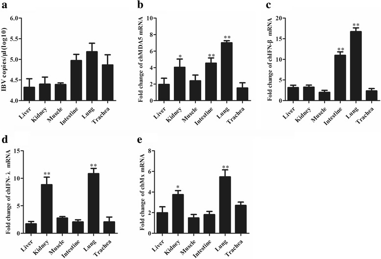
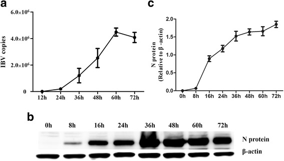
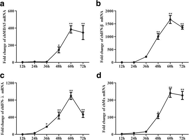
Background: Melanoma differentiation associated gene 5 (MDA5) and retinoic acid-inducible gene-I (RIG-I) selectively sense cytoplasmic viral RNA to induce an antiviral immune response. Infectious bronchitis virus (IBV) is one of the most important infectious agents in chickens, and in chicken cells, it can be recognized by MDA5 to activate interferon production. RIG-I is considered to be absent in chickens. However, the absence of RIG-I in chickens raises the question of whether this protein influences the antiviral immune response against IBV infection.

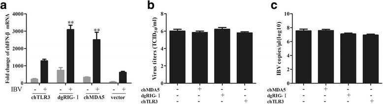
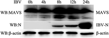
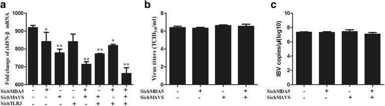
Results: Here, we showed that chicken cells transfected with domestic goose RIG-I (dgRIG-I) exhibited increased IFN-β activity after IBV infection. We also found that IBV can cleave MAVS, an adaptor protein downstream of RIG-I and MDA5 that acts as a platform for antiviral innate immunity at an early stage of infection.

Conclusions: Although chicken MDA5 (chMDA5) is functionally active during IBV infection, the absence of RIG-I may increase the susceptibility of chickens to IBV infection, and IBV may disrupt the activation of the host antiviral response through the cleavage of MAVS.

Keywords: Infectious bronchitis virus; Mavs; Melanoma differentiation associated gene 5; Retinoic acid-inducible gene-I.


Conflict of interest statement

Ethics approval and consent to participate

The Jiangsu Administrative Committee for Laboratory Animals approved all animal studies (Permit number: SYXKSU-2007-0005) according to the guidelines of Jiangsu Laboratory Animal Welfare and Ethical of Jiangsu Administrative Committee of Laboratory Animals.

Consent for publication

Not applicable

Competing interests

All authors declare that they have no competing interests.

Publisher’s Note

Springer Nature remains neutral with regard to jurisdictional claims in published maps and institutional affiliations.

Figures

Fig. 1
Fig. 1
IBV induces chMDA5, chIFN-β, chIFN-λ and chMx expression in chicken embryos. In this experiment three embryos were inoculated with IBV, and three embryos were inoculated with PBS served as negative control; then, the trachea, intestine, kidney, lung, liver, and muscle tissues were collected from the embryos 72 h post-infection. a The IBV genome loads were quantified by RT-qPCR. b chMDA5, (c) chIFN-β, (d) chIFN-λ and (e) chMx were calculated as fold change of the infected group relative to the uninfected group and normalized against β-actin. Data are shown as the mean ± SD (n = 3, 3 embryos) (* P ≤ 0.05; ** P ≤ 0.01). The representatives of three independent experiments showed similar results. Values represent the average of the results from three independent experiments with standard error bars
Fig. 2
Fig. 2
IBV replication has a time-dependent activity in CEK cells. CEK cells were infected with IBV at an MOI of 1. At the indicated times post-infection, (a) viral RNA was quantified by RT-qPCR. Data are presented as the mean ± SD (* P ≤ 0.05; ** P ≤ 0.01); (b) The cellular IBV N proteins were quantified by Western blot. c The graph indicating the fold change of the N proteins. The fold change of N proteins is expressed as densitometric units (Image-Pro-plus 6.0) of bands normalized to the β-actin, results from three independent experiments
Fig. 3
Fig. 3
IBV induces high expression of chMDA5, chIFN-β, chIFN-λ and chMx in CEK cells. CEK cells were infected with IBV at an MOI of 1. At the indicated times post-infection, cells were harvested for RNA isolation, and virus-induced expression of chMDA5 (a), chIFN-β (b), chIFN-λ (c) and chMx (d) was determined by RT-qPCR. All gene expression was calculated as the fold change relative to mock cells uninfected in parallel and normalized against a housekeeping gene (β-actin). Data are presented as the mean ± SD (n = 3, 3 wells on same plate) (* P ≤ 0.05; ** P ≤ 0.01)
Fig. 4
Fig. 4
Dose-dependent antiviral cytokine potency of IBV. CEK cells were inoculated with 10−2, 10−1, 100, 101 and 102 MOI of IBV, then the cells were harvested at 36 h post-treatment for RNA extraction. The IBV genome load (a) was quantified by RT-qPCR. The mRNA levels of chMDA5 (b), chIFN-β (c), chIFN-λ (d) and chMx (e) were evaluated by RT-qPCR. All gene expression was calculated as the fold change relative to uninfected control cells and normalized against a housekeeping gene (β-actin). Data are presented as the mean ± SD (n = 3, 3 wells on same plate) (* P ≤ 0.05; ** P ≤ 0.01)
Fig. 5
Fig. 5
DgRIG- I and chMDA5 overexpression enhances IBV-induced IFN-β transcription. a DF1 cells were transfected with the indicated plasmid for 24 h and then infected with IBV for 24 h. The extracted RNA was used to measure the expression of IFN-β. The expression of IFN-β in the test group was compared to the mock control group that was transfected with the control vector and infected with IBV. The growth properties of IBV in chTLR3-, chMDA5- and dgRIG-I-overexpressed cell supernatants titrated onto CEK cells are expressed as TCID50/ml (b) or by RT-qPCR to determine IBV genome load (c). Experiments were performed in triplicate, and data are representative of three independent experiments (* P ≤ 0.05; ** P ≤ 0.01)
Fig. 6
Fig. 6
IBV cleaves MAVS at an early stage of infection. CEK cells were infected with IBV at an MOI of 1. At the indicated times post-infection, cells were lysed and the expression of MAVS proteins were analyzed by Western blot using an anti-MAVS antibody (top panel). The IBV-encoded N protein was detected with an anti-N antibody (second panel). β-actin was detected as a loading control (bottom panel)
Fig. 7
Fig. 7
chMDA5 and chMAVS knockdown influenced IBV-induced IFN-β transcription. a DF1 cells were transfected with siRNA specific for chMDA5, chTLR3 or chMAVS alone or in combination for 24 h, after which the cells were infected with IBV for 24 h. The expression levels in the silenced groups were compared to the control siRNA-treated cells. The growth properties of IBV in chMDA5- and chMAVS-silenced cell supernatants titrated onto CEK cells are presented as TCID50/ml (b) or by RT-qPCR to determine IBV genome load (c). Data are represented as the mean ± SD from three independent experiments (* P ≤ 0.05; ** P ≤ 0.01)

Similar articles

Cited by

References

    1. Cavanagh D. Coronavirus avian infectious bronchitis virus. Vet Res. 2007;38(2):281–297. doi: 10.1051/vetres:2006055. - DOI - PubMed
    1. Oudshoorn D, van der Hoeven B, Limpens RW, Beugeling C, Snijder EJ, Barcena M, Kikkert M. Antiviral Innate Immune Response Interferes with the Formation of Replication-Associated Membrane Structures Induced by a Positive-Strand RNA Virus. MBio. 2016;7(6):e01991–16. - PMC - PubMed
    1. Kint J, Dickhout A, Kutter J, Maier HJ, Britton P, Koumans J, Pijlman GP, Fros JJ, Wiegertjes GF, Forlenza M. Infectious bronchitis Coronavirus inhibits STAT1 signaling and requires accessory proteins for resistance to type I interferon activity. J Virol. 2015;89(23):12047–12057. doi: 10.1128/JVI.01057-15. - DOI - PMC - PubMed
    1. Kint J, Langereis MA, Maier HJ, Britton P, van Kuppeveld FJ, Koumans J, Wiegertjes GF, Forlenza M. Infectious bronchitis Coronavirus limits interferon production by inducing a host shutoff that requires accessory protein 5b. J Virol. 2016;90(16):7519–7528. doi: 10.1128/JVI.00627-16. - DOI - PMC - PubMed
    1. Takeuchi O, Akira S. Recognition of viruses by innate immunity. Immunol Rev. 2007;220:214–224. doi: 10.1111/j.1600-065X.2007.00562.x. - DOI - PubMed

MeSH terms

LinkOut - more resources