. 2017 Nov 13;14(1):220.
doi: 10.1186/s12974-017-0993-4.

EZH2 suppression in glioblastoma shifts microglia toward M1 phenotype in tumor microenvironment

Affiliations

EZH2 suppression in glioblastoma shifts microglia toward M1 phenotype in tumor microenvironment

Yatao Yin et al. J Neuroinflammation. .

Abstract

Background: Glioblastoma multiforme (GBM) induces tumor immunosuppression through interacting with tumor-infiltrating microglia or macrophages (TAMs) with an unclear pathogenesis. Enhancer of zeste homolog 2 (EZH2) is abundant in GBM samples and cell lines and is involved in GBM proliferation, cell cycle, and invasion, whereas its association with innate immune response is not yet reported. Herein, the aim of this study was to investigate the role of EZH2 in GBM immune.

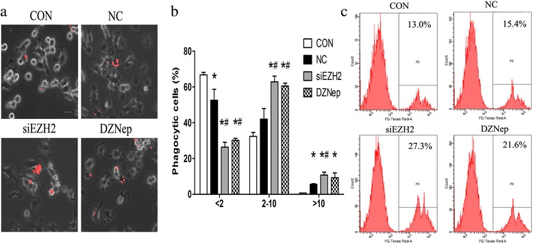
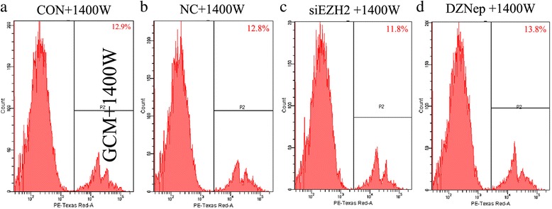
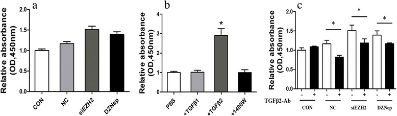
Methods: Co-culturing models of human/murine GBM cells with PBMC-derived macrophages/primary microglia were employed. EZH2 mRNAs and function were suppressed by siEZH2 and DZNep. Real-time PCR and flow cytometry were used to determine levels of microglia/macrophages markers. The fluorescence-labeled latex beads and flow cytometry were utilized to evaluate phagocytic abilities of microglia. CCK8 assay was performed to assess microglia proliferation.

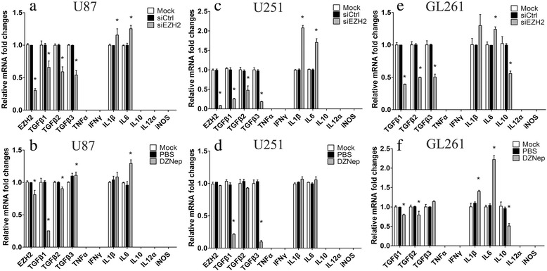
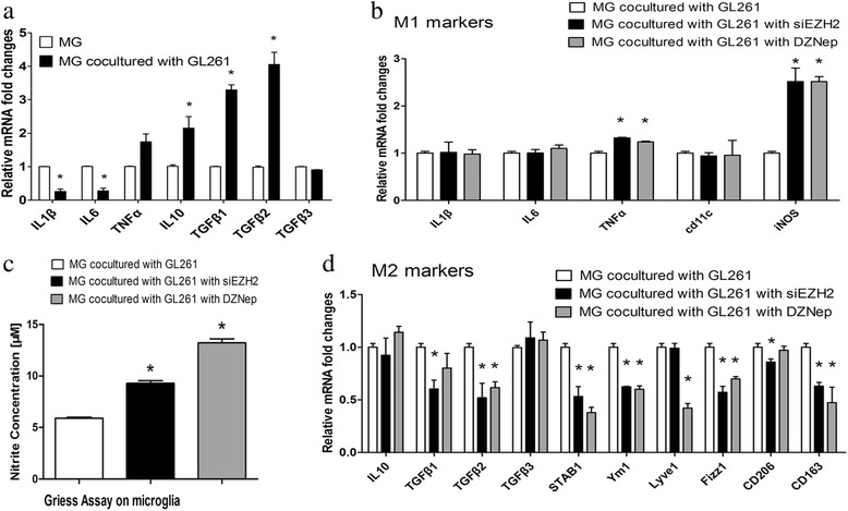
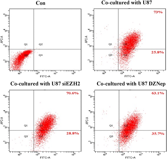
Results: EZH2 inhibition led to significant reduction of TGFβ1-3 and IL10 and elevation of IL1β and IL6 in human and murine GBM cells. More importantly, EZH2 suppression in GBM cells resulted in significant increase of M1 markers (TNFα and iNOS) and decrease of a pool of M2 markers in murine microglia. The proportion of CD206+ cells was decreased in PBMC-derived macrophages as co-incubated with EZH2-inhibited GBM cells. Functional researches showed that phagocytic capacities of microglia were significantly ameliorated after EZH2 inhibition in co-culturing GBM cells and microglia proliferation was declined after addition of TGFβ2 antibodies to co-incubated GBM cells with EZH2 inhibition. Besides, we found that EZH2 suppression in GBM cells enhanced co-culturing microglia engulfment through activation of iNOS.

Conclusions: Our data demonstrates that EZH2 participates in GBM-induced immune deficient and EZH2 suppression in GBM can remodel microglia immune functions, which is beneficial for understanding GBM pathogenesis and suggests potential targets for therapeutic approaches.

Keywords: Enhancer of zeste homolog 2 (EZH2); Glioblastoma; Microglia; Polarization.


Conflict of interest statement

Ethics approval and consent to participate

This study was approved by the ethics committee at Sun Yat-sen Memorial Hospital, Sun Yat-sen University.

Consent for publication

Not applicable.

Competing interests

The authors declare that they have no competing interests.

Publisher’s Note

Springer Nature remains neutral with regard to jurisdictional claims in published maps and institutional affiliations.

Figures

Fig. 1
Fig. 1
EZH2 inhibition induces mRNAs changes of inflammatory cytokines in GBM cells. a and b Human U87 cells were treated with siEZH2 and DZNep for 24 h, and then, mRNA levels of several cytokines were determined by quantitative real-time PCR. c and d Human U251 cells were treated similar to U87, and mRNA expression of several cytokines were determined by qPCR. e and f Murine GL261 cells were treated similar to U87, and mRNA expression of several cytokines were determined by real time PCR. NC and PBS were set as controls, respectively, and nothing were added to mock group. All these experiments were repeated thrice. *p < 0.05, compared with corresponding control group
Fig. 2
Fig. 2
EZH2 suppression in GBM cells switches microglia polarization toward M1 phenotypes. a Murine primary microglia (PM) were co-cultured with GL261 cells for 24 h, and mRNA expression of multiple cytokines of microglia were determined by real-time PCR. b GL261 cells were treated with siEZH2 and DZNep for 24 h and then co-cultured with PM cells for another 24 h. Next, mRNAs of several M1 markers of microglia were tested by qPCR. c GL 261 cells were treated similar to b. Then, NO production in supernatant of the co-culture system was defined using the Griess assay. The amount of nitrite was normalized to untreated PM. d GL261 cells were treated similar to b, and mRNAs of multiple M2 markers were tested by qPCR. NC and PBS were set as controls, respectively, and nothing were added to mock group. All these experiments were repeated thrice. *p < 0.05, compared with corresponding control group
Fig. 3
Fig. 3
EZH2 inhibition in GBM cells weakens M2 phenotype of PBMC-derived macrophages. Human U87 GBM cells were incubated with siEZH2 and DZnep for 24 h and then co-cultured with PBMC-derived macrophages of M2 phenotype prior induced by cytokines M-CSF. Another 24 h later, macrophages were harvested for evaluation of CD206 (an M2 marker) by flow cytometry. Right half quadrant shows macrophages, whereas right upper shows M2 phenotype of macrophages. NC and PBS were set as controls and just medium were added to mock group. This experiment was repeated twice, and one representative result was illustrated
Fig. 4
Fig. 4
Downregulation of EZH2 in GBM enhances microglia phagocytosis. a Murine GL261 cells were pre-treaded with siEZH2 and DZNep for 24 h and then co-cultured with primary microglia (PM) for another 24 h. The fluorescent latex beads were added to microglia for 90 min. Then, the phagocytosis of microglia, indicated as red fluorescence, was observed by confocal microscope. Representative pictures of phagocytic microglia were illustrated; scale bar = 20 μm. b Quantification of PM amounts with phagocytic activity as shown in a. Microglia were divided into three groups in virtue of fluorescent beads contained within PM (< 2 beads/cell, 2–10 beads/cell, and > 10 beads/cell) in 20 randomly selected fields. Data are presented as the mean ± SD from three experiments performed on independently derived microglia cultures. c Murine GL261 cells were pre-treaded with siEZH2 and DZNep for 24 h and then co-cultured with primary microglia for another 24 h. The fluorescent latex beads were added to microglia for 90 min. Amounts of latex beads phagocytized by microglia were detected by flow cytometry. NC and PBS were set as controls, respectively, and nothing were added to Con group. All these experiments were repeated thrice. *p < 0.05 vs. MG group; #p < 0.05 vs. GCM group
Fig. 5
Fig. 5
EZH2 inhibition in GBM stimulates phagocytosis in microglia in an iNOS-dependent manner. a-d Murine GL261 cells were pre-treated with siEZH2 and DZNep for 24 h, and then, GL261 were removed and 1400 W, a specific iNOS inhibitor, was added to microglia for another 24 h with a working concentration of 500 μM. Next, fluorescent latex beads were added to microglia for 90 min. Amounts of latex beads phagocytized by microglia were detected by flow cytometry. NC was used as controls, and nothing was added to CON group. The experiments were repeated thrice, and one representative experiment was shown here
Fig. 6
Fig. 6
GBM cells with EZH2 inhibition can promote microglia proliferation with TGFβ2 dependent. a Murine GL261 cells were treated with siEZH2 and DZNep for 24 h and then co-incubated with primary microglia for another 24 h. Next, microglia proliferation was determined by CCK-8 assays. b Primary microglia were incubated with cytokines TGFβ1, TGFβ2, and 1400 W with working concentrations of 10 μg/ml, 10 μg/ml, and 500 uM. Twenty-four hours later, cell proliferation was analyzed by CCK-8 assays. c Murine GL261 cells were treated with siEZH2 and DZNep for 24 h and then co-incubated with primary microglia for another 24 h. Next, GBM were removed and TGFβ2 antibodies were added into microglia at the concentration of 10 μg/ml for 24 h. Next, microglia proliferation was analyzed by CCK-8 assays. PBS and NC siRNAs were set as controls for TGFβ2 antibodies and siEZH2, respectively, and nothing were added to CON group. All these experiments were repeated thrice. *p < 0.05

References

    1. Hambardzumyan D, Gutmann DH, Kettenmann H. The role of microglia and macrophages in GBM maintenance and progression. Nat Neurosci. 2016;19:20–27. doi: 10.1038/nn.4185. - DOI - PMC - PubMed
    1. Arcuri C, Fioretti B, Bianchi R, Mecca C, Tubaro C, Beccari T, Franciolini F, Giambanco I, Donato R. Microglia-GBM cross-talk: a two way approach to new strategies against GBM. Front Biosci (Landmark Ed) 2017;22:268–309. doi: 10.2741/4486. - DOI - PubMed
    1. Mosser DM, Edwards JP. Exploring the full spectrum of macrophage activation. Nat Rev Immunol. 2008;8:958–969. doi: 10.1038/nri2448. - DOI - PMC - PubMed
    1. Wei J, Gabrusiewicz K, Heimberger A. The controversial role of microglia in malignant GBMs. Clin Dev Immunol. 2013;2013:285246. doi: 10.1155/2013/285246. - DOI - PMC - PubMed
    1. Margueron R, Reinberg D. The Polycomb complex PRC2 and its mark in life. Nature. 2011;469:343–349. doi: 10.1038/nature09784. - DOI - PMC - PubMed

Substances