Review
. 2017 Nov 13;15(1):81.
doi: 10.1186/s12951-017-0311-4.

Aptamer selection and applications for breast cancer diagnostics and therapy

Affiliations
Review

Aptamer selection and applications for breast cancer diagnostics and therapy

Mei Liu et al. J Nanobiotechnology. .

Abstract

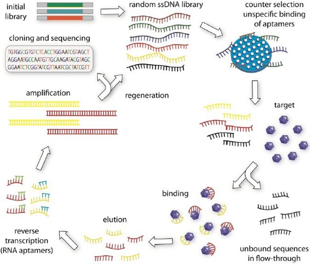
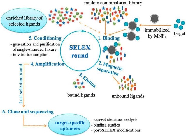
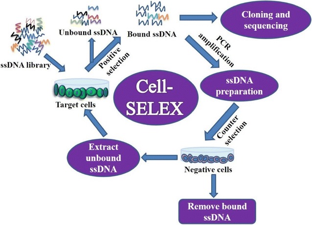
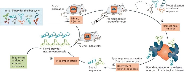
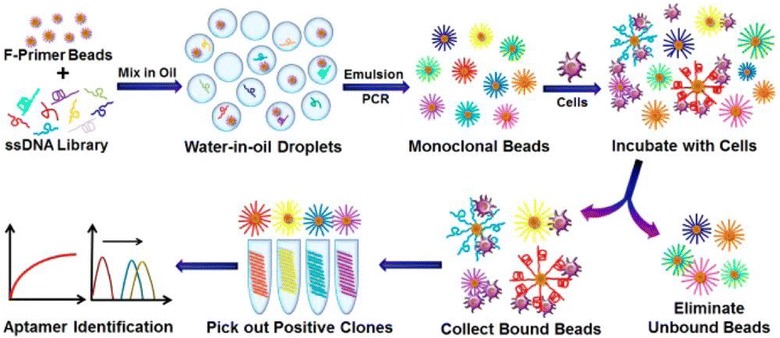
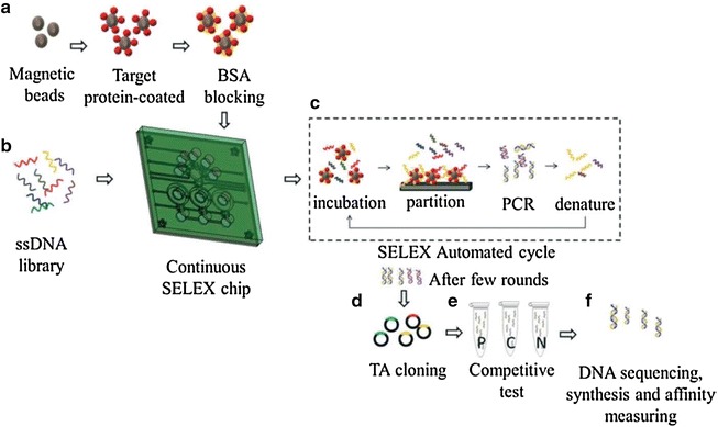
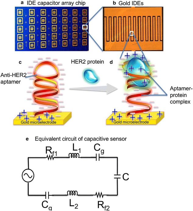
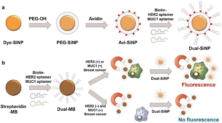
Aptamers are short non-coding, single-stranded oligonucleotides (RNA or DNA) developed through Systematic Evolution of Ligands by Exponential enrichment (SELEX) in vitro. Similar to antibodies, aptamers can bind to specific targets with high affinity, and are considered promising therapeutic agents as they have several advantages over antibodies, including high specificity, stability, and non-immunogenicity. Furthermore, aptamers can be produced at a low cost and easily modified, and are, therefore, called "chemical antibodies". In the past years, a variety of aptamers specifically bound to both breast cancer biomarkers and cells had been selected. Besides, taking advantage of nanomaterials, there were a number of aptamer-nanomaterial conjugates been developed and widely investigated for diagnostics and targeted therapy of breast cancer. In this short review, we first present a systematical review of various aptamer selection methods. Then, various aptamer-based diagnostic and therapeutic strategies of breast cancer were provided. Finally, the current problems, challenges, and future perspectives in the field were thoroughly discussed.

Keywords: Aptamer; Breast cancer; Diagnosis; SELEX; Targeted therapy.


Figures

Fig. 1
Fig. 1
Schematic illustration of conventional SELEX. Usually, an initial random library should be synthesized for selection. Then, counter selection is always needed before incubate the library with the targets to remove non-specific binding sequences. After incubation the target-bound sequeneces are collected and amplificated by PCR method. For selecting DNA aptamers, general PCR is enough for the amplification of the secondary library. While, as for selecting RNA aptamers, reverse transcription PCR is introduced before amplification and preparation of secondary library. After several successive rounds of selection, the library is cloned and sequenced followed by evaluation and identification of the enriched aptamers (Reprinted with permission from Ref. [51]. Copyright © 2015, Elsevier)
Fig. 2
Fig. 2
Schematic illustration of purified-target based SELEX procedures using magnetic particles as solid phase carrier for immobilization of targets (Reprinted with permission from Ref. [65]. Copyright © 2015, American Chemical Society)
Fig. 3
Fig. 3
Schematic illustration of Cell-SELEX procedures
Fig. 4
Fig. 4
Schematic illustration of in vivo SELEX procedures (Reprinted with permission from Ref. [61]. Copyright © 2017, Nature Publishing Group)
Fig. 5
Fig. 5
Schematic illustration of monoclonal surface display SELEX (MSD-SELEX) procedures (Reprinted with permission from Ref. [82]. Copyright © 2014, American Chemical Society)
Fig. 6
Fig. 6
Schematic illustration of microfluidic SELEX procedures (Reprinted with permission from Ref. [85]. Copyright © 2014, Royal Society of Chemistry)
Fig. 7
Fig. 7
Schematic illustration of label-free capacitive aptasensor for HER2 protein detection in serum. a capacitor array chip, b capacitor, c gold IDE functionalized with anti-HER2 aptamer whose phosphodiester backbone contributes abundant negatively charged species on capacitors, d formation of aptamer-HER2 complex that induce charge distribution and thus influence specific changes in capacitance that can be measured and e equivalent circuit of the interdigitated capacitor sensor (Reprinted with permission from Ref. [93]. Copyright © 2015, Elsevier)
Fig. 8
Fig. 8
a Modification processes of the silica nanoparticles. Dye-SiNPs were synthesized via the reverse microemulsion technique, followed by being coated with PEG and avidin immobilization. Then, dual aptamer-functionalized dye-doped silica nanoparticles (Dual-SiNPs) were synthesized by modifying MUC1 aptamer and HER2 aptamer onto Dye-SiNPs through biotin-avidin interactions. b Schematic illustration of Dual-SiNPs for highly sensitive detection of MUC1(+) and HER2(+) breast cancer cells. MUC1(+) or HER2(+) breast cancer cells were selectively enriched and separated by dual aptamer-modified magnetic beads followed by being detected with Dual-SiNPs (Reprinted with permission from Ref. [106]. Copyright © 2015, Elsevier)
Fig. 9
Fig. 9
Schematic illustration of the preparation procedure of the targeted Apt-pD-DTX/NPs (Adapted with permission from Ref. [126]. Copyright © 2016, Ivyspring International Publisher)
Fig. 10
Fig. 10
Schematic illustration of the synthesis and use of aptamer functionalized gold nanorods (Apt-GNRs) for targeted photothermal therapy of breast cancer. First, cetyl trimethylammonium bromide-GNRs (CTAB-GNRs) were synthesized via a seed-mediated approach (1). Secondly, the CTAB was replaced by PEG (2) for functionalizing aptamer (3). Then, the Apt-GNRs were incubated with the targeted breast cancer cells followed by photothermal ablation in near infrared range (4), which eventually caused targeted cell death specifically (5) (Reprinted with permission from Ref. [130]. Copyright © 2015, Royal Society of Chemistry)

Similar articles

Cited by

References

    1. DeSantis C, Ma J, Bryan L, Jemal A. Breast cancer statistics, 2013. CA Cancer J Clin. 2014;64:52–62. doi: 10.3322/caac.21203. - DOI - PubMed
    1. Liu M, Li Z, Yang J, Jiang Y, Chen Z, Ali Z, He N, Wang Z. Cell-specific biomarkers and targeted biopharmaceuticals for breast cancer treatment. Cell Prolif. 2016;49:409–420. doi: 10.1111/cpr.12266. - DOI - PMC - PubMed
    1. Torre LA, Bray F, Siegel RL, Ferlay J, Lortet-Tieulent J, Jemal A. Global cancer statistics, 2012. CA Cancer J Clin. 2015;65:87–108. doi: 10.3322/caac.21262. - DOI - PubMed
    1. Azim HA, Ibrahim AS. Breast cancer in Egypt, China and Chinese: statistics and beyond. J Thorac Dis. 2014;6:864–866. - PMC - PubMed
    1. DeSantis CE, Fedewa SA, Goding Sauer A, Kramer JL, Smith RA, Jemal A. Breast cancer statistics, 2015: convergence of incidence rates between black and white women. CA Cancer J Clin. 2016;66:31–42. doi: 10.3322/caac.21320. - DOI - PubMed

MeSH terms