. 2017 Nov 13;15(1):46.
doi: 10.1186/s12964-017-0201-2.

A transwell assay that excludes exosomes for assessment of tunneling nanotube-mediated intercellular communication

Affiliations

A transwell assay that excludes exosomes for assessment of tunneling nanotube-mediated intercellular communication

Venugopal Thayanithy et al. Cell Commun Signal. .

Abstract

Background: Tunneling nanotubes (TNTs) are naturally-occurring filamentous actin-based membranous extensions that form across a wide spectrum of mammalian cell types to facilitate long-range intercellular communication. Valid assays are needed to accurately assess the downstream effects of TNT-mediated transfer of cellular signals in vitro. We recently reported a modified transwell assay system designed to test the effects of intercellular transfer of a therapeutic oncolytic virus, and viral-activated drugs, between cells via TNTs. The objective of the current study was to demonstrate validation of this in vitro approach as a new method for effectively excluding diffusible forms of long- and close-range intercellular transfer of intracytoplasmic cargo, including exosomes/microvesicles and gap junctions in order to isolate TNT-selective cell communication.

Methods: We designed several steps to effectively reduce or eliminate diffusion and long-range transfer via these extracellular vesicles, and used Nanoparticle Tracking Analysis to quantify exosomes following implementation of these steps.

Results: The experimental approach outlined here effectively reduced exosome trafficking by >95%; further use of heparin to block exosome uptake by putative recipient cells further impeded transfer of these extracellular vesicles.

Conclusions: This validated assay incorporates several steps that can be taken to quantifiably control for extracellular vesicles in order to perform studies focused on TNT-selective communication.

Keywords: Exosomes; Extracellular vesicles; Intercellular communication; Intercellular transfer; Membrane nanotubes; Microvesicles; Transwell assay; Tunneling nanotubes.


Conflict of interest statement

Consent for publication

All authors have reviewed the final manuscript prior to submission and consented to its publication.

Competing interests

The authors declare that they have no competing interests.

Publisher’s Note

Springer Nature remains neutral with regard to jurisdictional claims in published maps and institutional affiliations.

Figures
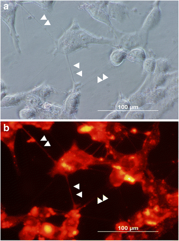
Fig. 1
Fig. 1
Representative images of TNT formation of MSTO-211H malignant pleural mesothelioma cells in open culture (6-well standard culture plates). TNTs connecting DiI-stained MSTO cells are marked by white arrowheads. MSTO cells were first grown using standard passage medium (10% FCS RPMI-1640), then switched to a low-serum, hyperglycemic RPMI-1640 culture medium, stained with lipophilic DiI, and imaged after 48 h on a Olympus X70 fluorescent microscope at 600× magnification using phase contrast (a) and fluorescent (b) settings
Fig. 2
Fig. 2
Confocal microscopy demonstrating TNTs traversing the pores of a transwell polyester membrane filter. MSTO cells were primed in TNT medium (low-serum, hyperglycemic medium) for 7 days prior to plating. To reduce exosomal trafficking, culture medium was removed 24 h prior to the experiment; cells were washed with PBS, and serum-free medium was added. Cells were stained with DiI prior to placement for culture on the top and bottom filters of a modified Boyden chamber encompassing a polyester filter containing pores measuring 400 nm in diameter. a DiI-stained cells with TNTs are shown on the left-hand panel. Comparison of MSTO cells and 400 nm pores is shown in the right-hand panel, with demonstration of Transmitted Detection (TD) imaging. Cells are marked by yellow cross bars and a pore is marked by an arrow head. b XZ view of Z-stacked confocal images of MSTO cells and associated TNTs/TNT-like structures crossing the membrane filter (indicated by arrowheads)
Fig. 3
Fig. 3
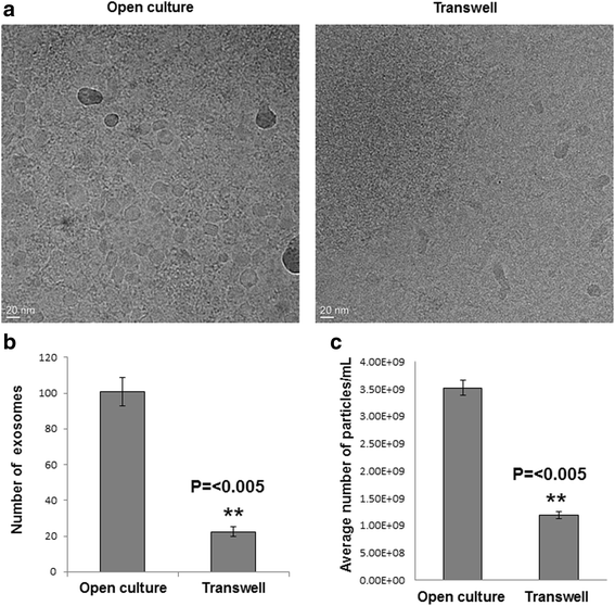
Transwell polyester membrane filters containing 400 nm-sized pores form a physical barrier that significantly reduces transfer of exosomes in the transwell assay. a Cryo-transmission electron microscopic (TEM) examination of exosomal transfer across a transwell assay membrane filter. TEM was performed on exosomes isolated in open culture wells (positive control, left) and the bottom transwell chamber (right) after 48 h of culture in serum-free media using the modifications described. b Quantification of exosomes transmitted to the bottom well of transwell chamber experiments, compared to exosomes in the open culture control. Exosomes were counted from 3 representative images per experiment and averaged. The relative reduction of exosomal trafficking using this transwell filter was ~ 80%, when assessed by using this method. c Nanoparticle tracking analysis of exosomes from above mentioned transwell and open culture experiments, quantifying the relative reduction at 66%. For statistical analysis, Student’s t-test was conducted, with a p-value of ≤0.05
Fig. 4
Fig. 4
Modifications to culture conditions can further reduce exosome carryover to the lower transwell chamber, beyond the physical barrier provided by the transmembrane filter. “Restrictive media” conditions refer to washing cells and adding basal, serum-free (mTeSR) medium to avoid addition of exogenous serum-based exosomes. Nanoparticle Tracking Analysis (NTA) was used to determine the concentration of exosomes isolated following usual culture and passage conditions (a) as compared to concentration of exosomes isolated from cells subjected to restrictive medium conditions (b). Mean values from 5 independent runs are shown ± SD. The restrictive conditions reduced exosome contamination by ~75% compared to cells in normal culture medium conditions (c)
Fig. 5
Fig. 5
Assessment of trafficking of exogenously added exosomes: the transwell polyester membrane containing 400 nm-sized pores serves as an effective physical barrier to exosomal trafficking as compared to open culture without barriers separating cells. MSTO cells were prepared under restrictive conditions (using exosome-depleted serum); and then plated and incubated with exogenously added VAMT-derived exosomes (2 × 109) in triplicate for 48 h. After incubation, medium from the bottom chamber was collected and subjected to exosome isolation and NTA as explained in the Methods section. NTA-assessed exosome concentrations (reported as number of particles/ml × 106) after open culture in 6-well plates (a); and after culture in the Boyden chamber transwell experiments (b). In the latter experiment, VAMT-derived exosomes were added to the top of the transwell chamber, and medium was recovered after 48 h from the lower chamber for NTA. The transwell filter with 400 nm pore size can significantly block exosome transfer, independently of other steps (c). Mean values from 5 independent runs are shown ± SD
Fig. 6
Fig. 6
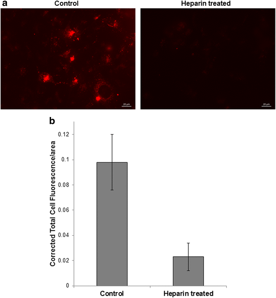
Exosome uptake is effectively blocked by pharmacologic treatment with heparin. a PKH26 (red dye)-labeled VAMT-derived exosomes were added to MSTO cells on the top of the transwell/Boyden chamber; and to the lower chamber, we added MSTO cells pre-treated with or without 10 μg/mL heparin. Extent of exosomal uptake of cells in the lower chamber was analyzed after 24 h for both conditions (scale bar = 20 μm). The reduction in red fluorescence in heparin pre-treated cells indicates efficient blocking of exosome uptake. b This experiment was performed in triplicate; representative images were taken from each replicate and subsequently used for calculations to compare the Corrected Total Cell Fluorescence (CTCF) per area of the control and heparin-treated cells. CTCF/area was calculated for 36 cells in the control group, and 72 cells in the heparin-treated group, and the results were averaged. Our results demonstrate that heparin treatment of recipient cells can significantly block uptake of the majority of remaining exosomes. Mean values are shown with ± standard error. p-value = 0.04
Fig. 7
Fig. 7
Scanning Electron Micrograph (SEM) of TNT-like structures penetrating the 400 nm pores of the transwell membrane. MSTO cells were grown on the transwell membrane culture inserts for 48 h and then fixed using PFA. On the bottom of the membrane, TNT-like structures were identified and imaged without staining. The image provides evidence that TNTs have the capacity to penetrate and traverse the transwell membrane via the 400 nm-sized pores of the polyester filter (scale bar 1000 nm). that formed through the membrane pores, and extended beyond the membrane are shown, marked by arrowheads. Several TNTs were disrupted by the fixation process, but are shown in cross-section (arrows)

References

    1. Biran A, et al. Senescent cells communicate via intercellular protein transfer. Genes Dev. 2015;29:791–802. doi: 10.1101/gad.259341.115. - DOI - PMC - PubMed
    1. Connor Y, et al. Physical nanoscale conduit-mediated communication between tumour cells and the endothelium modulates endothelial phenotype. Nat Commun. 2015;6:8671. doi: 10.1038/ncomms9671. - DOI - PMC - PubMed
    1. Costanzo M, et al. Transfer of polyglutamine aggregates in neuronal cells occurs in tunneling nanotubes. J Cell Sci. 2013;126:3678–3685. doi: 10.1242/jcs.126086. - DOI - PubMed
    1. Gousset K, et al. Prions hijack tunnelling nanotubes for intercellular spread. Nat Cell Biol. 2009;11:328–336. doi: 10.1038/ncb1841. - DOI - PubMed
    1. Langevin C, Gousset K, Costanzo M, Richard-Le Goff O, Zurzolo C. Characterization of the role of dendritic cells in prion transfer to primary neurons. Biochem J. 2010;431:189–198. doi: 10.1042/BJ20100698. - DOI - PubMed

LinkOut - more resources