. 2017 Nov 13;10(1):173.
doi: 10.1186/s13045-017-0540-x.

Kinase profiling of liposarcomas using RNAi and drug screening assays identified druggable targets

Affiliations

Kinase profiling of liposarcomas using RNAi and drug screening assays identified druggable targets

Deepika Kanojia et al. J Hematol Oncol. .

Abstract

Background: Liposarcoma, the most common soft tissue tumor, is understudied cancer, and limited progress has been made in the treatment of metastatic disease. The Achilles heel of cancer often is their kinases that are excellent therapeutic targets. However, very limited knowledge exists of therapeutic critical kinase targets in liposarcoma that could be potentially used in disease management.

Methods: Large RNAi and small-molecule tyrosine kinase inhibitor screens were performed against the proliferative capacity of liposarcoma cell lines of different subtypes. Each small molecule inhibitor was either FDA approved or in a clinical trial.

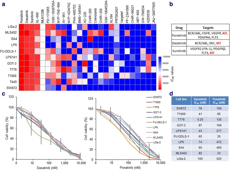
Results: Screening assays identified several previously unrecognized targets including PTK2 and KIT in liposarcoma. We also observed that ponatinib, multi-targeted tyrosine kinase inhibitor, was the most effective drug with anti-growth effects against all cell lines. In vitro assays showed that ponatinib inhibited the clonogenic proliferation of liposarcoma, and this anti-growth effect was associated with apoptosis and cell cycle arrest at the G0/G1 phase as well as a decrease in the KIT signaling pathway. In addition, ponatinib inhibited in vivo growth of liposarcoma in a xenograft model.

Conclusions: Two large-scale kinase screenings identified novel liposarcoma targets and a FDA-approved inhibitor, ponatinib with clear anti-liposarcoma activity highlighting its potential therapy for treatment of this deadly tumor.

Keywords: Kinase inhibitor; Liposarcoma; Ponatinib; Therapeutics.


Conflict of interest statement

Ethics approval

All animal experiments were performed according to the ethical regulations of Institutional Animal Care and Use Committee of the National University of Singapore.

Consent for publication

Not applicable

Competing interests

The authors have declared a conflict of interest. Research support for JWT receive from Aptose, Array, AstraZeneca, Constellation, Genentech, Gilead, Incyte, Janssen, Seattle Genetics, Syros, Takeda, and the Scientific Advisory Board for Leap Oncology.

Publisher’s Note

Springer Nature remains neutral with regard to jurisdictional claims in published maps and institutional affiliations.

Figures

Fig. 1
Fig. 1
Kinase siRNA screen of liposarcoma cell lines and kinase target validation. a Cell viability plot of LPS141 and MLS402 cells after 96 h post-transfection with the siRNA library. Results represent mean values compared to non-specific control. Blue bars represent viability more than 70% of control and P > 0.05. Red bars indicate viability less than 70% of control and P < 0.05. Black bars are non-specific pooled siRNA controls. b Validation of PTK2 and KIT as target kinase using siRNA and shRNA knockdown in LPS141 and MLS402 cells. Cell viability evaluated by MTT assay at different time points. Western blotting showed reduced protein levels of PTK2 and KIT after 72 h of siRNA transfection and shRNA infection. c Cell viability analysis at different time points of PTK2 and KIT CRISPR knockout LPS141 and MLS402 cells, respectively, along with LacZ sgRNA knockout control cells. Western blotting confirmed that CRISPR sgRNA1 and sgRNA2 significantly silenced protein expression in the respective cells compared to LacZ sgRNA (α-tubulin, loading control). Experiments were done in triplicates, 3 times and shown as mean ± SE. *P value < 0.01
Fig. 2
Fig. 2
Heat map of sensitivity of liposarcoma cell lines to small molecule inhibitors. Liposarcoma cells are grouped according to histotypes (UDLPS undifferentiated liposarcoma, WDLPS well-differentiated liposarcoma, DDLPS dedifferentiated liposarcoma, MLPS myxoid liposarcoma, and PLPS pleomorphic liposarcoma), and inhibitors are grouped into related target families. Median IC50 values (0–10,000 nM) for every drug and each cell line is represented from most sensitive (dark red) to most resistant (dark blue) as shown in color bars. Solid arrow demarks ponatinib
Fig. 3
Fig. 3
Anti-proliferative effects of receptor tyrosine kinase inhibitors against liposarcoma cells. a Heat map of IC50 values of receptor tyrosine kinase inhibitors arranged according to their cytotoxic efficacy (results derived from Fig. 1). Color bars indicate most sensitive (dark red) to most resistant (dark blue) cell lines. b The table shows top three most potent inhibitors (ponatinib, dasatinib, and sunitinib) and their known primary targets indicating KIT as a common target (highlighted in red). c Individual dose-response curves (MTT assay) of liposarcoma cell lines to dasatinib and ponatinib at different concentrations for 3 days. Experiments were done in triplicates, repeated 3 times and represent the mean ± SE. d IC50 values were calculated from graphs shown in c
Fig. 4
Fig. 4
Ponatinib treatment inhibits KIT signaling and clonogenic growth of liposarcoma cells. a Ponatinib (250–1000 nM, 16 h)-treated liposarcoma cell lines LPS141 (well-differentiated) and MLS402 (myxoid) were analyzed for KIT signaling pathway by western blotting. β-actin and GAPDH served as loading controls. b Anchorage-dependent colony formation assay of LPS141 and MLS402 cells treated with either ponatinib (250–1000 nM, 10 days) or diluent control. Colonies were stained with crystal violet (microscope images in left panel) and quantification of staining intensity is shown by bar graphs (right panel). c Soft agar colony formation (anchorage-independent) of LPS141 and MLS402 cells cultured with either ponatinib (250–1000 nM, 21 days) or diluent control. Number of colonies shown as bar graphs. Experiments were done in triplicates and repeated 3 times. *P value ≤ 0.01
Fig. 5
Fig. 5
Ponatinib causes cell cycle arrest and apoptosis of liposarcoma cells. a Flow cytometric cell cycle analysis of ponatinib (1000 nM, 16 h)-treated LPS141 and MLS402 liposarcoma cells compared to diluent control. Bar graphs show the percentage of cells in different phases of the cell cycle. Experiments were done in triplicates, repeated 3 times and results represent mean ± SE. b Western blot analysis of cell cycle-related proteins in LPS141 and MLS402 cells treated with ponatinib (250–1000 nM, 16 h) or diluent control (β-actin, loading control). c Flow cytometric Annexin V-APC/Propidium iodide staining of LPS141 and MLS402 cells treated with ponatinib (1000 nM, 16 h). Percentage of Annexin V+/PI+ cells (apoptotic cells) of ponatinib- and diluent control-treated cells shown in bar graphs. Experiments were done in triplicates, repeated 3 times and results represent mean ± SE. *P value ≤ 0.01. d Proteins associated with growth and apoptosis were analyzed by western blotting in liposarcoma cells treated with either ponatinib or diluent control (GAPDH, loading control)
Fig. 6
Fig. 6
Ponatinib inhibits growth of liposarcoma xenografts. a LPS141 cells (2 × 106) mixed with equal volume of matrigel were injected subcutaneously in the flank of NSG mice. When tumors reached ~ 100 mm3, mice were randomly divided into two groups [experimental (n = 9) and control (n = 0)]. Experimental group received daily ponatinib (10 mg/kg), and control group received an equal volume of the vehicle, both by oral gavage for 21 days. Images of dissected tumors from mice of both groups are shown. b Bar graphs of tumor weights from vehicle- and ponatinib-treated mice. Results represent mean ± standard deviation. *P value ≤ 0.01. c Immunohistochemical staining for Ki-67 (proliferation marker) on tumor sections of mice treated with either vehicle or ponatinib. d Western blot analysis of phosphorylated and total protein levels of KIT and FGFR in the xenograft tumors of vehicle- and ponatinib-treated mice (α-tubulin, loading control). e Apoptotic- and growth-associated proteins examined by western blotting from tumor lysates of the xenografts treated with either ponatinib or vehicle (α-tubulin, loading control)

Similar articles

Cited by

References

    1. Clark MA, Fisher C, Judson I, Thomas JM. Soft-tissue sarcomas in adults. N Engl J Med. 2005;353:701–711. doi: 10.1056/NEJMra041866. - DOI - PubMed
    1. Fletcher CD. The evolving classification of soft tissue tumours—an update based on the new 2013 WHO classification. Histopathology. 2014;64:2–11. doi: 10.1111/his.12267. - DOI - PubMed
    1. Conyers R, Young S, Thomas DM. Liposarcoma: molecular genetics and therapeutics. Sarcoma. 2011;2011:483154. doi: 10.1155/2011/483154. - DOI - PMC - PubMed
    1. Dalal KM, Antonescu CR, Singer S. Diagnosis and management of lipomatous tumors. J Surg Oncol. 2008;97:298–313. doi: 10.1002/jso.20975. - DOI - PubMed
    1. Demetri GD, von Mehren M, Jones RL, Hensley ML, Schuetze SM, Staddon A, Milhem M, Elias A, Ganjoo K, Tawbi H, et al. Efficacy and safety of trabectedin or dacarbazine for metastatic liposarcoma or leiomyosarcoma after failure of conventional chemotherapy: results of a phase III randomized multicenter clinical trial. J Clin Oncol. 2016;34:786–793. doi: 10.1200/JCO.2015.62.4734. - DOI - PMC - PubMed