

Prefrontal and Striatal Gamma-Aminobutyric Acid Levels and the Effect of Antipsychotic Treatment in First-Episode Psychosis Patients
- PMID: 29132653
- PMCID: PMC5809278
- DOI: 10.1016/j.biopsych.2017.09.028
Prefrontal and Striatal Gamma-Aminobutyric Acid Levels and the Effect of Antipsychotic Treatment in First-Episode Psychosis Patients
Abstract
Background: Abnormally elevated levels of gamma-aminobutyric acid (GABA) in the medial prefrontal cortex (mPFC) have been reported in antipsychotic-free patients with schizophrenia. Whether such GABA elevations are also present in other brain regions and persist after antipsychotic treatment has not been previously investigated.
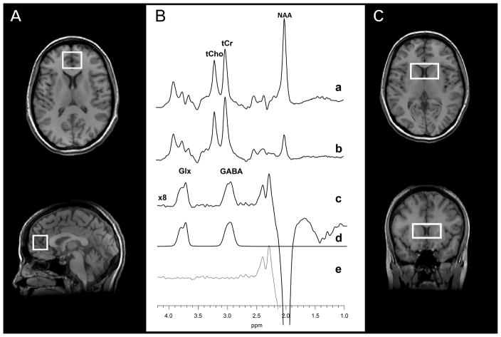
Methods: Twenty-eight antipsychotic-naïve patients with first-episode psychosis (FEP) and 18 healthy control subjects completed the study. Following baseline proton magnetic resonance spectroscopy scans targeting the mPFC and a second region, the dorsal caudate, patients with FEP were treated with oral risperidone for 4 weeks at an initial dose of 1 mg/day that was titrated as necessary based on clinical judgment. After the 4-week treatment period, both groups were brought back to undergo outcome magnetic resonance spectroscopy scans, which were identical to the scans conducted at baseline.
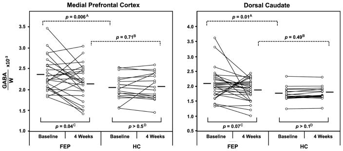
Results: At baseline, higher GABA levels were found both in the mPFC and in the dorsal caudate of patients with FEP compared with healthy control subjects. Following 4 weeks of antipsychotic treatment, GABA levels in patients with FEP decreased relative to baseline in the mPFC, but decreased only at the trend level relative to baseline in the dorsal caudate. For either brain region, GABA levels at 4 weeks or posttreatment did not differ between patients with FEP and healthy control subjects.
Conclusions: The results of the present study documented elevations of GABA levels both in the mPFC and, for the first time, in the dorsal caudate of antipsychotic-naïve patients with FEP, which normalized in both regions following 4 weeks of antipsychotic treatment.
Keywords: Antipsychotic treatment; First-episode; GABA; Magnetic resonance spectroscopy; Psychosis; Schizophrenia.
Copyright © 2017 Society of Biological Psychiatry. Published by Elsevier Inc. All rights reserved.
Figures
Comment in
-
Magnetic Resonance Spectroscopy Gamma-Aminobutyric Acid: A Promising Biomarker for Antipsychotic Treatment?Biol Psychiatry. 2018 Mar 15;83(6):468-469. doi: 10.1016/j.biopsych.2018.01.004. Biol Psychiatry. 2018. PMID: 29429499 No abstract available.
References
-
- Akbarian S, Kim JJ, Potkin SG, Hagman JO, Tafazzoli A, Bunney WE, Jr, et al. Gene expression for glutamic acid decarboxylase is reduced without loss of neurons in prefrontal cortex of schizophrenics. Arch Gen Psychiatry. 1995;52:258–266. - PubMed
-
- Benes FM, Berretta S. GABAergic interneurons: implications for understanding schizophrenia and bipolar disorder. Neuropsychopharmacology. 2001;25:1–27. - PubMed
-
- Tse MT, Piantadosi PT, Floresco SB. Prefrontal cortical gamma-aminobutyric acid transmission and cognitive function: drawing links to schizophrenia from preclinical research. Biol Psychiatry. 2015;77:929–939. - PubMed
Publication types
MeSH terms
Substances
Grants and funding
LinkOut - more resources
Full Text Sources
Other Literature Sources
Medical
