. 2017:82:111-121.
doi: 10.1101/sqb.2017.82.033712. Epub 2017 Nov 13.

Taming the Beast: Control of APC/CCdc20-Dependent Destruction

Affiliations

Taming the Beast: Control of APC/CCdc20-Dependent Destruction

Pablo Lara-Gonzalez et al. Cold Spring Harb Symp Quant Biol. 2017.

Abstract

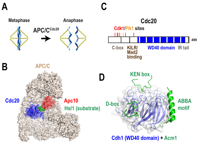
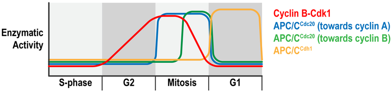
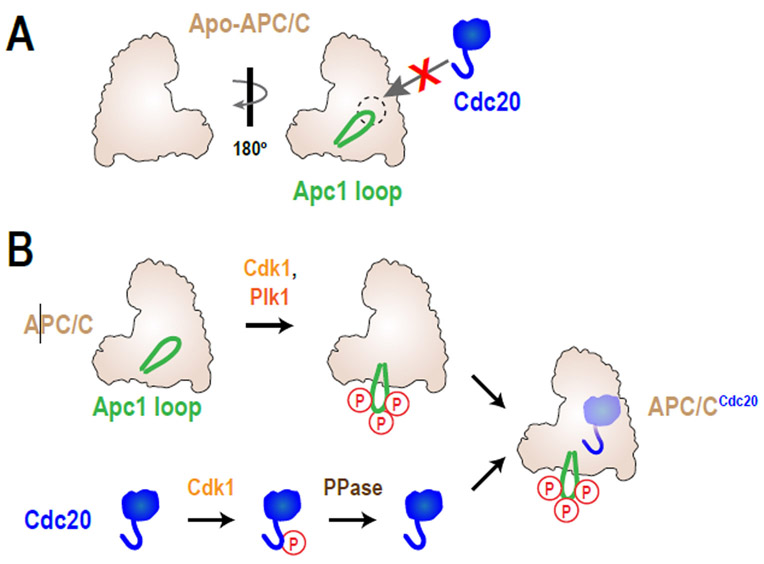
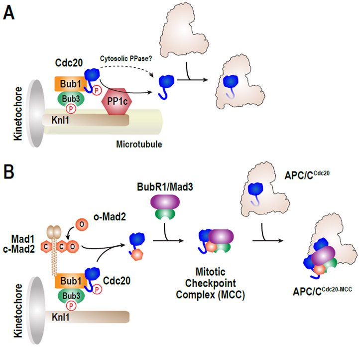
The anaphase-promoting complex/cyclosome (APC/C) is a large multisubunit ubiquitin ligase that triggers the metaphase-to-anaphase transition in the cell cycle by targeting the substrates cyclin B and securin for destruction. APC/C activity toward these two key substrates requires the coactivator Cdc20. To ensure that cells enter mitosis and partition their duplicated genome with high accuracy, APC/CCdc20 activity must be tightly controlled. Here, we discuss the mechanisms that regulate APC/CCdc20 activity both before and during mitosis. We focus our discussion primarily on the chromosomal pathways that both accelerate and delay APC/C activation by targeting Cdc20 to opposing fates. The findings discussed provide an overview of how cells control the activation of this major cell cycle regulator to ensure both accurate and timely cell division.


Figures

Figure 1.
Figure 1.. APC/C structure and mechanism of substrate recognition.
(A) Cartoon illustrating the metaphase-to-anaphase transition, which is promoted by APC/CCdc20 activity. Microtubules are in yellow, chromosomes in blue and kinetochores in grey. (B) Structure of APC/CCdc20 bound to a D-box-containing substrate, Hsl1 (Zhang et al. 2016). The substrate binds to the interphase between Cdc20 and the APC/C subunit Apc10 (adapted from Corbett 2017). (C) Schematic illustrating the domains in human Cdc20. The C-box, KILR and IR tail motifs contribute to APC/C binding, whereas the WD40 domain is involved in substrate recognition. Inhibitory Cdk1 phosphorylation sites are shown in red, whereas S92, which is phosphorylated by Plk1, is in orange. Note that the KILR motif is also the Mad2 interacting motif. (D) Structure of the WD40 domain of S.cerevisiae Cdh1 bound to an inhibitor, Acm1 (He et al. 2013). The structure shows the interaction sites for the three APC/C degrons: D-box, KEN box and ABBA motif (adapted from Corbett 2017).
Figure 2.
Figure 2.. Regulation of APC/C activity during the cell cycle.
Schematic illustrates the current model for the temporal regulation of the activities of Cdk1-Cyclin B (red), APC/CCdh1 (orange) and APC/CCdc20 towards cyclin A (blue) or cyclin B (green).
Figure 3.
Figure 3.. Control of APC/CCdc20 activity by phosphorylation.
(A) Schematic of apo-APC/C, showing auto-inhibition by the Apc1 loop. (B) The Cdk1 and Plk1 kinases phosphorylate the Apc1 loop, which releases the APC/C auto-inhibition mechanism. At the same time, Cdk1 phosphorylates the N-terminal tail of Cdc20, which prevents its interaction with the APC/C. De-phosphorylation of Cdc20 by phosphatases (PPase) would cause its activation and binding to the APC/C.
Figure 4.
Figure 4.. The two fates of Cdc20 at kinetochores.
During mitosis, Cdc20 is recruited to kinetochores by Bub1/Bub3, which is bound to phospho-Knl1. (A) When kinetochores are attached by microtubules, kinetochores promote Cdc20 de-phosphorylation by kinetochore-localized PP1c, which allows its activation. Cdc20 may also be dephosphorylated at the cytosol, likely through PP2A-B56. (B) When microtubules are unattached, signal from the spindle assembly checkpoint catalyzes the incorporation of Cdc20 into the mitotic checkpoint complex (MCC), which binds and inhibits APC/CCdc20 activity. See text for more details.

Similar articles

Cited by

References

    1. Alfieri C, Chang L, Zhang Z, Yang J, Maslen S, Skehel M, Barford D. 2016. Molecular basis of APC/C regulation by the spindle assembly checkpoint. Nature 536: 431–436. - PMC - PubMed
    1. Araki M, Yu H, Asano M. 2005. A novel motif governs APC-dependent degradation of Drosophila ORC1 in vivo. Genes Dev 19: 2458–2465. - PMC - PubMed
    1. Aravamudhan P, Goldfarb AA, Joglekar AP. 2015. The kinetochore encodes a mechanical switch to disrupt spindle assembly checkpoint signalling. Nat Cell Biol 17: 868–879. - PMC - PubMed
    1. Aristarkhov A, Eytan E, Moghe A, Admon A, Hershko A, Ruderman JV. 1996. E2-C, a cyclin-selective ubiquitin carrier protein required for the destruction of mitotic cyclins. Proc Natl Acad Sci U S A 93: 4294–4299. - PMC - PubMed
    1. Barford D 2015. Understanding the structural basis for controlling chromosome division. Philos Trans A Math Phys Eng Sci 373. - PMC - PubMed