. 2018 Jan 22;86(2):e00387-17.
doi: 10.1128/IAI.00387-17. Print 2018 Feb.

Mycobacterium tuberculosis GroEL2 Modulates Dendritic Cell Responses

Affiliations

Mycobacterium tuberculosis GroEL2 Modulates Dendritic Cell Responses

Maria Georgieva et al. Infect Immun. .

Abstract

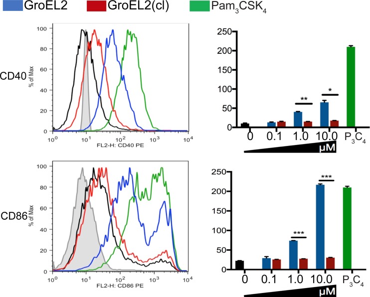
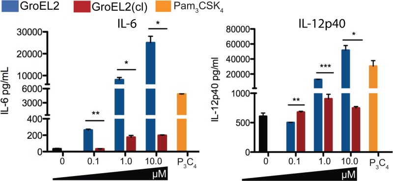
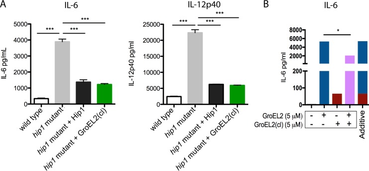
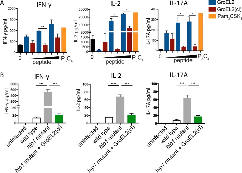
Mycobacterium tuberculosis successfully subverts the host immune response to promote disease progression. In addition to its known intracellular niche in macrophages, M. tuberculosis interferes with the functions of dendritic cells (DCs), which are the primary antigen-presenting cells of the immune system. We previously showed that M. tuberculosis dampens proinflammatory responses and impairs DC functions through the cell envelope-associated serine protease Hip1. Here we present data showing that M. tuberculosis GroEL2, a substrate of Hip1, modulates DC functions. The full-length GroEL2 protein elicited robust proinflammatory responses from DCs and promoted DC maturation and antigen presentation to T cells. In contrast, the cleaved form of GroEL2, which predominates in M. tuberculosis, was poorly immunostimulatory and was unable to promote DC maturation and antigen presentation. Moreover, DCs exposed to full-length, but not cleaved, GroEL2 induced strong antigen-specific gamma interferon (IFN-γ), interleukin-2 (IL-2), and IL-17A cytokine responses from CD4+ T cells. Moreover, the expression of cleaved GroEL2 in the hip1 mutant restored the robust T cell responses to wild-type levels, suggesting that proteolytic cleavage of GroEL2 allows M. tuberculosis to prevent optimal DC-T cell cross talk during M. tuberculosis infection.

Keywords: Mycobacterium tuberculosis; dendritic cells.


Figures

FIG 1
FIG 1
Expression of costimulatory molecules CD40 and CD86 on DCs in response to full-length GroEL2 and GroEL2(cl). We stimulated C57BL/6 BMDCs with recombinant GroEL2 or GroEL2(cl) for 24 h and analyzed the cell surface expression of CD40 and CD86. Representative histograms and mean fluorescence intensity values for the CD11c+ DC subpopulation are shown. Isotype and Pam3CSK4 controls are shown as gray and green outlines, respectively. Data are shown as means ± SD of results of one representative experiment from three independent experiments.
FIG 2
FIG 2
Differential stimulation of proinflammatory cytokines from dendritic cells by GroEL2 and GroEL2(cl). We measured levels of the IL-6 and IL-12p40 cytokines produced by C57BL/6 BMDCs 24 h after stimulation with various levels of recombinant GroEL2 or GroEL2(cl). Data are shown as means ± SD of results from one representative experiment of three independent experiments.
FIG 3
FIG 3
GroEL2(cl) restores wild-type levels of proinflammatory cytokine responses in dendritic cells and dampens the stimulatory capacity of full-length GroEL2. (A) We measured levels of the IL-6 and IL-12p40 cytokines produced by C57BL/6 BMDCs 24 h after infection with live M. tuberculosis strains. (B) We incubated C57BL/6 BMDCs with recombinant GroEL2 and GroEL2(cl) either alone (5 μM) or together (5 μM each) for 24 h. The calculated additive effect of GroEL2 and GroEL2(cl) is represented as a sum of the cytokine levels for each protein alone. Data are shown as means ± SD of results of one representative experiment from three independent experiments.
FIG 4
FIG 4
GroEL2 proteolysis modulates DC antigen presentation and T cell polarization. (A) We pulsed DCs with various amounts of the OVA323–339 peptide (1 μg/ml, 10 μg/ml, and 50 μg/ml) for 6 h and then stimulated cells with either recombinant GroEL2 or GroEL2(cl) for 24 h. Following coculture with antigen-specific TCR-Tg CD4+ T cells for 72 h, we assayed cells for the cytokines IFN-γ, IL-2, and IL-17A by an ELISA. (B) We infected DCs with live M. tuberculosis strains and then cocultured them with purified ESAT-6-specific TCR-Tg CD4+ T cells. After 80 h, cell-free supernatants were collected and assessed for the cytokines IFN-γ, IL-2, and IL-17A by an ELISA. Data are shown as means ± SD of results of one representative experiment from three independent experiments.

Similar articles

Cited by

References

    1. Ehrt S, Schnappinger D. 2009. Mycobacterial survival strategies in the phagosome: defence against host stresses. Cell Microbiol 11:1170–1178. doi:10.1111/j.1462-5822.2009.01335.x. - DOI - PMC - PubMed
    1. Philips JA, Ernst JD. 2012. Tuberculosis pathogenesis and immunity. Annu Rev Pathol 7:353–384. doi:10.1146/annurev-pathol-011811-132458. - DOI - PubMed
    1. Russell DG. 2001. Mycobacterium tuberculosis: here today, here tomorrow. Nat Rev Microbiol 2:569–577. doi:10.1038/35084527. - DOI - PubMed
    1. Wolf AJ, Linas B, Trevejo-Nunez GJ, Kincaid E, Tamura T, Takatsu K, Ernst JD. 2007. Mycobacterium tuberculosis infects dendritic cells with high frequency and impairs their function in vivo. J Immunol 179:2509–2519. doi:10.4049/jimmunol.179.4.2509. - DOI - PubMed
    1. Hanekom WA, Mendillo M, Manca C, Haslett PAJ, Siddiqui MR, Barry C III, Kaplan G. 2003. Mycobacterium tuberculosis inhibits maturation of human monocyte-derived dendritic cells in vitro. J Infect Dis 188:257–266. doi:10.1086/376451. - DOI - PubMed

Publication types

LinkOut - more resources