. 2018 Jan;176(1):552-565.
doi: 10.1104/pp.17.00755. Epub 2017 Nov 13.

A Mucin-Like Protein of Planthopper Is Required for Feeding and Induces Immunity Response in Plants

Affiliations

A Mucin-Like Protein of Planthopper Is Required for Feeding and Induces Immunity Response in Plants

Xinxin Shangguan et al. Plant Physiol. 2018 Jan.

Abstract

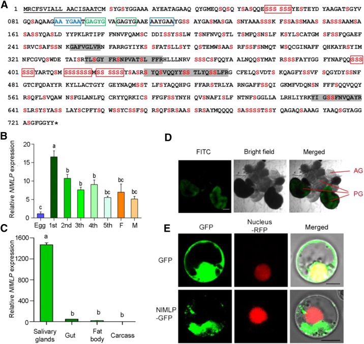
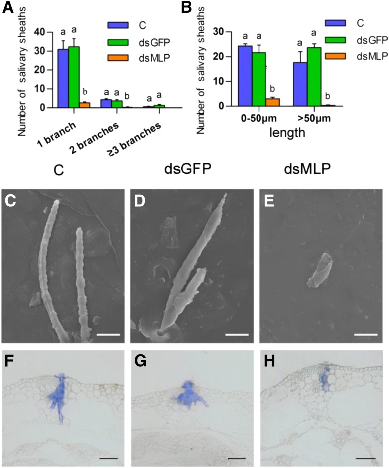
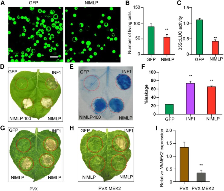
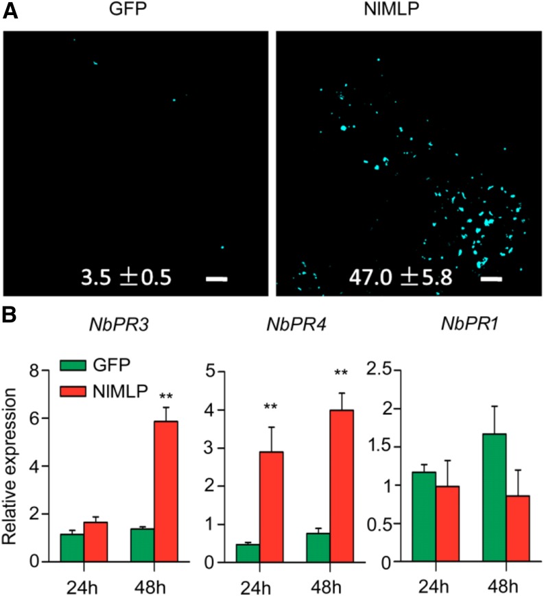
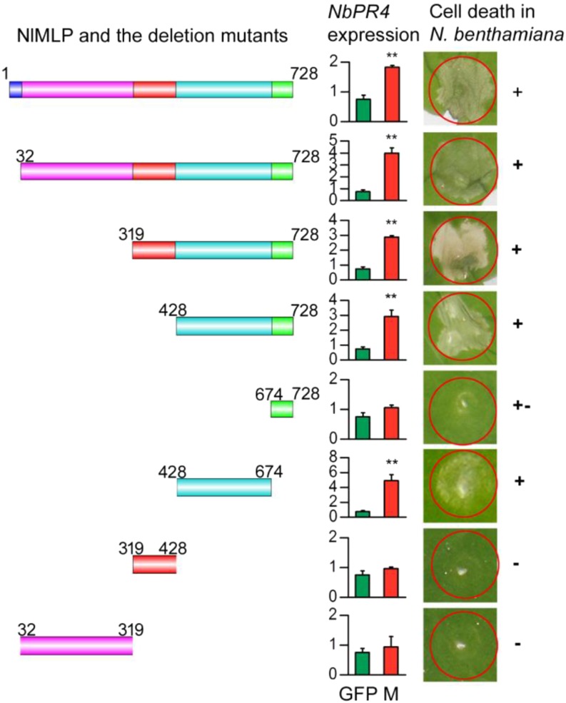
The brown planthopper, Nilaparvata lugens, is a pest that threatens rice (Oryza sativa) production worldwide. While feeding on rice plants, planthoppers secrete saliva, which plays crucial roles in nutrient ingestion and modulating plant defense responses, although the specific functions of salivary proteins remain largely unknown. We identified an N. lugens-secreted mucin-like protein (NlMLP) by transcriptome and proteome analyses and characterized its function, both in brown planthopper and in plants. NlMLP is highly expressed in salivary glands and is secreted into rice during feeding. Inhibition of NlMLP expression in planthoppers disturbs the formation of salivary sheaths, thereby reducing their performance. In plants, NlMLP induces cell death, the expression of defense-related genes, and callose deposition. These defense responses are related to Ca2+ mobilization and the MEK2 MAP kinase and jasmonic acid signaling pathways. The active region of NlMLP that elicits plant responses is located in its carboxyl terminus. Our work provides a detailed characterization of a salivary protein from a piercing-sucking insect other than aphids. Our finding that the protein functions in plant immune responses offers new insights into the mechanism underlying interactions between plants and herbivorous insects.


Figures

Figure 1.
Figure 1.
Molecular characterization of NlMLP. A, Amino acid sequence of NlMLP. The solid underline indicates the signal peptide predicted by SignalP. The asterisk indicates the stop codon. The shaded amino acid residues indicate the peptides detected in BPH-infested rice tissue by liquid chromatography-tandem mass spectrometry analyses. Ser is shown in red. The repeat region is boxed. B and C, Expression patterns of NlMLP at different developmental stages (B) and in different tissues (C). Relative levels of NlMLP expression at the indicated developmental stages (1st to 5th, first to fifth instar; F, female adult; M, male adult) and in different tissues (salivary gland, gut, fat body, and remaining carcass) were normalized against β-actin gene expression, as determined by qRT-PCR. Data represent means ± se of three repeats; n = 30. Different letters above the bars indicate significant differences, as determined by Tukey’s honestly significant difference test (P < 0.05). D, mRNA in situ hybridization of NlMLP in salivary glands. Signals from anti-digoxigenin-fluorescein isothiocyanate (FITC; shown in green) bound to a digoxigenin-labeled antisense riboprobe used for hybridization to NIMLP transcripts were detected by confocal laser-scanning microscopy. AG, Accessory gland; PG, principal gland. Bar = 100 μm. E, NlMLP localizes to the cytoplasm of rice cells when transiently expressed. GFP or NlMLP-GFP fusion protein was expressed in rice protoplasts by polyethylene glycol-mediated transformation. Confocal laser-scanning microscopy was used to investigate fusion protein distribution 16 h after transformation. Bars = 5 μm.
Figure 2.
Figure 2.
Effects of NlMLP silencing on BPH feeding and performance. A, The survival rates of BPH insects after injection were monitored daily. The survival rates of BPHs injected with dsMLP were reduced significantly compared with those of BPHs with no injection and those injected with dsGFP after 2 d of treatment (Student’s t test). C, BPHs with no injection; dsGFP, BPHs injected with GFP-dsRNA; dsMLP, BPHs injected with NlMLP-dsRNA. The experiment was repeated five times with 10 BPHs per replicate. Data represent means ± sd of five repeats. B, Honeydew excretion by BPH insects on filter paper. The intensity of the honeydew color and area of the honeydew correspond to BPH feeding activity. The experiment was repeated three times with 10 filter papers per replicate. Data represent means ± se of three repeats. Different letters above the bars indicate significant differences, as determined by Tukey’s honestly significant difference test (P < 0.05). C and D, BPH weight gain (C) and BPH weight gain ratio (D) of BPHs after injection with dsGFP or dsMLP. Data represent means ± se of five independent experiments with 10 BPHs per replicate. Different letters above the bars indicate significant differences, as determined by Tukey’s honestly significant difference test (P < 0.05). E, Survival rates of BPH insects feeding on NlMLP-dsRNA-transgenic plants (SG33-2) and wild-type plants (WT). The experiment was repeated five times with 20 BPHs per replicate. Data represent means ± sd of five repeats. Asterisks above the columns indicate significant differences compared with wild-type plants (*, P < 0.05 and **, P < 0.01, Student's t test). F, BPH weight after feeding for 10 d on NlMLP-dsRNA-transgenic plants (SG33-2) and wild-type plants. The experiment was repeated five times with 10 BPHs per replicate. Data represent means ± se of five repeats. The asterisk above one column indicates a significant difference compared with wild-type plants (*, P < 0.05, Student’s t test).
Figure 3.
Figure 3.
Effects of NlMLP silencing on salivary sheath formation. A and B, Distribution of branch number (A) and length (B) of salivary sheaths formed by BPHs on an artificial diet. BPH insects were fed with dietary Suc for 48 h, and the salivary sheaths were collected and counted using a fluorescence microscope. C, Noninjected BPHs; dsGFP, BPHs injected with GFP-dsRNA; dsMLP, BPHs injected with NlMLP-dsRNA. Data represent means ± se of three repeats. Different letters above the bars indicate significant differences, as determined by Tukey’s honestly significant difference test (P < 0.05). C to E, Scanning electron micrographs showing the morphology of salivary sheaths formed by BPHs on an artificial diet. Salivary sheaths from noninjected BPHs (C) show a complete structure. BPHs injected with GFP-dsRNA (D) formed similar types of sheaths. BPHs injected with NlMLP-dsRNA (E) produced abnormal salivary sheaths. Bars = 20 μm. F to H, Fluorescence microscopy images showing the morphology of salivary sheaths in rice plants. Rice plants were infested with BPH insects for 48 h and investigated by paraffin sectioning. Sheaths produced by noninjected BPHs (F), BPHs injected with GFP-dsRNA (G), and BPHs injected with NlMLP-dsRNA (H) are shown. Bars = 50 μm.
Figure 4.
Figure 4.
NlMLP causes cell death in rice protoplasts and N. benthamiana leaves. A, Images of FDA-stained viable rice protoplasts transformed with GFP or NlMLP. Living cells were visualized using confocal laser-scanning microscopy, and images were taken 20 h after transformation. GFP is a control protein that does not induce cell death. Bar = 25 μm. B, Number of viable rice protoplasts transformed with GFP or NlMLP determined after FDA staining. Average values and se were calculated from three independent experiments, and 10 randomly selected microscopy fields were counted per experiment. Asterisks above one column indicate a significant difference compared with GFP (**, P < 0.01, Student’s t test). C, LUC activity in rice protoplasts coexpressing LUC and NlMLP. Data represent means ± se of three independent experiments. Asterisks above one column indicate a significant difference compared with GFP (**, P < 0.01, Student’s t test). D and E, Leaves of N. benthamiana were infiltrated with A. tumefaciens carrying GFP, INF1, and NlMLP. The leaves were photographed 5 d after agroinfiltration (D), and the treated leaves were stained with Trypan Blue (E). INF1 is a control protein that induces cell death. GFP, INF1, and NlMLP were transiently expressed with the YFP-HA epitope tag; NlMLP-100, NlMLP with no epitope tag. F, Quantification of cell death by measuring electrolyte leakage in N. benthamiana leaves. Electrolyte leakage from the infiltrated leaf discs was measured as a percentage of leakage from boiled discs 4 d after agroinfiltration. Data represent means ± se of four repeats. Asterisks above the columns indicate significant differences compared with GFP (**, P < 0.01, Student’s t test). G and H, Silencing of NbMEK2 in N. benthamiana leaves compromises NlMLP-induced cell death but not INF1-induced cell death. GFP, INF1, and NlMLP were transiently expressed in N. benthamiana leaves expressing PVX vector (G) and leaves in which NbMEK2 had been silenced by VIGS (H). The leaves were photographed 5 d after agroinfiltration. I, NbMEK2 transcript abundance in silenced N. benthamiana leaves as measured by qRT-PCR. Data represent means ± se of three repeats. Asterisks above one column indicate a significant difference compared with GFP (**, P < 0.01, Student’s t test).
Figure 5.
Figure 5.
NlMLP affects plant defense responses during transfection. A, Aniline Blue staining of N. benthamiana leaves showing callose deposition spots in areas transfected with GFP or NlMLP. Photographs were taken 48 h after inoculation. Numbers indicate means ± se of the number of callose spots in four individual leaf discs. Bars = 100 mm. B, Expression of the defense-related genes NbPR3, NbPR4, and NbPR1 following transient expression of GFP and NlMLP in N. benthamiana leaves. Data represent means ± se of three repeats. Asterisks above the columns indicate significant differences compared with GFP (**, P < 0.01, Student’s t test).
Figure 6.
Figure 6.
Deletion analysis of NlMLP. Left column, Schematic diagrams of NlMLP and the deletion mutants. Middle column, Relative expression of NbPR4 in N. benthamiana leaves agroinfiltrated with GFP or NlMLP deletion mutants, as determined by qRT-PCR. Data represent means ± se of three repeats. M, NlMLP or the deletion mutants. Asterisks above the columns indicate significant differences compared with GFP (**, P < 0.01, Student’s t test). Right column, Cell death lesions in N. benthamiana leaves expressing the NlMLP deletion mutants. +, +–, and – indicate obvious cell death, weak cell death, and no cell death, respectively. Each experiment was repeated at least three times with similar results.

Similar articles

Cited by

References

    1. Asai T, Stone JM, Heard JE, Kovtun Y, Yorgey P, Sheen J, Ausubel FM (2000) Fumonisin B1-induced cell death in Arabidopsis protoplasts requires jasmonate-, ethylene-, and salicylate-dependent signaling pathways. Plant Cell 12: 1823–1836 - PMC - PubMed
    1. Atamian HS, Chaudhary R, Cin VD, Bao E, Girke T, Kaloshian I (2013) In planta expression or delivery of potato aphid Macrosiphum euphorbiae effectors Me10 and Me23 enhances aphid fecundity. Mol Plant Microbe Interact 26: 67–74 - PubMed
    1. Axelsson MA, Asker N, Hansson GC (1998) O-Glycosylated MUC2 monomer and dimer from LS 174T cells are water-soluble, whereas larger MUC2 species formed early during biosynthesis are insoluble and contain nonreducible intermolecular bonds. J Biol Chem 273: 18864–18870 - PubMed
    1. Backus EA, Serrano MS, Ranger CM (2005) Mechanisms of hopperburn: an overview of insect taxonomy, behavior, and physiology. Annu Rev Entomol 50: 125–151 - PubMed
    1. Bonaventure G. (2012) Perception of insect feeding by plants. Plant Biol (Stuttg) 14: 872–880 - PubMed

Publication types