. 2018 Feb;364(2):170-178.
doi: 10.1124/jpet.117.243865. Epub 2017 Nov 13.

Restoration of Rostral Ventrolateral Medulla Cystathionine- γ Lyase Activity Underlies Moxonidine-Evoked Neuroprotection and Sympathoinhibition in Diabetic Rats

Affiliations

Restoration of Rostral Ventrolateral Medulla Cystathionine- γ Lyase Activity Underlies Moxonidine-Evoked Neuroprotection and Sympathoinhibition in Diabetic Rats

Mohamed A Fouda et al. J Pharmacol Exp Ther. 2018 Feb.

Abstract

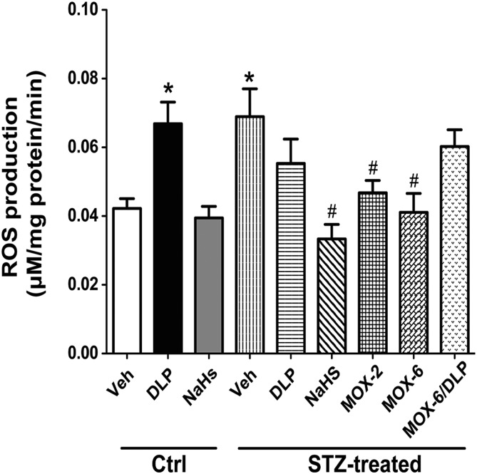
We recently demonstrated a fundamental role for cystathionine-γ lyase (CSE)-derived hydrogen sulfide (H2S) in the cardioprotective effect of the centrally acting drug moxonidine in diabetic rats. Whether a downregulated CSE/H2S system in the rostral ventrolateral medulla (RVLM) underlies neuronal oxidative stress and sympathoexcitation in diabetes has not been investigated. Along with addressing this question, we tested the hypothesis that moxonidine prevents the diabetes-evoked neurochemical effects by restoring CSE/H2S function within its major site of action, the RVLM. Ex vivo studies were performed on RVLM tissues of streptozotocin (55 mg/kg, i.p.) diabetic rats treated daily for 3 weeks with moxonidine (2 or 6 mg/kg; gavage), H2S donor sodium hydrosulfide (NaHS) (3.4 mg/kg, i.p.), CSE inhibitor DL-propargylglycine (DLP) (37.5 mg/kg, i.p.), a combination of DLP with moxonidine, or their vehicle. Moxonidine alleviated RVLM oxidative stress, neuronal injury, and increased tyrosine hydroxylase immunoreactivity (sympathoexcitation) by restoring CSE expression/activity as well as heme oxygenase-1 (HO-1) expression. A pivotal role for H2S in moxonidine-evoked neuroprotection is supported by the following: 1) NaHS replicated the moxonidine-evoked neuroprotection, and the restoration of RVLM HO-1 expression in diabetic rats; and 2) DLP abolished moxonidine-evoked neuroprotection in diabetic rats, and caused RVLM neurotoxicity, reminiscent of a diabetes-evoked neuronal phenotype, in healthy rats. These findings suggest a novel role for RVLM CSE/H2S/HO-1 in moxonidine-evoked neuroprotection and sympathoinhibition, and as a therapeutic target for developing new drugs for alleviating diabetes-evoked RVLM neurotoxicity and cardiovascular anomalies.


Figures

Fig. 1.
Fig. 1.
FluoroJade C (FJC) positive cells examined in the RVLM of rats showing neurodegeneration. (A–I) Representative images of FJC-positive cells in male rats treated with STZ (55 mg/kg, i.p. for 4 weeks) or its vehicle (buffer) receiving NaHS (H2S donor, 3.4 mg/kg per day, i.p, for 3 weeks after diabetes induction), DLP (CSE inhibitor, 37.5 mg/kg, i.p., for 3 weeks after diabetes induction), moxonidine (MOX) (2 or 6 mg/kg per day for 3 weeks after diabetes induction, gavage), a combination of MOX and DLP, or their vehicle (for 3 weeks after diabetes induction). (J) Group data showing the neurodegeneration expressed as the mean number of FJC-positive cells measured using National Institutes of Health ImageJ analysis of confocal images. Values are expressed as mean ± S.E.M. (n = 5 rats/group). *P < 0.05 vs. corresponding control/vehicle (Ctrl/Veh) values; #P < 0.05 vs. corresponding STZ/Veh values; $P < 0.05 vs. STZ/MOX-6 values.
Fig. 2.
Fig. 2.
Immunohistochemical detection of caspase-3 examined in the RVLM of rats. (A–I) Representative images of caspase-3 expression in male rats treated with STZ (55 mg/kg, i.p., for 4 weeks) or its vehicle (buffer) receiving NaHS (H2S donor for 3 weeks after diabetes induction; 3.4 mg/kg per day), DLP (CSE inhibitor, 37.5 mg/kg, i.p., for 3 weeks after diabetes induction), moxonidine (MOX) (2 or 6 mg/kg per day for 3 weeks after diabetes induction, gavage), a combination of MOX and DLP, or their vehicle (for 3 weeks after diabetes induction). (J) Group data showing the mean number of caspase-3 expression measured using National Institutes of Health ImageJ analysis of confocal images. Values are expressed as mean ± S.E.M. (n = 5 rats/group). *P < 0.05 vs. corresponding control/vehicle (Ctrl/Veh) values; #P < 0.05 vs. corresponding STZ/Veh values; $P < 0.05 vs. STZ/MOX-6 values.
Fig. 3.
Fig. 3.
The DCF biochemical assay of the generation of ROS showing the slopes (regression coefficients) of the regression lines representing the rate of ROS production in the RVLM of male rats treated with STZ (55 mg/kg, i.p., for 4 weeks) or its vehicle (buffer) receiving NaHS (H2S donor for 3 weeks after diabetes induction; 3.4 mg/kg per day), DLP (CSE inhibitor, 37.5 mg/kg, i.p., for 3 weeks after diabetes induction), moxonidine (MOX) (2 or 6 mg/kg per day for 3 weeks after diabetes induction, gavage), a combination of MOX and DLP, or their vehicle (for 3 weeks after diabetes induction). Values are expressed as mean ± S.E.M. (n = 5 rats/group). *P < 0.05 vs. corresponding control/vehicle (Ctrl/Veh) values; #P < 0.05 vs. corresponding STZ/Veh values.
Fig. 4.
Fig. 4.
(A–I) Confocal images showing superoxide level indicated by DHE staining (red) in the RVLM of male rats treated with STZ (55 mg/kg, i.p., for 4 weeks) or its vehicle (buffer) receiving NaHS (H2S donor for 3 weeks after diabetes induction; 3.4 mg/kg per day), DLP (CSE inhibitor, 37.5 mg/kg, i.p., for 3 weeks after diabetes induction), moxonidine (MOX) (2 or 6 mg/kg per day for 3 weeks after diabetes induction; gavage), a combination of MOX and DLP, or their vehicle (for 3 weeks after diabetes induction). (J) Values are expressed as mean ± S.E.M. (n = 5 rats/group). *P < 0.05 vs. corresponding control/vehicle (Ctrl/Veh) values; #P < 0.05 vs. corresponding STZ/Veh values; $P < 0.05 vs. STZ/MOX-6 values.
Fig. 5.
Fig. 5.
Western blot analyses showing the protein expression in the RVLM of male rats treated with STZ (55 mg/kg, i.p., for 4 weeks) or its vehicle (buffer) receiving NaHS (H2S donor for 3 weeks after diabetes induction; 3.4 mg/kg per day), DLP (CSE inhibitor; 37.5 mg/kg, i.p., for 3 weeks after diabetes induction), moxonidine (MOX) (2 or 6 mg/kg per day for 3 weeks after diabetes induction, gavage), a combination of MOX and DLP, or their vehicle (for 3 weeks after diabetes induction). (A) TH ratio to glyceraldehyde-3-phosphate dehydrogenase (GAPDH) protein (housekeeping protein) and western bands depicting the protein expression are shown below the bar graphs. (B) CSE ratio to GAPDH protein (housekeeping protein) and western bands depicting the protein expression are shown below the bar graphs. (C) HO-1 ratio to β-actin protein (housekeeping protein) and western bands depicting the protein expression are shown below the bar graphs. Values are expressed as mean ± S.E.M. (n = 5 rats/group). *P < 0.05 vs. corresponding control/vehicle (Ctrl/Veh) values; #P < 0.05 vs. corresponding STZ/Veh values; $P < 0.05 vs. STZ/MOX-6 values.
Fig. 6.
Fig. 6.
H2S synthesizing enzyme activity in the RVLM of male rats treated with STZ (55 mg/kg, i.p., for 4 weeks) or its vehicle (buffer) receiving NaHS (H2S donor for 3 weeks after diabetes induction; 3.4 mg/kg per day), DLP (CSE inhibitor, 37.5 mg/kg, i.p., for 3 weeks after diabetes induction), moxonidine (MOX) (2 or 6 mg/kg per day for 3 weeks after diabetes induction, gavage), a combination of MOX and DLP, or their vehicle (for 3 weeks after diabetes induction). Values are expressed as mean ± S.E.M. (n = 5 rats/group). *P < 0.05 vs. corresponding control/vehicle (Ctrl/Veh) values; #P < 0.05 vs. corresponding STZ/Veh values; $P < 0.05 vs. STZ/MOX-6 values.

Similar articles

Cited by

References

    1. Abdel Moneim AE. (2015) The neuroprotective effect of berberine in mercury-induced neurotoxicity in rats. Metab Brain Dis 30:935–942. - PubMed
    1. Bahniwal M, Little JP, Klegeris A. (2017) High glucose enhances neurotoxicity and inflammatory cytokine secretion by stimulated human astrocytes. Curr Alzheimer Res 14:731–741. - PubMed
    1. Bakuridze K, Savli E, Gongadze N, Baş DB, Gepdiremen A. (2009) Protection in glutamate-induced neurotoxicity by imidazoline receptor agonist moxonidine. Int J Neurosci 119:1705–1717. - PubMed
    1. Biessels GJ, van der Heide LP, Kamal A, Bleys RL, Gispen WH. (2002) Ageing and diabetes: implications for brain function. Eur J Pharmacol 441:1–14. - PubMed
    1. Burke WJ, Li SW, Chung HD, Ruggiero DA, Kristal BS, Johnson EM, Lampe P, Kumar VB, Franko M, Williams EA, et al. (2004) Neurotoxicity of MAO metabolites of catecholamine neurotransmitters: role in neurodegenerative diseases. Neurotoxicology 25:101–115. - PubMed

Publication types

MeSH terms

LinkOut - more resources