. 2017 Nov 28;114(48):E10319-E10328.
doi: 10.1073/pnas.1707992114. Epub 2017 Nov 13.

Specific inhibition of GPCR-independent G protein signaling by a rationally engineered protein

Affiliations

Specific inhibition of GPCR-independent G protein signaling by a rationally engineered protein

Anthony Leyme et al. Proc Natl Acad Sci U S A. .

Abstract

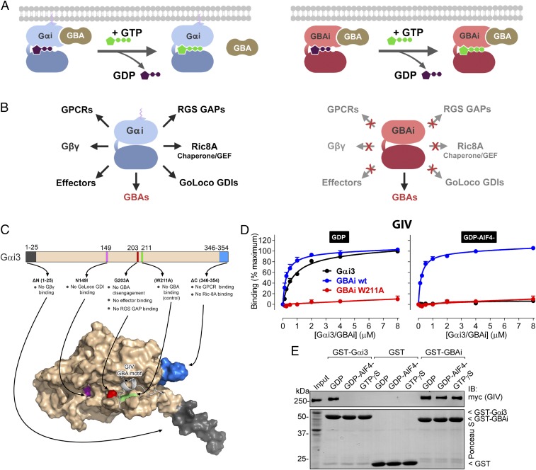
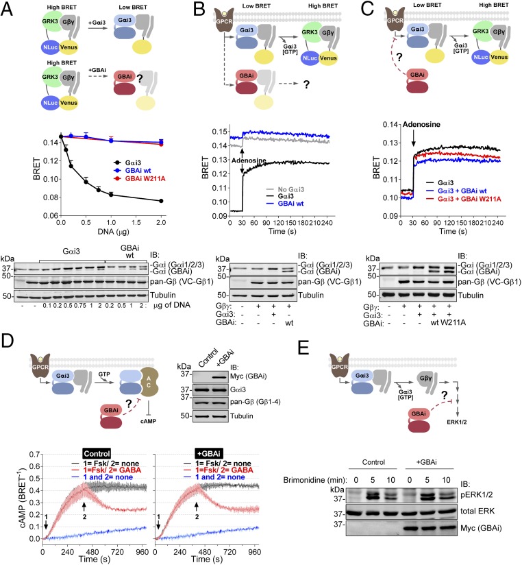
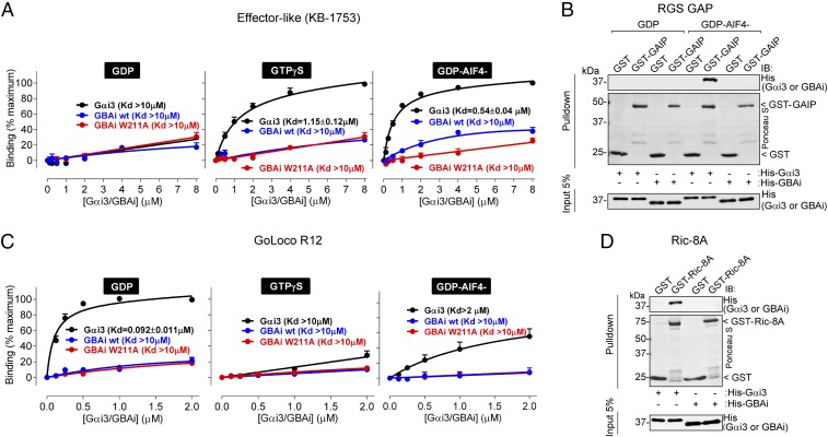
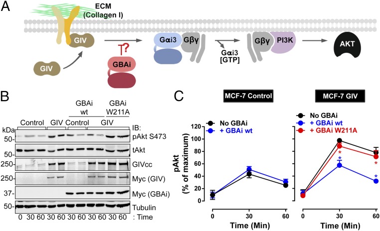
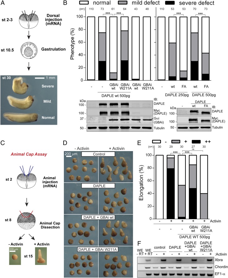
Activation of heterotrimeric G proteins by cytoplasmic nonreceptor proteins is an alternative to the classical mechanism via G protein-coupled receptors (GPCRs). A subset of nonreceptor G protein activators is characterized by a conserved sequence named the Gα-binding and activating (GBA) motif, which confers guanine nucleotide exchange factor (GEF) activity in vitro and promotes G protein-dependent signaling in cells. GBA proteins have important roles in physiology and disease but remain greatly understudied. This is due, in part, to the lack of efficient tools that specifically disrupt GBA motif function in the context of the large multifunctional proteins in which they are embedded. This hindrance to the study of alternative mechanisms of G protein activation contrasts with the wealth of convenient chemical and genetic tools to manipulate GPCR-dependent activation. Here, we describe the rational design and implementation of a genetically encoded protein that specifically inhibits GBA motifs: GBA inhibitor (GBAi). GBAi was engineered by introducing modifications in Gαi that preclude coupling to every known major binding partner [GPCRs, Gβγ, effectors, guanine nucleotide dissociation inhibitors (GDIs), GTPase-activating proteins (GAPs), or the chaperone/GEF Ric-8A], while favoring high-affinity binding to all known GBA motifs. We demonstrate that GBAi does not interfere with canonical GPCR-G protein signaling but blocks GBA-dependent signaling in cancer cells. Furthermore, by implementing GBAi in vivo, we show that GBA-dependent signaling modulates phenotypes during Xenopus laevis embryonic development. In summary, GBAi is a selective, efficient, and convenient tool to dissect the biological processes controlled by a GPCR-independent mechanism of G protein activation mediated by cytoplasmic factors.

Keywords: DAPLE; GEF; GPCR; Girdin; integrin.


Conflict of interest statement

The authors declare no conflict of interest.

Figures

Fig. 1.
Fig. 1.
Rational design of GBAi, a specific inhibitor of GBA motifs. (A, Left) GBA motifs bind to Gαi in its inactive conformation (bound to GDP, purple), but not in its active conformation (bound to GTP, green). (A, Right) Desired property of Gαi-derived GBAi is that its binding to GBA motifs is not reversed upon nucleotide exchange of GDP for GTP, thereby resulting in constitutive high-affinity binding to GBA motifs in cells. (B) Desired property of Gαi-derived GBAi is a lack of binding to any other Gαi binding partner (GPCRs, Gβγ, effectors, RGS GAPs, GoLoco GDIs, or the chaperone/GEF Ric-8A; Left), thereby resulting in specific binding and inhibition of only GBA motifs (Right). (C, Top) Schematic of the modifications engineered into Gαi3 to generate GBAi. The G203A mutation disrupts a conformational change of the SwII region that normally occurs upon GTP binding and is required to bind effectors and RGS GAPs and, at the same time, precludes GBA binding. Additional mutations/deletions were introduced to prevent binding of Gβγ (ΔN 1–25), GPCRs (ΔC 346–354), GoLoco GDIs (N149I), and Ric-8A (ΔC 346–354). The W211A mutation is known to inhibit binding of Gαi3 to all GBA motifs described to date (i.e., from GIV, DAPLE, Calnuc, NUCB2) and is used as a negative control throughout this study. (C, Bottom) Localization of each modification is shown in a previously described model of Gαi3 bound to the GBA motif of GIV (33). (D) GBAi binds to the GBA motif of GIV with the same affinity as Gαi3, and GBAi-GIV binding is not affected by the activation mimetic GDP-AlF4. Binding of a fluorescently labeled GIV peptide corresponding to its GBA motif (residues 1,671–1,701) to the indicated concentrations of purified His-tagged Gαi3 (black), GBAi wt (blue), or GBAi W211A (red) was determined by FP in the presence of GDP (Left) or GDP-AlF4 (Right). Data were normalized to maximal binding and fitted to a one-site binding model (solid lines). Results from three independent experiments are expressed as mean ± SEM. (E) GBAi binds the same to full-length GIV in the presence of GDP, GDP-AlF4, or GTPγS, whereas Gαi3 binds GIV only in the presence of GDP. Lysates of HEK293T cells expressing full-length, myc-tagged GIV were incubated with GST, GST-Gαi3, or GST-GBAi immobilized on glutathione-agarose beads in the presence of GDP, GDP-AlF4, or GTPγS (as indicated). Resin-bound proteins were eluted, separated by SDS/PAGE, and analyzed by Ponceau S-staining and immunoblotting (IB) as indicated. Input = 10% of the amount of lysate used in each pulldown. One experiment of two with very similar results is shown.
Fig. 2.
Fig. 2.
GBAi does not interfere with GPCR-mediated activation of G proteins. (A) GBAi does not associate with Gβγ in mammalian cells as determined by BRET. (Top) Schematic of the BRET assay used to monitor the association of Gαi3 or GBAi with Gβγ. In the absence of Gαi3, Venus-tagged Gβγ (V-Gβγ, BRET acceptor) associates with mas-GRK3ct-NLuc (GRK3, BRET donor) inducing a high BRET signal. Expression of Gαi3 competitively displaces V-Gβγ from mas-GRK3ct-NLuc and decreases BRET. (Middle) HEK293T cells were transfected with the indicated amounts of plasmids encoding for Gαi3, GBAi wt, and GBAi W211A, and equal amounts of the BRET donor (mas-GRK3ct-NLuc) and acceptor (V-Gβγ). BRET was measured under resting (unstimulated) conditions and is presented as mean ± SEM of four independent experiments. (Bottom) Protein expression of Gαi3, GBAi wt, and V-Gβγ was assessed by immunoblotting (IB) with the indicated antibodies. The upper band detected by the Gαi antibody corresponds to exogenous Gαi3 plus endogenous Gαi1, Gαi2, and Gαi3, whereas the lower band corresponds to GBAi. (B) GBAi does not couple to GPCRs in mammalian cells as determined by BRET. (Top) Schematic depicting the BRET assay used to determine the coupling of Gαi3 or GBAi to GPCRs. Under resting conditions, V-Gβγ associates with Gαi3 and BRET is low. Upon GPCR stimulation, the Gαi3:V-Gβγ heterotrimer dissociates and V-Gβγ interacts with mas-GRK3ct-NLuc, leading to an increase in BRET. (Middle) HEK293T cells were transfected with plasmids encoding for Gαi3 (0.5 μg) or GBAi wt (2 μg), along with plasmids for the BRET donor and acceptor (mas-GRK3ct-NLuc and V-Gβγ), as well as for the adenosine 1 receptor. BRET was measured every second. After 30 s of measurement under resting conditions, cells were stimulated with adenosine (10 μM). One representative experiment of three is shown. (Bottom) Protein expression of Gαi3, GBAi wt, and V-Gβγ was assessed by IB with the indicated antibodies. (C) GBAi does not interfere with GPCR-mediated activation of Gαi3 as determined by BRET. (Top) Schematic depicting the BRET experiment used to assess the possible interference of GBAi with Gαi3 coupling to GPCRs. The experimental design is as in B except that GBAi is coexpressed, along with Gαi3 and the rest of the BRET assay components, to test if it could impair the G protein-dependent BRET increase observed upon GPCR stimulation. (Middle) Experiments were carried out exactly as in B except that GBAi (wt or W211A, 2 μg) and Gαi3 (0.5 μg) were expressed simultaneously in the same cells. One representative experiment of three is shown. (Bottom) Protein expression of Gαi3, GBAi, and V-Gβγ was assessed by IB with the indicated antibodies. (D) GBAi does not interfere with GPCR-mediated regulation of cAMP by Gαi. (Top Left) Schematic depicting the experiment used to assess the possible interference of GBAi with GPCR-mediated regulation of cAMP levels. (Top Right) Protein expression of GBAi, Gαi3, and Gβγ was assessed by IB with the indicated antibodies. (Bottom) HEK293T cells were transfected with plasmids encoding for Nluc-EPAC-VV (0.05 μg) and GABABRs (0.2 μg) in the presence (+GBAi) or absence (control) of GBAi wt (2 μg). BRET was measured every 4 s. Forskolin (Fsk, 1 μM) and GABA (1 μM) were added (sequentially) at the indicated times. The blue trace corresponds to unstimulated control cells and is duplicated in both panels as a visual reference of the baseline. Results from three independent experiments are expressed as mean ± SEM. (E) GBAi does not interfere with GPCR-mediated regulation of ERK1/2 by Gβγ. (Top) Schematic depicting the experiment used to assess the possible interference of GBAi with GPCR-mediated regulation of ERK1/2. (Bottom) HEK293T cells were transfected with plasmids encoding for α2A-AR (0.2 μg) in the presence (+GBAi) or absence (control) of GBAi wt (2 μg). Cells were serum-starved overnight and then stimulated with brimonidine (5 μM) for the indicated times. Cell lysates were immunoblotted with the indicated antibodies. One representative experiment of three is shown.
Fig. 3.
Fig. 3.
GBAi does not bind to Gαi effectors or the Gαi regulators RGS GAPs, GoLoco GDIs, and Ric-8A. (A) GBAi does not bind to the effector-like peptide KB-1753. Binding of a fluorescently labeled KB-1753 peptide to the indicated concentrations of purified His-tagged Gαi3 (black), GBAi wt (blue), or GBAi W211A (red) was determined by FP in the presence of GDP, GTPγS, or GDP-AlF4 as indicated. Data were normalized to maximal binding and fitted to a one-site binding model (solid lines) to calculate the indicated Kds. KB-1753 binds Gαi3 with high affinity in the presence of GDP-AlF4 or GTPγS and low affinity in the presence of GDP, whereas GBAi does not bind in any of the three conditions. Results from three independent experiments are expressed as mean ± SEM. (B) GBAi does not bind to the RGS GAP protein GAIP. Purified GST or GST-GAIP was immobilized on glutathione-agarose beads and incubated with purified His-Gαi3 or His-GBAi in the presence of GDP or GDP-AlF4 as indicated. Resin-bound proteins were eluted, separated by SDS/PAGE, and analyzed by Ponceau S-staining and immunoblotting (IB) as indicated. GST-GAIP binds His-Gαi3 in the presence of GDP-AlF4, but not GDP, whereas it does not bind to GBAi either in the presence of GDP or GDP-AlF4. One representative experiment of three is shown. (C) GBAi does not bind to the GoLoco GDI motif of RGS12 (GoLoco R12). Binding of a fluorescently labeled peptide corresponding to GoLoco R12 (RGS12 residues 1,185–1,221) to the indicated concentrations of purified His-tagged Gαi3 (black), GBAi wt (blue), or GBAi W211A (red) was determined by FP in the presence of GDP (Left), GTPγS (Center), or GDP-AlF4 (Right). Data were normalized to maximal binding and fitted to a one-site binding model (solid lines) to calculate the indicated Kds. GoLoco R12 binds Gαi3 with high affinity in the presence of GDP and low affinity in the presence of GTPγS or GDP-AlF4, whereas GBAi binding is almost or completely absent under the same conditions. Results from three independent experiments are expressed as mean ± SEM. (D) GBAi does not bind to the chaperone/GEF protein Ric-8A. Experiments were carried out exactly as in B except that GST–Ric-8A was used instead of GST–KB-1753 and all conditions were tested in the presence of GDP. GST–Ric-8A binds to His-Gαi3 but not to His-GBAi. One representative experiment of three is shown.
Fig. 4.
Fig. 4.
GBAi inhibits GIV-mediated potentiation of PI3K-Akt signaling upon integrin stimulation. (A) Schematic of a previously described mechanism (18) by which GIV potentiates PI3K-Akt signaling in response to integrin stimulation via its GBA motif. Stimulation of cells with extracellular matrix (ECM) proteins (e.g., collagen I) triggers the recruitment of GIV to the intracellular tail of integrins, which, in turn, leads to GBA-dependent G protein signaling. PI3K-Akt activation is achieved via free Gβγ subunits released from Gi heterotrimers upon GIV GBA action. (B and C) GBAi wt, but not GBAi W211A, inhibits GIV-mediated potentiation of Akt activation upon collagen I stimulation of MCF-7 cells. MCF-7 cells stably expressing a vector control or full-length GIV and transfected with myc-GBAi wt (blue) or myc-GBAi W211A (red), as indicated, were lifted from culture dishes; kept in suspension for 1 h in serum-free media (time 0); and stimulated by plating on collagen I-coated culture dishes for 30 and 60 min. One representative immunoblot result from four independent experiments is shown in A, and the results for the quantification of Akt activation [as determined by levels of phosphorylated Akt (pAkt)] expressed as mean ± SEM are shown in C. *P < 0.05, using the Student’s t test (blue, compared with no GBAi; red, compared with GBAi wt). tAkt, total Akt.
Fig. 5.
Fig. 5.
GBAi inhibits DAPLE-mediated convergent extension defects in X. laevis embryos. (A and B) GBAi wt or the F1675A mutation in DAPLE inhibits developmental defects induced by DAPLE. (A) Schematic depicting the assay to assess DAPLE-induced developmental defects. mRNAs are injected equatorially in both dorsal blastomeres of two- to four-cell embryos [stage (st) 2–3], and phenotypes are assessed at st 30. Representative phenotypes are shown: normal or dorsally bent embryos (mild or severe). (B, Left) DAPLE (500 pg), GBAi wt (1 ng), or GBAi W211A (1 ng) mRNAs were injected as indicated, and the frequency of phenotypes is assessed at st 30. The total number of embryos analyzed from three independent experiments for each group is indicated on the top of the graph. (Bottom) Protein expression of DAPLE and GBAi was assessed by immunoblotting (IB) with the indicated antibodies. (B, Right) DAPLE wt or DAPLE FA mRNAs (250 pg or 500 pg) were injected, and phenotypes were analyzed as in the left graph. (Bottom) Protein expression of DAPLE wt and DAPLE FA (250 pg of mRNA) was assessed by IB with the indicated antibodies. (CF) GBAi wt, but not GBAi W211A, blocks DAPLE-mediated inhibition of convergent extension movements. (C) DAPLE (500 pg) and GBAi (wt or W211A, 1 ng) were coinjected into the animal hemisphere of both blastomeres of two-cell embryos. Animal caps were dissected at st 8, treated (or not treated) with activin to induce elongation, and analyzed for elongation at st 15. Representative pictures of activin-treated versus untreated caps injected with the indicated mRNAs (D) and the frequency of different elongation phenotypes [E; −, none, +, mild, or ++, strong elongation] are shown. The total number of embryos analyzed from three independent experiments for each group is indicated on the top of the graph. (F) RT-PCR of activin-mediated gene induction from caps at st 10.5 injected with the indicated mRNAs and treated or not treated with activin. Whole embryos (WE) with (+RT) or without (−RT) the reverse transcriptase reaction are shown on the left lanes as positive and negative controls, respectively. ***P < 0.001, χ2 test.

Similar articles

Cited by

References

    1. Gilman AG. G proteins: Transducers of receptor-generated signals. Annu Rev Biochem. 1987;56:615–649. - PubMed
    1. Sato M, Blumer JB, Simon V, Lanier SM. Accessory proteins for G proteins: Partners in signaling. Annu Rev Pharmacol Toxicol. 2006;46:151–187. - PubMed
    1. Willard FS, Kimple RJ, Siderovski DP. Return of the GDI: The GoLoco motif in cell division. Annu Rev Biochem. 2004;73:925–951. - PubMed
    1. Ross EM, Wilkie TM. GTPase-activating proteins for heterotrimeric G proteins: Regulators of G protein signaling (RGS) and RGS-like proteins. Annu Rev Biochem. 2000;69:795–827. - PubMed
    1. Tall GG. Ric-8 regulation of heterotrimeric G proteins. J Recept Signal Transduct Res. 2013;33:139–143. - PMC - PubMed

Publication types

MeSH terms

Substances

LinkOut - more resources