. 2017 Nov 28;114(48):12815-12820.
doi: 10.1073/pnas.1711536114. Epub 2017 Nov 13.

Phasic inhibition as a mechanism for generation of rapid respiratory rhythms

Affiliations

Phasic inhibition as a mechanism for generation of rapid respiratory rhythms

Jared M Cregg et al. Proc Natl Acad Sci U S A. .

Abstract

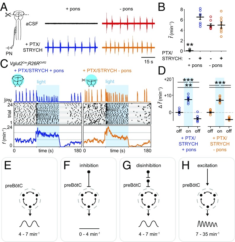
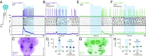
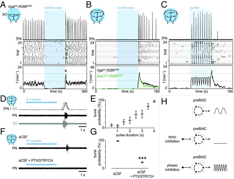
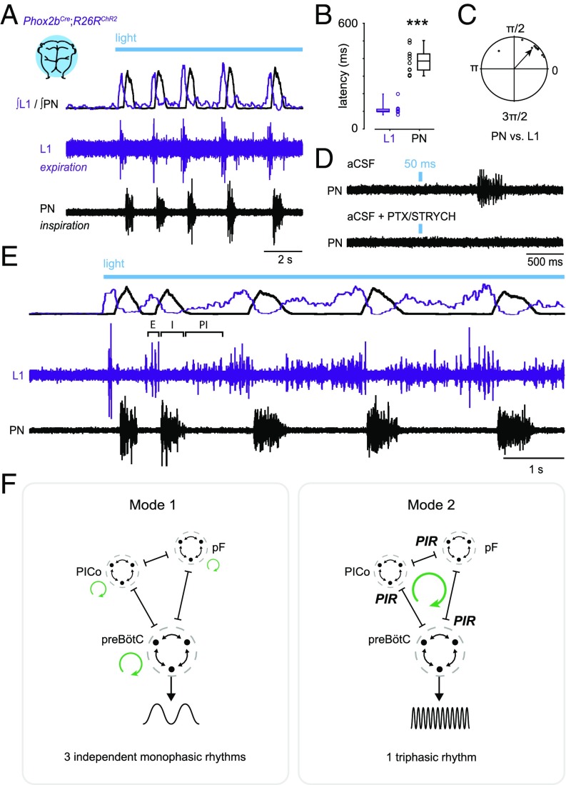
Central neural networks operate continuously throughout life to control respiration, yet mechanisms regulating ventilatory frequency are poorly understood. Inspiration is generated by the pre-Bötzinger complex of the ventrolateral medulla, where it is thought that excitation increases inspiratory frequency and inhibition causes apnea. To test this model, we used an in vitro optogenetic approach to stimulate select populations of hindbrain neurons and characterize how they modulate frequency. Unexpectedly, we found that inhibition was required for increases in frequency caused by stimulation of Phox2b-lineage, putative CO2-chemosensitive neurons. As a mechanistic explanation for inhibition-dependent increases in frequency, we found that phasic stimulation of inhibitory neurons can increase inspiratory frequency via postinhibitory rebound. We present evidence that Phox2b-mediated increases in frequency are caused by rebound excitation following an inhibitory synaptic volley relayed by expiration. Thus, although it is widely thought that inhibition between inspiration and expiration simply prevents activity in the antagonistic phase, we instead propose a model whereby inhibitory coupling via postinhibitory rebound excitation actually generates fast modes of inspiration.

Keywords: breathing; optogenetics; oscillator; respiration.


Conflict of interest statement

The authors declare no conflict of interest.

Figures

Fig. 1.
Fig. 1.
Excitation increases inspiratory frequency. (A, Top Left) Pontomedullary preparations do not exhibit spontaneous inspiration. (Top Right) Transection at the pontomedullary boundary initiates fictive inspiration. (Bottom Left) PTX/STRYCH application to pontomedullary preparations also initiates fictive inspiration. (Bottom Right) Fictive inspiration initiated via transection at the pontomedullary boundary is not affected by application of PTX/STRYCH. (B) Quantification of average f. **P < 0.01, artificial CSF (aCSF) + pons vs. all other conditions. (C, Top) After application of PTX/STRYCH, stimulation of excitatory neurons resulted in high-frequency inspiratory bursting (fmax + pons = 24.8 min−1; fmax − pons = 35.5 min−1). (Middle) Raster plots were constructed from eight biological replicates (each highlighted by gray shading), with three technical replicates each. (Bottom) f averaged over 24 trials relative to light onset. (D) Change in average f during and after light stimulation relative to baseline (off). PTX/STRYCH + pons: baseline vs. photostimulation, ***P = 1.3 × 10⁻⁵. Photostimulation vs. after, ***P = 1.0 × 10⁻⁷. Baseline vs. after, **P = 0.0012. PTX/STRYCH − pons: baseline vs. photostimulation, ***P = 3.2 × 10⁻⁶. Photostimulation vs. after, ***P = 1.5 × 10⁻⁷. Before vs. after, ***P = 2.1 × 10⁻⁶. Welch’s ANOVA with Bonferroni correction. n = 8 for each condition. Data are mean ± SEM. (EH, Top and Middle) Illustration of tested hypothesis and output of preBötC. (Bottom) Summary of finding. (E) Baseline f. (F) Inhibition decreases f (A and B). (G) Disinhibition can return f to baseline frequency but does not increase f above baseline (A and B). (H) Excitation increases f above baseline (C and D).
Fig. 2.
Fig. 2.
Inhibition is implicated in Phox2b and Atoh1 modulation of inspiratory frequency. (A) Stimulation of Phox2b-lineage neurons dramatically increased f (fmax = 68.6 min−1). (B) Phox2b-mediated increases in f were blocked by bath application of PTX/STRYCH. (C) Anatomical identification of ventral Phox2b-lineage neurons likely stimulated in the brainstem. Arrow indicates rostral and caudal directions. (D) Change in average f during and after light stimulation relative to baseline. Baseline vs. photostimulation, **P = 0.002. Photostimulation vs. after, **P = 0.002. Mann–Whitney U test with Bonferroni correction. PTX/STRYCH blocked the effect of photostimulation (Mann–Whitney U test). (E) Stimulation of Atoh1-lineage neurons resulted in a transient increase in f after the termination of the photostimulus. (F) Atoh1-mediated increases in f were blocked by application of PTX/STRYCH. (G) Position of ventral Atoh1-lineage neurons likely stimulated in the brainstem. (H) Change in average f during and after light stimulation relative to baseline. Baseline vs. after, **P = 0.002. Photostimulation vs. after, **P = 0.002. Mann–Whitney U test with Bonferroni correction. PTX/STRYCH blocked the effect of photostimulation (one-way ANOVA). n = 8 for each condition; data are mean ± SEM.
Fig. 3.
Fig. 3.
Phasic inhibition can drive increases in inspiratory frequency. (A) Photostimulation of Vgat+ neurons during fictive inspiration suppressed burst initiation for the duration of the photostimulus. (Bottom) Analysis of f indicated a reset-like response upon light off (asterisk, half-life of t1/2 ∼ 4 s). (B) In a silent preparation, stimulation of Vgat+ neurons evoked inspiratory bursting via a rebound-like mechanism (half-life of t1/2 ∼ 23 s). (C) Phasic inhibition evoked inspiratory bursting at high frequencies. (D) Rebound bursts recruit both phrenic and hypoglossal motor neurons (n = 5), indicating that rebound bursts arise from the preBötC. (E) The probability of observing a rebound burst in response to stimulation of inhibitory neurons was directly proportional to the photostimulus duration. n = 8; data are mean ± SEM. (F and G) PTX/STRYCH application blocked rebound inspiratory bursts caused by stimulation of inhibitory neurons. The pulse of light in the Bottom was performed between two spontaneous bursts (outside trace). ***P = 5.5 × 10⁻⁴. Mann–Whitney U test. n = 8; data are mean ± SEM. (H) Whereas tonic inhibition suppresses inspiratory burst initiation, phasic inhibition can actually increase f via postinhibitory rebound.
Fig. 4.
Fig. 4.
Phox2b-lineage neurons evoke expiration, inspiration, and postinspiration. (A) Stimulation of Phox2b-lineage neurons evoked phasic expiratory (L1, abdominal) motor activity preceding inspiratory bursts. (Top) Integrated and rectified traces. (Bottom) Raw traces. (B) Latency to first response. n = 10 preparations; ***P = 0.0002, Mann–Whitney U test. (C) Coupling between PN and L1 exhibited a phase separation of 0.85 ± 0.17 rad (49 ± 10°; n = 10). (D) The 50-ms Phox2bCre;R26RChR2 photostimulation was sufficient to cause an inspiratory burst several hundreds of milliseconds later. This inspiratory response was blocked by application of PTX/STRYCH (Fig. 2B). The pulse of light in the Bottom was performed between two spontaneous bursts (outside trace). (E) Stimulation of Phox2b-lineage neurons evoked all three phases of respiration—expiration (E), inspiration (I), and postinspiration (PI), forming a continuous triphasic rhythm. (F) Illustration of proposed coupling between E, I, and PI. In mode 1, inhibition does not couple E/I/PI and instead causes antiphasic patterning (38). Here, rhythmic oscillations (represented by green circular arrows) in motor output are the consequence of excitatory mechanisms. In mode 2, inhibition causes postinhibitory rebound, which acts to couple E, I, and PI. Here, rhythmic oscillation (represented by a single green circular arrow) is generated as a consequence of inhibitory synaptic coupling between E, I, and PI. pF, parafacial oscillator (E); preBötC (I); PICo, postinspiratory complex (PI); PIR, postinhibitory rebound excitation.

References

    1. Dutschmann M, Dick TE. Pontine mechanisms of respiratory control. Compr Physiol. 2012;2:2443–2469. - PMC - PubMed
    1. Rose MF, et al. Math1 is essential for the development of hindbrain neurons critical for perinatal breathing. Neuron. 2009;64:341–354. - PMC - PubMed
    1. Mulkey DK, et al. Respiratory control by ventral surface chemoreceptor neurons in rats. Nat Neurosci. 2004;7:1360–1369. - PubMed
    1. Jones SE, Dutschmann M. Testing the hypothesis of neurodegeneracy in respiratory network function with a priori transected arterially perfused brain stem preparation of rat. J Neurophysiol. 2016;115:2593–2607. - PMC - PubMed
    1. Smith JC, Ellenberger HH, Ballanyi K, Richter DW, Feldman JL. Pre-Bötzinger complex: A brainstem region that may generate respiratory rhythm in mammals. Science. 1991;254:726–729. - PMC - PubMed

Publication types

MeSH terms