Case Reports
. 2017 Nov 28;114(48):E10475-E10483.
doi: 10.1073/pnas.1714801114. Epub 2017 Nov 13.

Intact hemisphere and corpus callosum compensate for visuomotor functions after early visual cortex damage

Affiliations
Case Reports

Intact hemisphere and corpus callosum compensate for visuomotor functions after early visual cortex damage

Alessia Celeghin et al. Proc Natl Acad Sci U S A. .

Abstract

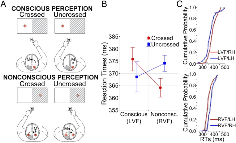
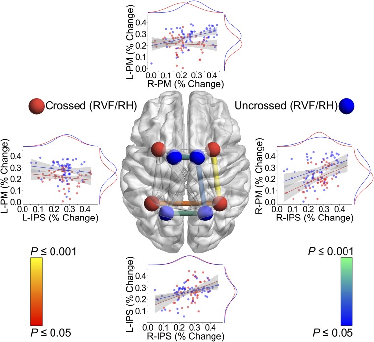
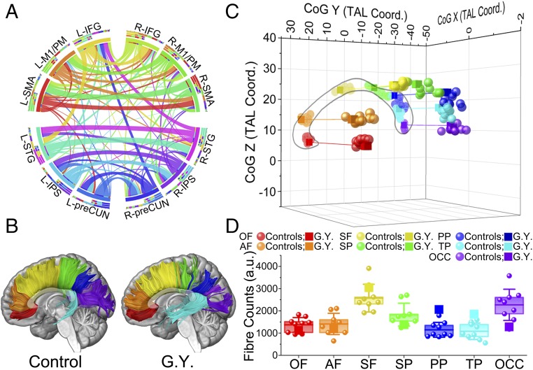
Unilateral damage to the primary visual cortex (V1) leads to clinical blindness in the opposite visual hemifield, yet nonconscious ability to transform unseen visual input into motor output can be retained, a condition known as "blindsight." Here we combined psychophysics, functional magnetic resonance imaging, and tractography to investigate the functional and structural properties that enable the developing brain to partly overcome the effects of early V1 lesion in one blindsight patient. Visual stimuli appeared in either the intact or blind hemifield and simple responses were given with either the left or right hand, thereby creating conditions where visual input and motor output involve the same or opposite hemisphere. When the V1-damaged hemisphere was challenged by incoming visual stimuli, or controlled manual responses to these unseen stimuli, the corpus callosum (CC) dynamically recruited areas in the visual dorsal stream and premotor cortex of the intact hemisphere to compensate for altered visuomotor functions. These compensatory changes in functional brain activity were paralleled by increased connections in posterior regions of the CC, where fibers connecting homologous areas of the parietal cortex course.

Keywords: Poffenberger; blindsight; corpus callosum; plasticity; tractography.


Conflict of interest statement

The authors declare no conflict of interest.

Figures

Fig. 1.
Fig. 1.
Illustration of interhemispheric transfer and behavioral results. (A) Model proposed to interpret the CUD differences observed in RTs during conscious and nonconscious perception. (B) Mean RTs (± SEM) during conscious (LVF) and nonconscious (RVF) perception as a function of the crossed and uncrossed response conditions. (C) CDFs of RTs during conscious (Upper Inset) and nonconscious (Lower Inset) perception, as a function of the crossed (red lines) and uncrossed (blue lines) response conditions showing opposite patterns throughout the whole distributions.
Fig. 2.
Fig. 2.
Brain activity during conscious and nonconscious perception. The areas are significantly activated for each specified contrast at P < 0.05 corrected for FDR. Areas colored from red to yellow are significantly more active for crossed vs. uncrossed conditions during conscious perception (Upper), for nonconscious perception irrespective of response hand (Middle), and for the crossed vs. uncrossed conditions during nonconscious perception (Lower). The opposite holds for areas colored from blue to green. The lesion in the left V1 of G.Y. is visible and represented in dark red.
Fig. 3.
Fig. 3.
Correlations in fMRI percent signal change during nonconscious perception. All possible correlations between left (L-) and right (R-) IPS and left and right premotor cortex (PM) as a function of crossed and uncrossed response conditions are displayed in the central transparent brain. The four areas are symbolized by spheres of different colors for the crossed (red) and uncrossed (blue) conditions. Edges diameter represents Pearson r value, whereas edges color represents P value. Nonsignificant correlations are shown in transparent gray. Lateral insets report scatterplots for the eight most relevant correlations. Marginal curves show the distribution of the data along X and Y axes, internal lines represent fitted correlations, and gray areas show 95% confidence limits.
Fig. 4.
Fig. 4.
Connectional, topographic, and microstructural properties of the CC. (A) Circular representation of the connectional fingerprint associated with cortical areas coactivated with the CC. Segments represent different areas and ribbons fiber tracts. Connections between areas in opposite hemispheres that did not course through the activated clusters in the CC are not displayed. (B) Tractographic segmentation of callosal fibers based on cortical projections in patient G.Y. and one representative control. Color codes for the seven callosal subregions are the same as those reported in the legends of C and D. (C) Three-dimensional representation of the CoG for each callosal compartment in the control subjects (circles) and G.Y. (squares). (D) Fiber counts for each callosal sector in the controls and G.Y. Box represents 25th and 75th percentiles, internal line represents controls mean, and whiskers represent SD. AF, anterior frontal; OCC, occipital; OF, orbitofrontal; PP, posterior parietal; SF, superior frontal; SP superior parietal; TP, temporal.

References

    1. Payne BR, Lomber SG. Reconstructing functional systems after lesions of cerebral cortex. Nat Rev Neurosci. 2001;2:911–919. - PubMed
    1. Voytek B, et al. Dynamic neuroplasticity after human prefrontal cortex damage. Neuron. 2010;68:401–408. - PMC - PubMed
    1. Johansen-Berg H, et al. The role of ipsilateral premotor cortex in hand movement after stroke. Proc Natl Acad Sci USA. 2002;99:14518–14523. - PMC - PubMed
    1. Takatsuru Y, et al. Neuronal circuit remodeling in the contralateral cortical hemisphere during functional recovery from cerebral infarction. J Neurosci. 2009;29:10081–10086. - PMC - PubMed
    1. Edelman GM, Gally JA. Degeneracy and complexity in biological systems. Proc Natl Acad Sci USA. 2001;98:13763–13768. - PMC - PubMed

Publication types