. 2018 Jan 3;38(1):74-92.
doi: 10.1523/JNEUROSCI.1483-17.2017. Epub 2017 Nov 13.

Locus Coeruleus Ablation Exacerbates Cognitive Deficits, Neuropathology, and Lethality in P301S Tau Transgenic Mice

Affiliations

Locus Coeruleus Ablation Exacerbates Cognitive Deficits, Neuropathology, and Lethality in P301S Tau Transgenic Mice

Termpanit Chalermpalanupap et al. J Neurosci. .

Abstract

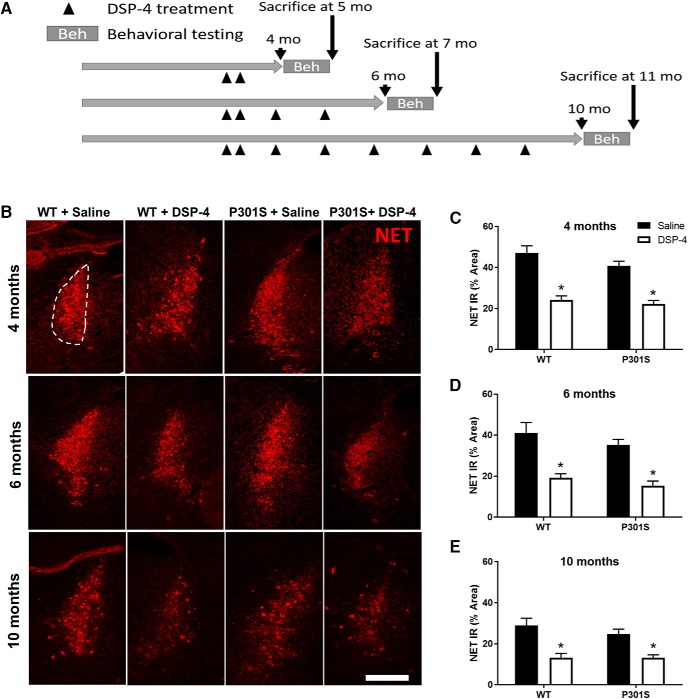
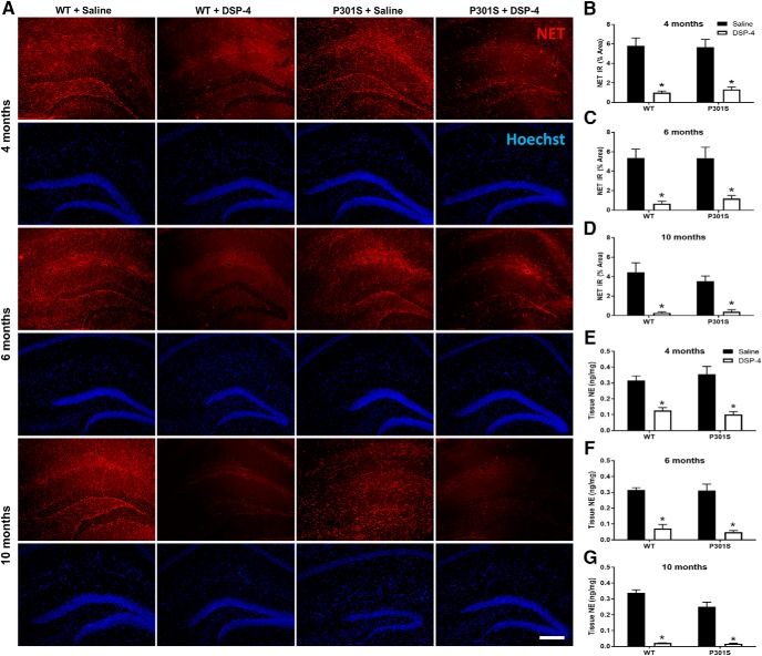
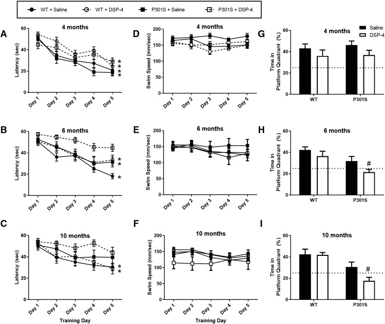
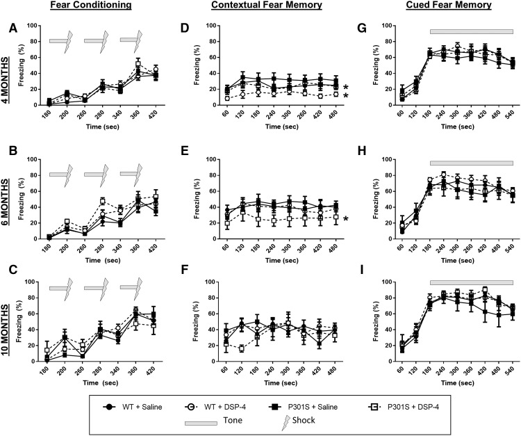
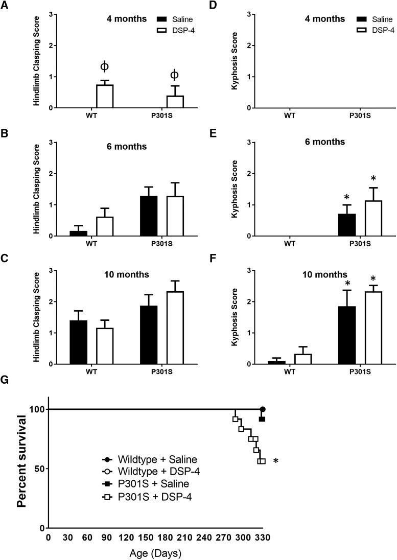
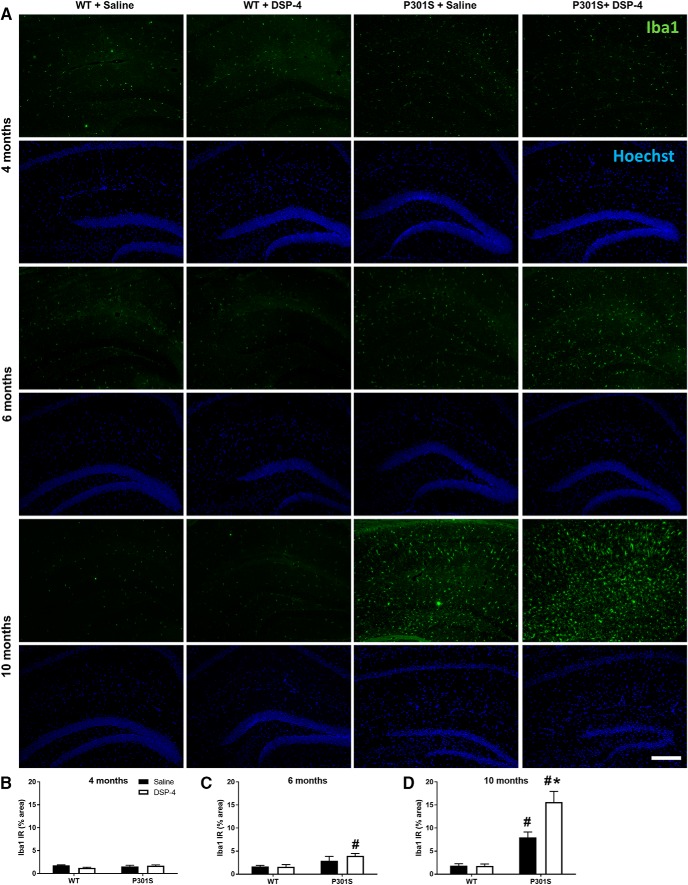
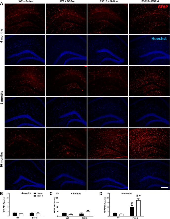
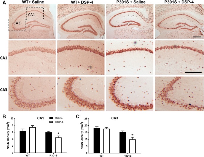
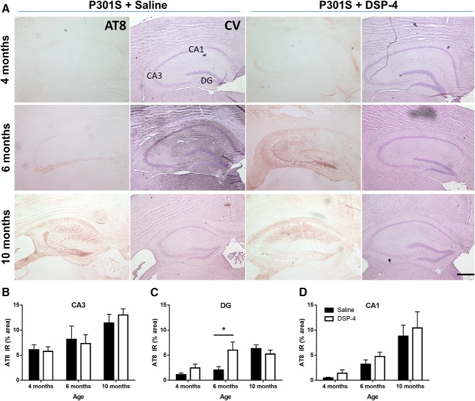
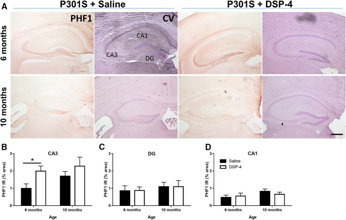
The brainstem locus coeruleus (LC) supplies norepinephrine to the forebrain and degenerates in Alzheimer's disease (AD). Loss of LC neurons is correlated with increased severity of other AD hallmarks, including β-amyloid (Aβ) plaques, tau neurofibrillary tangles, and cognitive deficits, suggesting that it contributes to the disease progression. Lesions of the LC in amyloid-based transgenic mouse models of AD exacerbate Aβ pathology, neuroinflammation, and cognitive deficits, but it is unknown how the loss of LC neurons affects tau-mediated pathology or behavioral abnormalities. Here we investigate the impact of LC degeneration in a mouse model of tauopathy by lesioning the LC of male and female P301S tau transgenic mice with the neurotoxin N-(2-chloroethyl)-N-ethyl-bromobenzylamine (DSP-4) starting at 2 months of age. By 6 months, deficits in hippocampal-dependent spatial (Morris water maze) and associative (contextual fear conditioning) memory were observed in lesioned P301S mice while performance remained intact in all other genotype and treatment groups, indicating that tau and LC degeneration act synergistically to impair cognition. By 10 months, the hippocampal neuroinflammation and neurodegeneration typically observed in unlesioned P301S mice were exacerbated by DSP-4, and mortality was also accelerated. These DSP-4-induced changes were accompanied by only a mild aggravation of tau pathology, suggesting that increased tau burden cannot fully account for the effects of LC degeneration. Combined, these experiments demonstrate that loss of LC noradrenergic neurons exacerbates multiple phenotypes caused by pathogenic tau, and provides complementary data to highlight the dual role LC degeneration has on both tau and Aβ pathologies in AD.SIGNIFICANCE STATEMENT Elucidating the mechanisms underlying AD is crucial to developing effective diagnostics and therapeutics. The degeneration of the LC and loss of noradrenergic transmission have been recognized as ubiquitous events in AD pathology, and previous studies demonstrated that LC lesions exacerbate pathology and cognitive deficits in amyloid-based mouse models. Here, we reveal a complementary role of LC degeneration on tau-mediated aspects of the disease by using selective lesions of the LC and the noradrenergic system to demonstrate an exacerbation of cognitive deficits, neuroinflammation, neurodegeneration in a transgenic mouse model of tauopathy. Our data support an integral role for the LC in modulating the severity of both canonical AD-associated pathologies, as well as the detrimental consequences of LC degeneration during disease progression.

Keywords: DSP-4; hippocampus; locus coeruleus; neuroinflammation; norepinephrine; tau.


Figures

Figure 1.
Figure 1.
DSP-4 reduces NET IR in the LC of both WT and P301S mice. WT and P301S mice were administered multiple doses of saline or DSP-4 (50 mg/kg, i.p.) and then assessed for behavior and pathology at various time points. A, Timeline of experiments. B, Representative immunofluorescence images. C–E, Quantification of the NET IR in the LC as percentage of total LC area. Two-way ANOVA (treatment × genotype) with Sidak's post hoc tests. Data are mean ± SEM; n = 8–12 per group. Scale bar, 100 μm. *p < 0.05, compared with saline for that genotype.
Figure 2.
Figure 2.
DSP-4 reduces NET IR and tissue NE content in the hippocampus. A, Representative immunofluorescence images. B–D, Quantification of NET IR in the hippocampus following saline or DSP-4 treatment in WT and P301S mice. E–G, Tissue NE concentrations in the hippocampus following saline or DSP-4 treatment in WT and P301S transgenic mice. Two-way ANOVA (treatment × genotype) with Sidak's post hoc tests. Data are mean ± SEM; n = 8–12 per group. Scale bar, 200 μm. *p < 0.05, compared with saline for that genotype. Hoechst, neuronal counterstain.
Figure 3.
Figure 3.
Neither LC degeneration nor P301S impacts circadian locomotor activity. Data are mean ± SEM ambulations over 24 h in WT and P301S mice treated with saline or DSP-4; n = 7–12 per group. A, 4 months; B, 6 months; C, 10 months.
Figure 4.
Figure 4.
Neither LC degeneration nor P301S impacts anxiety-like behavior in the open field test. Data are mean ± SEM (A–C) percentage time spent in center of the arena and (D–F) latency to leave the center of the arena in WT and P301S mice treated with saline or DSP-4; n = 7–12 per group.
Figure 5.
Figure 5.
LC degeneration exacerbates spatial learning and memory in P301S mice in the Morris water maze. Data are mean ± SEM (A–C) latency to find the platform and (D–F) swim speed during 5 d of initial training in WT and P301S mice treated with saline or DSP-4. N = 8–12 per group. Two-way repeated-measures ANOVA (group × training day) with Dunnett's post hoc tests. *p < 0.05, compared with day 1 for that group. G–I, Data are mean ± SEM percentage time spent swimming during the probe trial in the quadrant that previously contained the platform. Dotted line indicates the percentage time an animal would spend swimming in the correct quadrant by chance (25%). N = 8–12 per group. Two-way ANOVA (treatment × genotype) with Sidak's post hoc tests. #p < 0.05, compared with WT + saline.
Figure 6.
Figure 6.
LC degeneration impairs contextual, but not cued fear memory in P301S mice. Following 3 tone-shock pairings (A–C), fear behavior was recorded as percentage time spent freezing (mean ± SEM) following reexposure to the shock-associated context (D–F) or tone (G–I) in WT and P301S treated with saline or DSP-4. N = 8–12 per group. Two-way ANOVA (treatment × genotype) with Tukey's post hoc tests. *p < 0.05, compared with WT + saline.
Figure 7.
Figure 7.
LC degeneration reduces survival in transgenic mice but does not alter physical neurodegenerative phenotypes. Quantification (mean ± SEM) of (A–C) hindlimb clasping and (D–F) kyphosis. N = 6–12 per group. Two-way ANOVA (treatment × genotype) with Tukey's post hoc tests. *p < 0.05, compared with WT group with same treatment. ϕp < 0.05, compared with unlesioned group of the same genotype. G, Survival curve of WT and P301S mice treated with saline or DSP-4. N = 12 per group. Log-rank (Mantel–Cox) test. *p < 0.05, compared with all other groups.
Figure 8.
Figure 8.
LC degeneration increases activated microglia in the hippocampus of P301S mice. A, Representative immunofluorescence images. B–D, Quantification of Iba1 IR in the hippocampus of WT and P301S mice following saline or DSP-4 treatment. Data are mean ± SEM; n = 8–12 per group. Scale bar, 200 μm. Two-way ANOVA (treatment × genotype) with Tukey's post hoc tests. #p < 0.05, compared with WT + saline. *p < 0.05, compared with P301S + saline. Iba1, microglia gene; Hoechst, neuronal counterstain.
Figure 9.
Figure 9.
LC degeneration increases activated astrocytes in the hippocampus of P301S mice. A, Representative immunofluorescence images. B–D, Quantification of GFAP IR in the hippocampus of WT and P301S mice following saline or DSP-4 treatment. Two-way ANOVA (treatment × genotype) with Tukey's post hoc tests. Data are mean ± SEM; n = 8–12 per group. Scale bar, 200 μm. #p < 0.05, compared with WT + saline. *p < 0.05, compared with P301S + saline. Hoechst, neuronal counterstain.
Figure 10.
Figure 10.
LC lesions promote neurodegeneration in the hippocampus of 10 month P301S mice. Representative images (A) and quantification of NeuN IR in the (B) CA1 and (C) CA3 regions of the hippocampus of WT and P301S mice treated with saline or DSP-4. Two-way ANOVA (treatment × genotype) with Sidak's post hoc tests. Data are mean ± SEM; n = 8–12 per group. Scale bars: Top, 200 μm; Bottom, 100 μm. *p < 0.05, compared with P301S + saline.
Figure 11.
Figure 11.
LC degeneration exacerbates hyperphosphorylated tau pathology in the dentate gyrus of P301S mice. Representative images of AT8 IR in the hippocampus of P301S mice following saline or DSP-4 treatment (A). Quantification of AT8 IR in CA3 (B), DG (C), and CA1 (D). Two-way ANOVA (treatment × genotype) with Sidak's post hoc tests. Data are mean ± SEM; n = 8–12 per group. Scale bar, 200 μm. *p < 0.05. AT8, Phospho-tau (Ser202, Thr205), hyperphosphorylated tau; CV, cresyl violet, neuronal counterstain.
Figure 12.
Figure 12.
LC degeneration exacerbates paired helical tau pathology in CA3 of P301S mice. Representative images of PHF1 IR in hippocampus of P301S mice following saline or DSP-4 treatment (A). Quantification of PHF1 IR in CA3 (B), DG (C), and CA1 (D). Two-way ANOVA (treatment × genotype) with Sidak's post hoc tests. Data are mean ± SEM; n = 8–12 per group. Scale bar, 200 μm. *p < 0.05. PHF1, Phospho-tau (Ser396, Ser404), paired-helical filaments; CV, cresyl violet, neuronal counterstain.

Comment in

Similar articles

Cited by

References

    1. Allison SL, Fagan AM, Morris JC, Head D (2016) Spatial navigation in preclinical Alzheimer's disease. J Alzheimers Dis 52:77–90. 10.3233/JAD-150855 - DOI - PMC - PubMed
    1. Alzheimer's Association (2016) Alzheimer's disease facts and figures. Alzheimers Dement 12:459–509. - PubMed
    1. Andrés-Benito P, Fernández-Dueñas V, Carmona M, Escobar LA, Torrejón-Escribano B, Aso E, Ciruela F, Ferrer I (2017) Locus coeruleus at asymptomatic early and middle Braak stages of neurofibrillary tangle pathology. Neuropathol Appl Neurobiol 43:373–392. 10.1111/nan.12386 - DOI - PubMed
    1. Bliwise DL. (2004) Sleep disorders in Alzheimer's disease and other dementias. Clin Cornerstone 6 [Suppl 1A]:S16–S28. - PubMed
    1. Bondareff W, Mountjoy CQ, Roth M, Rossor MN, Iversen LL, Reynolds GP, Hauser DL (1987) Neuronal degeneration in locus ceruleus and cortical correlates of Alzheimer disease. Alzheimer Dis Assoc Disord 1:256–262. 10.1097/00002093-198701040-00005 - DOI - PubMed

Publication types

MeSH terms

LinkOut - more resources