

Spontaneous anatomical and functional recovery of bilateral electric shock maculopathy
- PMID: 29133672
- PMCID: PMC5700614
- DOI: 10.4103/ijo.IJO_536_17
Spontaneous anatomical and functional recovery of bilateral electric shock maculopathy
Abstract
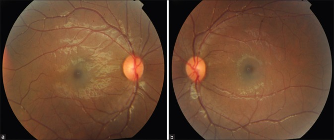
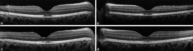
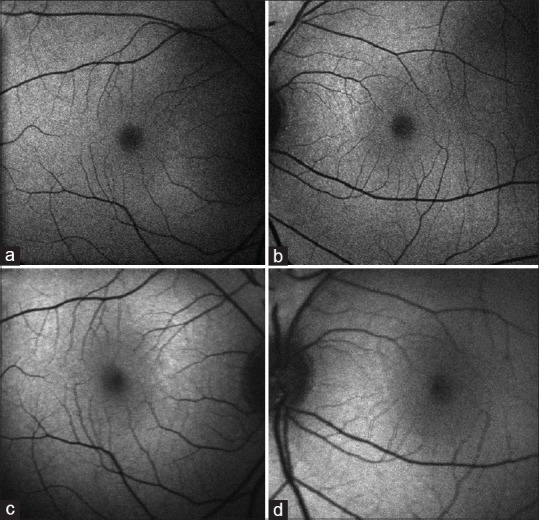
A 12-year-old boy presented with best-corrected visual acuity (BCVA) of 6/9 in both eyes following an episode of electric shock. Optical coherence tomography (OCT) showed disruption of the ellipsoid zone as well as retinal pigment epithelium (RPE) layer. Fundus autofluorescence (FAF) showed increased central hypoautofluorescence in both eyes. At 3-month follow-up, BCVA improved to 6/6 with OCT showing spontaneous resolution of maculopathy in both eyes with reorganized RPE layer and ellipsoid zone. To the best of our knowledge, this is the first case of bilateral electric shock maculopathy (ESM) with spontaneous anatomical as well as functional recovery. Ophthalmologists must be aware of various forms of ESM. OCT and FAF must be done in patients presenting with defective vision and history of electric shock for the diagnostic as well as prognostic evaluation.
Conflict of interest statement
There are no conflicts of interest.
Figures
References
-
- García-Sánchez V, Gomez Morell P. Electric burns: High-and low-tension injuries. Burns. 1999;25:357–60. - PubMed
-
- Boozalis GT, Purdue GF, Hunt JL, McCulley JP. Ocular changes from electrical burn injuries. A literature review and report of cases. J Burn Care Rehabil. 1991;12:458–62. - PubMed
-
- Miller BK, Goldstein MH, Monshizadeh R, Tabandeh H, Bhatti MT. Ocular manifestations of electrical injury: A case report and review of the literature. CLAO J. 2002;28:224–7. - PubMed
-
- Sony P, Venkatesh P, Tewari HK, Garg SP. Bilateral macular cysts following electric burn. Clin Exp Ophthalmol. 2005;33:78–80. - PubMed
-
- Bayar SA, Sarigul A, Yilmaz G, Pinarci EY. Development of optic neuropathy and foveal pseudocyst in a case of high-voltage electrical injury: A three-year follow-up. [Last accessed on 2017 Apr 02];Turkish J Opthalmol. 2014 44:410–2. Available from: http://wwwoftalmolojiorg/article_7387/Development-Of-Optic-Neuropathy-An... .
Publication types
MeSH terms
LinkOut - more resources
Full Text Sources
Other Literature Sources
Medical
