

Effects of Melatonin Levels on Neurotoxicity of the Medial Prefrontal Cortex in a Rat Model of Parkinson's Disease
- PMID: 29133763
- PMCID: PMC5695060
- DOI: 10.4103/0366-6999.218025
Effects of Melatonin Levels on Neurotoxicity of the Medial Prefrontal Cortex in a Rat Model of Parkinson's Disease
Abstract
Background: Damage of the medial prefrontal cortex (mPFC) results in similar characteristics to the cognitive deficiency seen with the progress of Parkinson's disease (PD). Since the course of mPFC damage is still unclear, our study aimed to investigate the effects of melatonin (MT) on neurotoxicity in the mPFC of a rat model of PD.
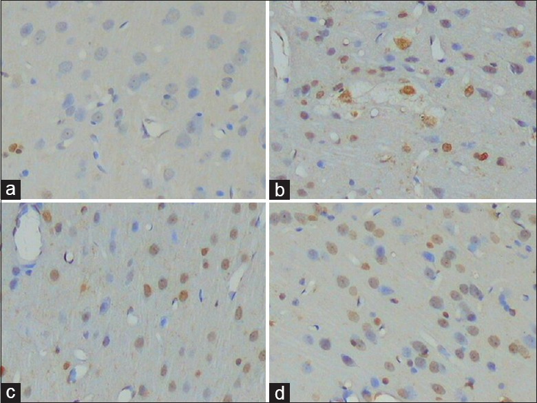
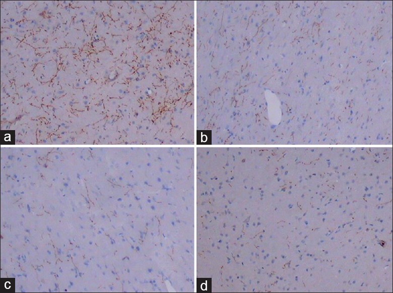
Methods: One hundred and fifty-four normal, male Wistar rats were randomly divided into the following five groups: normal + normal saline (NS), normal + 6-hydroxydopamine (6-OHDA), sham pinealectomy (PX) + 6-OHDA, PX + 6-OHDA, and MT + 6-OHDA. 6-OHDA was injected into the right substantia nigra pars compacta (SNc) and ventral tegmental area (VTA) of each group, except normal + NS, 60 days after the PX. In the MT treatment group, MT was administered immediately after the intraperitoneal injection at 4 p.m. every day, for 14 days. Neuronal apoptosis in the mPFC was examined using the TUNEL method, while the expression of tyrosine hydroxylase (TH), Bax,and Bcl-2 in this region was measured using immunohistochemistry. The concentration of malondialdehyde (MDA) in the mPFC was examined using the thiobarbituric acid method.
Results: Rats in the normal + 6-OHDA and sham PX + 6-OHDA groups were combined into one group (Group N + 6-OHDA) since there was no significant discrepancy between the groups for all the detected parameters. Apoptosis of cells in the NS, MT + 6-OHDA, N + 6-OHDA, and PX + 6-OHDA groups was successively significantly increased (Hc = 256.25, P < 0.001). The gray value of TH (+) fibers in the NS, MT + 6-OHDA, N + 6-OHDA, and PX + 6-OHDA groups was also successively significantly increased (F = 99.33, P < 0.001). The staining intensities of Bax and Bcl-2 were as follows: Group NS +/+, Group MT + 6-OHDA ++/+, Group N + 6-OHDA ++/+, and PX + 6-OHDA +++/+. The concentrations of MDA in the NS, MT + 6-OHDA, N + 6-OHDA, and PX + 6-OHDA groups were significantly increased in sequence (Hc = 296.309, P < 0.001).
Conclusions: Neuronal damage of the VTA by 6-OHDA might induce VTA-mPFC nerve fibers to undergo anterograde nerve damage, in turn inducing transneuronal damage of the mPFC. PX significantly exacerbated the neurotoxicity in the mPFC, which was induced by the neuronal injury of the VTA. However, MT replacement therapy significantly alleviated the neurotoxicity in the mPFC.
Conflict of interest statement
There are no conflicts of interest.
Figures
References
-
- Sgambato-Faure V, Buggia V, Gilbert F, Lévesque D, Benabid AL, Berger F, et al. Coordinated and spatial upregulation of arc in striatonigral neurons correlates with L-dopa-induced behavioral sensitization in dyskinetic rats. J Neuropathol Exp Neurol. 2005;64:936–47. doi: 10.1097/01.jnen.0000186922.42592.b7. - PubMed
-
- Kraus MF, Maki PM. Effect of amantadine hydrochloride on symptoms of frontal lobe dysfunction in brain injury: Case studies and review. J Neuropsychiatry Clin Neurosci. 1997;9:222–30. doi: 10.1176/jnp.9.2.222. - PubMed
-
- Chaudhuri KR, Prieto-Jurcynska C, Naidu Y, Mitra T, Frades-Payo B, Tluk S, et al. The nondeclaration of nonmotor symptoms of Parkinson's disease to health care professionals: An international study using the nonmotor symptoms questionnaire. Mov Disord. 2010;25:704–9. doi: 10.1002/mds.22868. - PubMed
MeSH terms
Substances
LinkOut - more resources
Full Text Sources
Other Literature Sources
Research Materials
Miscellaneous
