. 2017 Nov 13;8(1):1473.
doi: 10.1038/s41467-017-01728-5.

Unique phenotypes and clonal expansions of human CD4 effector memory T cells re-expressing CD45RA

Affiliations

Unique phenotypes and clonal expansions of human CD4 effector memory T cells re-expressing CD45RA

Yuan Tian et al. Nat Commun. .

Abstract

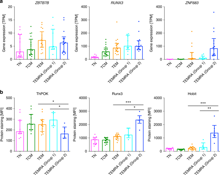
The expression of CD45RA is generally associated with naive T cells. However, a subset of effector memory T cells re-expresses CD45RA (termed TEMRA) after antigenic stimulation with unknown molecular characteristics and functions. CD4 TEMRA cells have been implicated in protective immunity against pathogens such as dengue virus (DENV). Here we show that not only the frequency but also the phenotype of CD4 TEMRA cells are heterogeneous between individuals. These cells can be subdivided into two major subsets based on the expression of the adhesion G protein-coupled receptor GPR56, and GPR56+ TEMRA cells display a transcriptional and proteomic program with cytotoxic features that is distinct from effector memory T cells. Moreover, GPR56+ TEMRA cells have higher levels of clonal expansion and contain the majority of virus-specific TEMRA cells. Overall, this study reveals the heterogeneity of CD4 TEMRA cells and provides insights into T-cell responses against DENV and other viral pathogens.


Conflict of interest statement

The authors declare no competing financial interests.

Figures

Fig. 1
Fig. 1
Gene expression data indicates that CD4 effector memory T cells re-expressing CD45RA (TEMRA) are highly variable between different donors. a Representative staining of the memory CD4 T-cell subsets. b PCA analysis of gene expression data (top 1000 variable genes) for different CD4 T-cell subsets (n = 6 for TN, TCM and TEM, and n = 12 for TEMRA). Note the dashed vertical line separates TEMRA from group 2 donors from all other cell subsets. c CD45 exon 4 abundance in TEMRA with respect to effector memory T (TEM) cells. The line connects TEM and TEMRA relative exon 4 expression for each individual donor (n = 2 and 4 for group 1 and group 2 donors, respectively). d The expression levels of HNRPLL in each of the different cell types (n = 17 for TN, TCM and TEM, and n = 9 and 14 for TEMRA cells from group 1 and group 2 donors, respectively). Error bars show median with interquartile range
Fig. 2
Fig. 2
Identification of specific transcriptomic profiles for CD4 effector memory T cells re-expressing CD45RA (TEMRA) from group 1 and group 2 donors. a Pairwise differential expression analysis between CD4 effector memory T (TEM) cells, CD4 TEMRA cells from group 1 donors, and CD4 TEMRA cells from group 2 donors. A total of 228 genes that are specific for TEMRA cells from group 2 donors were identified (n = 17, 9 and 14 for TEM, TEMRA cells from group 1 donors and TEMRA cells from group 2 donors, respectively). b Transcriptomics median and Padj values for the five markers significantly upregulated/downregulated in TEMRA cells from group 2 donors. c Bar graphs show gene expression values in transcripts per million (TPM) for various molecules in different cell subsets (n = 17 for naive (TN), central memory (TCM) and TEM, and n = 9 and 14 for TEMRA cells from group 1 and group 2 donors, respectively). d Bar graphs show protein abundance in mean fluorescence intensity (MFI) for various molecules in different cell subsets (n = 11 for TN, TCM and TEM, and n = 5 and 6 for TEMRA cells from group 1 and group 2 donors, respectively). Error bars show median with interquartile range. Statistical significance was determined by two-tailed Mann–Whitney test. *p < 0.05, **p < 0.01, ***p < 0.001
Fig. 3
Fig. 3
TEMRA from group 2 donors exhibit differential expression of key transcriptional factors that regulate the development of cytotoxic CD4 T cells in the protein level. a Bar graphs show gene expression values in transcripts per million (TPM) for ZBTB7B, RUNX3 and ZNF683 in different cell subsets (n = 17 for TN, TCM and TEM, and n = 9 and 14 for TEMRA cells from group 1 and group 2 donors, respectively). b Bar graphs show protein abundance in mean fluorescence intensity (MFI) for ThPOK, Runx3, and Hobit in different cell subsets (n = 11 for TN, TCM and TEM, and n = 5 and 6 for TEMRA cells from group 1 and group 2 donors, respectively). Error bars show median with interquartile range. Statistical significance was determined by two-tailed Mann–Whitney test. *p < 0.05, **p < 0.01, ***p < 0.001
Fig. 4
Fig. 4
Identification of TEMRA subtypes within a given individual. a Representative flow cytometry plots show the expression of GPR56 and perforin by CD4 TEMRA cells in group 1 and group 2 donors. b Bar graphs show the percentages of GPR56perforin (left panel) and GPR56+perforin+ (right panel) TEMRA cells in group 1 and group 2 donors (n = 5 and 9 or group 1 and group 2 donors, respectively). Error bars show median with interquartile range. Statistical significance was determined by two-tailed Mann–Whitney test. **p < 0.01. c Plot shows the percentages of GPR56perforin and GPR56+perforin+ TEMRA cells with respect to the total CD4 T cells in group 1 and group 2 donors (n = 11). Error bars show median with interquartile range. d Plot shows that the proportion of TEMRA cells that were GPR56perforin within the TEMRA population decreases exponentially as the TEMRA frequency within the CD4 T cells increases (n = 11)
Fig. 5
Fig. 5
High dimensional CyTOF analysis reveals that TEMRA are highly heterogeneous. a Gating strategy for the identification of CD4 TEMRA cells (CD14CD19CD3+CD8CD4+CD45RA+CCR7). b viSNE analysis arranged cells along tSNE1 and tSNE 2 axes based on the expression of 21 proteins. Plots show the expression of GPR56, KLRG1, CD244, granzyme B, T-bet, Runx3, and perforin in each individual cell (n = 4). c Representative manual gating of GPR56 and GPR56+ TEMRA cells on a viSNE plot showing the expression of granzyme B
Fig. 6
Fig. 6
CD4 GPR56+ TEMRA display increased clonality. a Bar graph shows the normalized clonality of various CD4 T-cell subsets (n = 17 for naive (TN), central memory (TCM) and effector memory (TEM), and n = 9 and 14 for TEMRA cells from group 1 and group 2 donors, respectively). Error bars show median with interquartile range. Statistical significance was determined by two-tailed Mann–Whitney test. ***p < 0.001. b Bar graph shows the normalized clonality of GPR56+ TEMRA and GPR56 TEMRA as well as TCM and TEM cells (n = 2) based on TCR-beta repertoire sequencing results. Error bars show median with interquartile range. Statistical significance was determined by two-tailed Mann–Whitney test. ***p < 0.001. c, d Bar graphs show the percentages of GPR56 TEMRA and GPR56+ TEMRA clonotypes that overlapped with c TEM and d TCM cells, respectively (n = 2). A clonotype was defined as highly represented if it was presented in at least 10 cells. This threshold was determined to have roughly the top percentile clonotypes (>90%)
Fig. 7
Fig. 7
DENV-specific CD4 TEMRA are predominantly GPR56+. DENV-specific TEMRA cells were identified by the production of IFN-γ after stimulation with DENV CD4 megapool in donors associated with secondary DENV infections. a Bar graph shows the percentages of IFN-γ-producing CD4 TEMRA cells (n = 10). Error bars show median with interquartile range. b Flow cytometry plots show the expression of GPR56 and CD244 (left panel) or perforin (right panel) by total TEMRA cells and IFN-γ+ TEMRA cells. c Graphs show the percentages of total and IFN-γ+ GPR56+ (left panel), GPR56+CD244+ (middle panel), or GPR56+perforin+ (right panel) TEMRA cells (n = 10). Statistical significance was determined by two-tailed Wilcoxon test. *p < 0.05, **p < 0.01. (d) Flow cytometry plots show the percentages of IFN-γ-producing cells among sorted GPR56 and GPR56+ TEMRA cells (n = 2)
Fig. 8
Fig. 8
CMV- specific and EBV-specific CD4 TEMRA display heterogeneity and enhanced expression of GPR56. CMV- and EBV-specific TEMRA cells were identified by the production of IFN-γ after stimulation with CMV and EBV CD4 T-cell peptide pools, respectively. a Bar graph shows the percentages of IFN-γ-producing CD4 TEMRA cells (n = 4 and 5 for CMV and EBV, respectively). Error bars show median with interquartile range. b Flow cytometry plots show the expression of GPR56 and perforin by CMV- and EBV-specific TEMRA cells. c Graphs show the percentages of GPR56Perforin, GPR56+Perforin, and GPR56+Perforin+ cells for CMV- (left panel) or EBV-specific (right panel) TEMRA cells (n = 4 and 5 for CMV and EBV, respectively). Error bars show median with interquartile range

References

    1. Sallusto F, Geginat J, Lanzavecchia A. Central memory and effector memory T cell subsets: function, generation, and maintenance. Annu. Rev. Immunol. 2004;22:745–763. doi: 10.1146/annurev.immunol.22.012703.104702. - DOI - PubMed
    1. Sallusto F, Lenig D, Forster R, Lipp M, Lanzavecchia A. Two subsets of memory T lymphocytes with distinct homing potentials and effector functions. Nature. 1999;401:708–712. doi: 10.1038/44385. - DOI - PubMed
    1. Okhrimenko A, et al. Human memory T cells from the bone marrow are resting and maintain long-lasting systemic memory. Proc. Natl Acad. Sci. USA. 2014;111:9229–9234. doi: 10.1073/pnas.1318731111. - DOI - PMC - PubMed
    1. Sathaliyawala T, et al. Distribution and compartmentalization of human circulating and tissue-resident memory T cell subsets. Immunity. 2013;38:187–197. doi: 10.1016/j.immuni.2012.09.020. - DOI - PMC - PubMed
    1. Harari A, Vallelian F, Pantaleo G. Phenotypic heterogeneity of antigen-specific CD4 T cells under different conditions of antigen persistence and antigen load. Eur. J. Immunol. 2004;34:3525–3533. doi: 10.1002/eji.200425324. - DOI - PMC - PubMed

Publication types

MeSH terms