. 2017 Nov 13;7(1):15434.
doi: 10.1038/s41598-017-15656-3.

Early Postnatal Cardiomyocyte Proliferation Requires High Oxidative Energy Metabolism

Affiliations

Early Postnatal Cardiomyocyte Proliferation Requires High Oxidative Energy Metabolism

Ana Elisa Teófilo Saturi de Carvalho et al. Sci Rep. .

Abstract

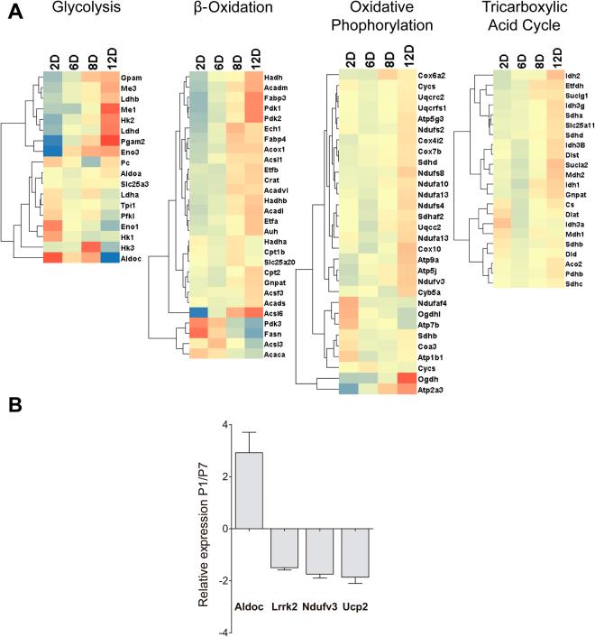
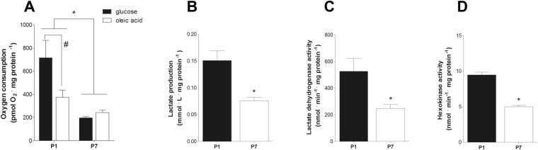
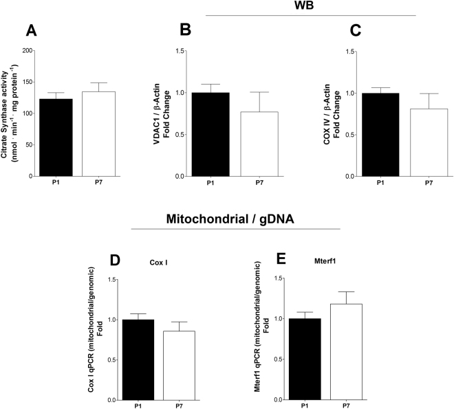
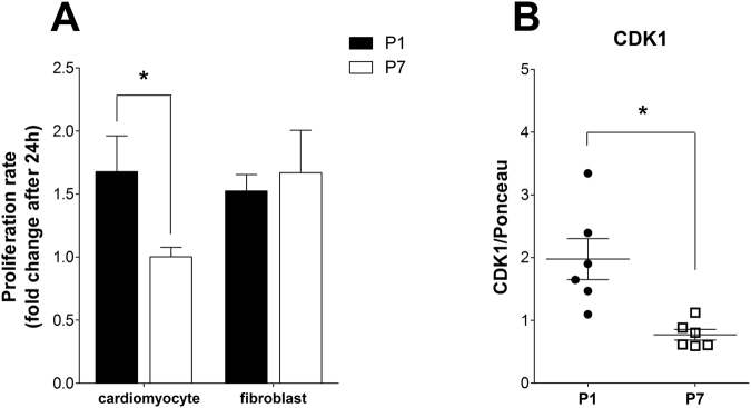
Cardiac energy metabolism must cope with early postnatal changes in tissue oxygen tensions, hemodynamics, and cell proliferation to sustain development. Here, we tested the hypothesis that proliferating neonatal cardiomyocytes are dependent on high oxidative energy metabolism. We show that energy-related gene expression does not correlate with functional oxidative measurements in the developing heart. Gene expression analysis suggests a gradual overall upregulation of oxidative-related genes and pathways, whereas functional assessment in both cardiac tissue and cultured cardiomyocytes indicated that oxidative metabolism decreases between the first and seventh days after birth. Cardiomyocyte extracellular flux analysis indicated that the decrease in oxidative metabolism between the first and seventh days after birth was mostly related to lower rates of ATP-linked mitochondrial respiration, suggesting that overall energetic demands decrease during this period. In parallel, the proliferation rate was higher for early cardiomyocytes. Furthermore, in vitro nonlethal chemical inhibition of mitochondrial respiration reduced the proliferative capacity of early cardiomyocytes, indicating a high energy demand to sustain cardiomyocyte proliferation. Altogether, we provide evidence that early postnatal cardiomyocyte proliferative capacity correlates with high oxidative energy metabolism. The energy requirement decreases as the proliferation ceases in the following days, and both oxidative-dependent metabolism and anaerobic glycolysis subside.


Conflict of interest statement

The authors declare that they have no competing interests.

Figures

Figure 1
Figure 1
Gene expression analysis. (A) Heat map showing gene expression assayed by RNASeq. The heat maps show gene clusters involved in glycolysis, β-oxidation, oxidative phosphorylation and the tricarboxylic acid cycle in rat hearts from 2, 6, 8, and 12 days after birth (n = 6 for both). (B) Quantitative RT-PCR analysis of genes involved in cell metabolism. Bars indicate the relative expression (2−ΔΔct) for P1 versus P7 hearts (n = 6 for both) of Aldolase c (a glycolysis enzyme) and mitochondrial Lrrk2, Ndufv3, and Ucp2; t-test, p < 0.05.
Figure 2
Figure 2
High oxygen consumption and glycolytic capacity in P1 heart tissue. (A) Bars indicate oxygen consumption (mean ± SEM) determined by use of an Oroboros O2k high-resolution respirometer in P1 (n = 5) and P7 (n = 6) hearts with glucose (5 mM) or oleic acid (100 µM) as substrates; two-way ANOVA, *p < 0.05 vs. P1, #p < 0.05 vs. glucose in P1. (B) Lactate levels (mean ± SEM) measured in the oxygen consumption conditioned buffer for P1 and P7 hearts (n = 6 for both); t-test, *p < 0.05 vs. P1. (C) Lactate dehydrogenase activity (mean ± SEM) in P1 and P7 hearts (n = 5 for both); t-test, *p < 0.05 vs. P1. (D) Hexokinase activity (mean ± SEM) in P1 and P7 hearts (n = 6 for both); t-test, *p < 0.05 vs. P1.
Figure 3
Figure 3
Postnatal metabolic maturation does not alter mitochondrial mass. (A) Citrate synthase activity (mean ± SEM) in P1 and P7 hearts (n = 6 for both); t-test, NS. (B) VDAC1 protein expression (mean ± SEM) in P1 and P7 hearts (n = 6 for both) measured by Western Blotting; t-test, NS. (C) COX IV protein expression (mean ± SEM) in P1 and P7 hearts (n = 6 for both) measured by Western Blotting; t-test, NS. (D) Mitochondrial Cox I and (E) Mterf1DNA quantification relative to genomic DNA in P1 and P7 heart tissue (n = 6 for both); t-test, NS.
Figure 4
Figure 4
P1 cardiomyocytes have a significant mitochondrial oxidative metabolism. (A) Time scan measurements of real-time oxygen consumption (OCR) using a Seahorse Flux Analyzer in cardiomyocyte cultures from P1 and P7 (n = 3 for both). Panels B-H show quantitative comparisons derived from experiments (mean ± SEM), such as those in panel (A,B) Basal OCR; t-test, *p < 0.05 vs. P1. (C) ATP-linked OCR; t-test, *p < 0.05 vs. P1. (D) Proton leak; t-test, *p < 0.05 vs. P1. (E) Maximal OCR; t-test, *p < 0.05 vs. P1. (F) Reserve capacity; t-test, NS. (G) Non-mitochondrial; t-test, *p < 0.05 vs. P1. (H) TMRM-stained the cell-integrated pixel area (mean ± SEM) in cardiomyocyte cultures from P1 and P7 pups (n = 3 for both); t-test, *p < 0.05 vs. P1.
Figure 5
Figure 5
Cardiac cell proliferation does not alter tissue metabolic profiles. (A) Bars indicate oxygen consumption (mean ± SEM) with glucose (5 mM) measured using Oroboros O2k high-resolution respirometry in P1, P7, and sham resected neonatal hearts 5 days (+5) after the procedure (S1 + 5d, n = 8 and R1 + 5d, n = 3; S7 + 5d, n = 7 and R7 + 5d, n = 4); two-way ANOVA, *p < 0.05 vs. 1d + 5. (B) Lactate production (mean ± SEM) measured in the oxygen consumption conditioned buffer from P1 and P7 sham and resected neonatal hearts five days after the procedure (S1 + 5d, n = 8 and R1 + 5d, n = 3; S7 + 5d, n = 7 and R7 + 5d, n = 4); two-way ANOVA, NS. (C) O2/CO2 rate (mean ± SEM) calculated from O2 and CO2 concentration in the oxygen consumption conditioned buffer from P1 and P7 sham and resected neonatal hearts 5 days after the procedure (S1 + 5d, n = 9 and R1 + 5d, n = 3; S7 + 5d, n = 7 and R7 + 5d, n = 4); two-way ANOVA, NS.
Figure 6
Figure 6
P1 cardiomyocytes maintain high proliferative rates in culture. (A) Bars indicate P1 (n = 7) and P7 (n = 4) cardiomyocyte and fibroblast proliferative rates (mean ± SEM) after 24 hours in culture; two-way ANOVA, *p < 0.05 vs. P1. (B) CDK1 protein expression (mean ± SD); t-test, *p < 0.05 vs. P1); t-test, *p < 0.05 vs. P1, in P1 and P7 hearts (n = 6).
Figure 7
Figure 7
Decreased oxygen consumption reduces P1 cardiomyocyte proliferative rates. (A) Cell viability (mean ± SEM) measured in P1 cardiomyocyte culture under control conditions and treated for 48 hours with 5 nM rotenone (n = 3 for both); t-test, NS. (B) Basal OCR (mean ± SEM) using a Seahorse Flux Analyzer in P1 cardiomyocyte cultures, control condition and treated with 5 nM rotenone for 48 hours (n = 3 for both); t-test, *p < 0.05 vs. control. (C) ATP-linked OCR (mean ± SEM) in P1 cardiomyocyte cultures, control condition and treated with 5 nM of rotenone for 48 hours (n = 3 for both); t-test, *p < 0.05 vs. control. (D) Cell cycle analysis (mean ± SEM) in P1 cardiomyocyte cultures under control conditions and treated for 48 hours with 5 nM rotenone (n = 3 for both); two-way ANOVA, *p < 0.05 vs. control. (E) Percentage of Ki67+ cardiomyocytes (mean ± SEM) in P1 cardiomyocyte cultures (n = 4 for both); t-test, *p < 0.05). (F) Bars indicate P1 cardiomyocyte and fibroblast proliferative rates (mean ± SEM) of after 24 hours in culture under control conditions (n = 7) or when treated with 5 nM rotenone (n = 6) for 48 hours; two-way ANOVA, *p < 0.05 vs. control.

Similar articles

Cited by

References

    1. Porrello E, et al. Transient regenerative potential of the neonatal mouse heart. Science. 2011;331:1078–1080. doi: 10.1126/science.1200708. - DOI - PMC - PubMed
    1. Zogbi C, Saturi de Carvalho AE, et al. Early postnatal rat ventricle resection leads to long-term preserved cardiac function despite tissue hypoperfusion. Physiological Reports. 2014;2:e12115. doi: 10.14814/phy2.12115. - DOI - PMC - PubMed
    1. Alkass K, et al. No evidence for cardiomyocyte number expansion in preadolescent mice. Cell. 2015;163:1026–1036. doi: 10.1016/j.cell.2015.10.035. - DOI - PubMed
    1. Senyo SE, et al. Mammalian heart renewal by pre-existing cardiomyocytes. Nature. 2013;493:433–436. doi: 10.1038/nature11682. - DOI - PMC - PubMed
    1. Bergmann O, et al. Evidence for cardiomyocyte renewal in humans. Science. 2009;324:98–102. doi: 10.1126/science.1164680. - DOI - PMC - PubMed

Publication types

LinkOut - more resources