. 2017 Nov 14;8(1):1488.
doi: 10.1038/s41467-017-01563-8.

NitroSynapsin therapy for a mouse MEF2C haploinsufficiency model of human autism

Affiliations

NitroSynapsin therapy for a mouse MEF2C haploinsufficiency model of human autism

Shichun Tu et al. Nat Commun. .

Abstract

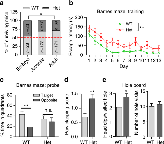
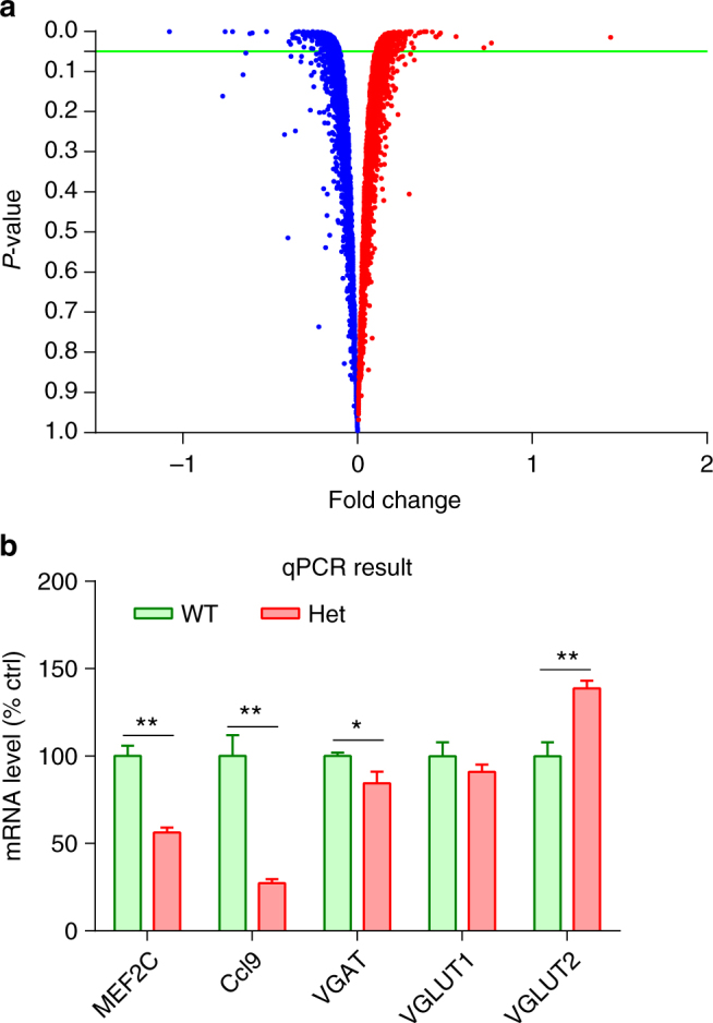
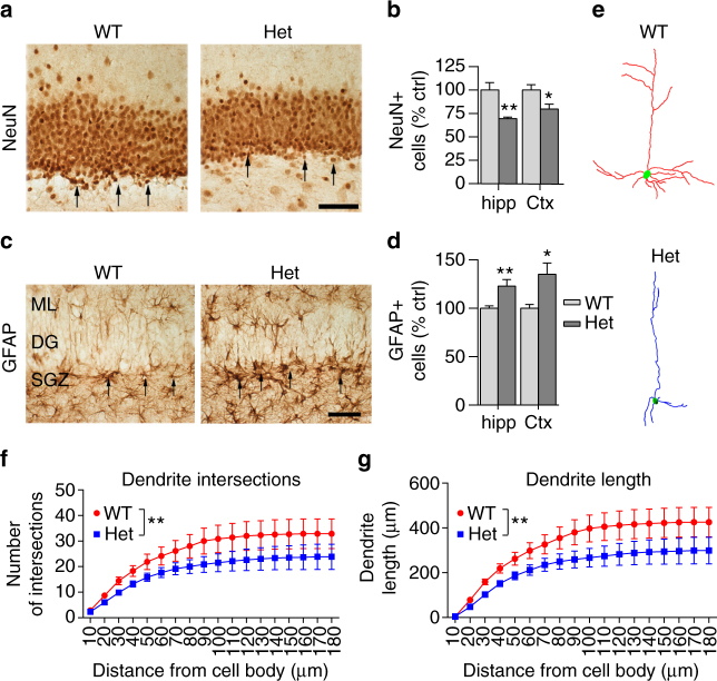
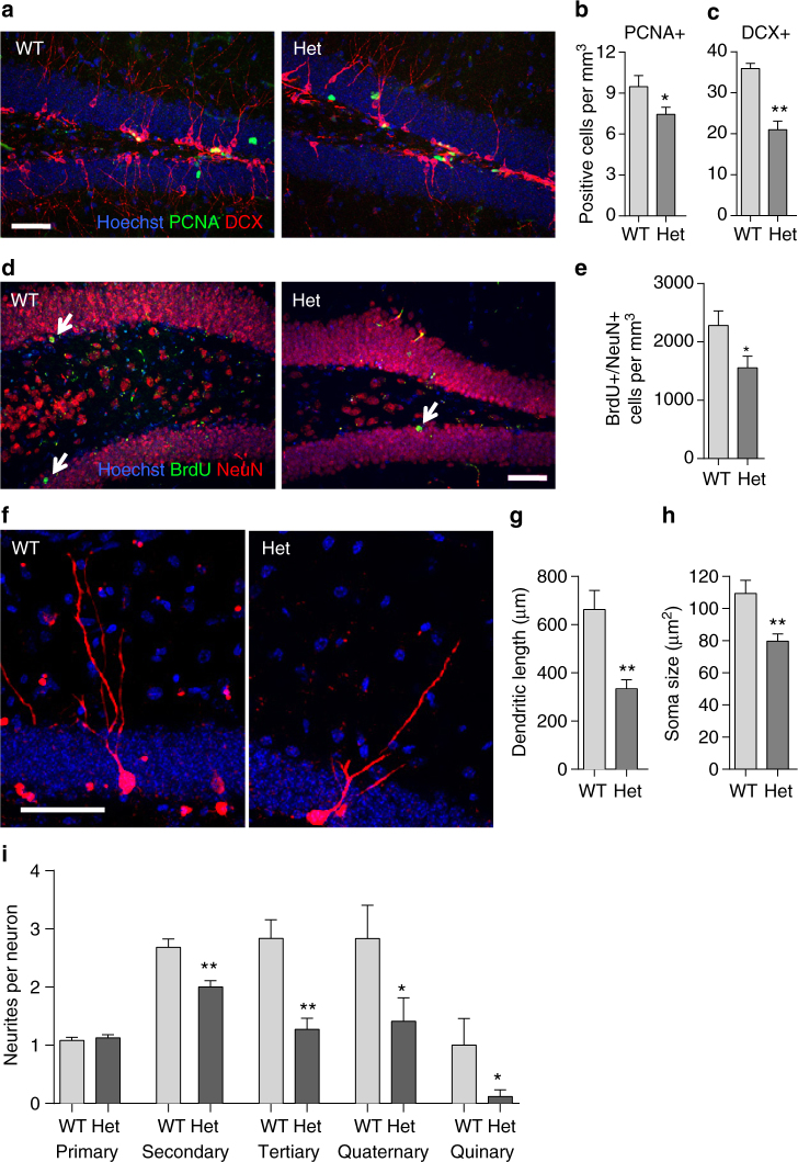
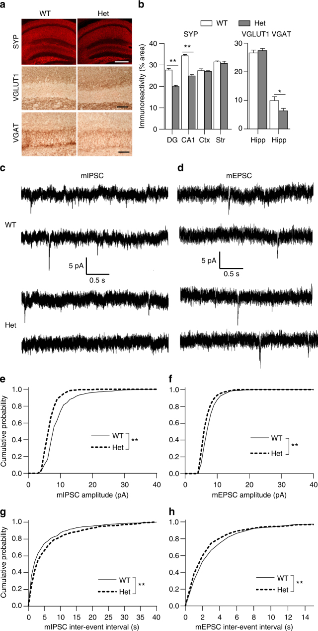
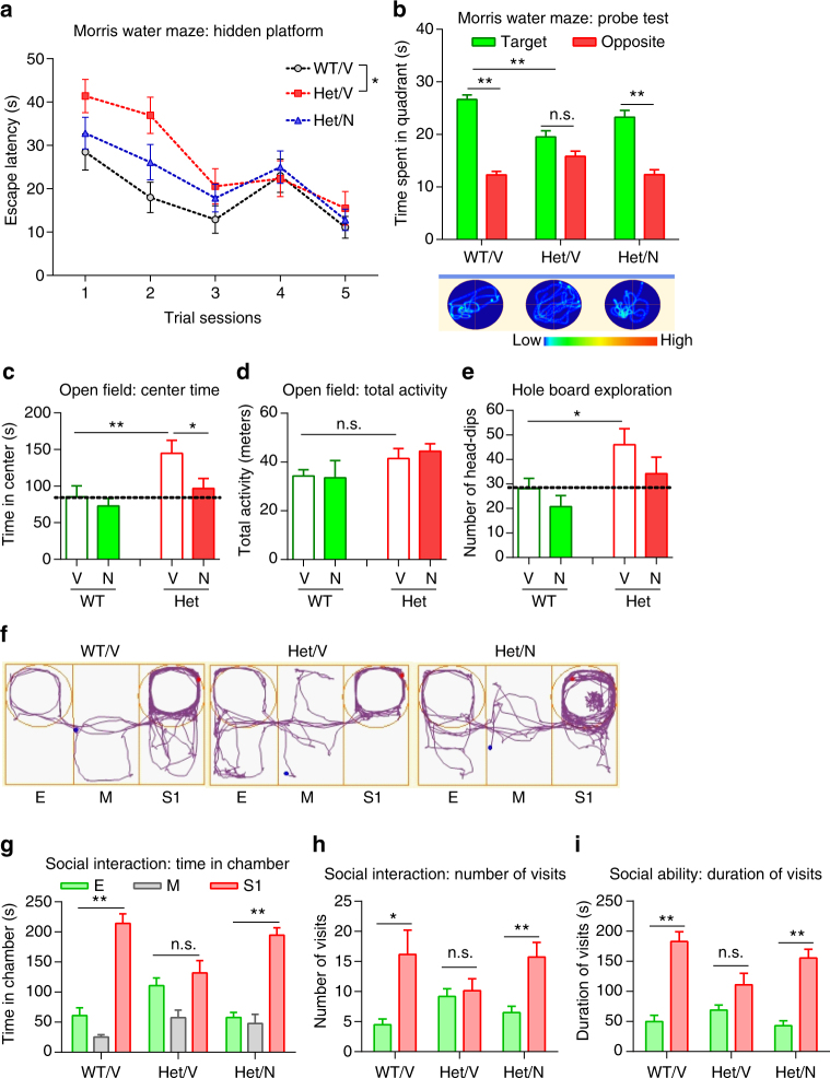
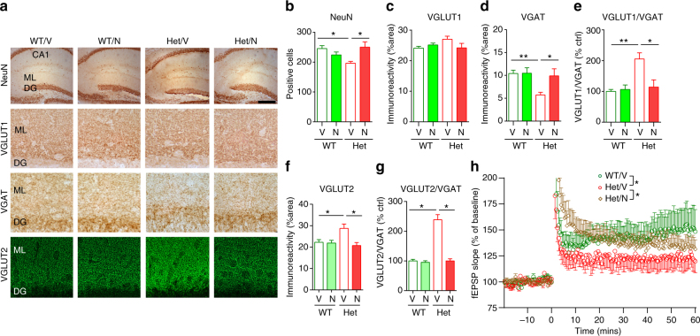
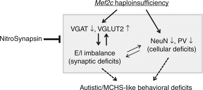
Transcription factor MEF2C regulates multiple genes linked to autism spectrum disorder (ASD), and human MEF2C haploinsufficiency results in ASD, intellectual disability, and epilepsy. However, molecular mechanisms underlying MEF2C haploinsufficiency syndrome remain poorly understood. Here we report that Mef2c +/-(Mef2c-het) mice exhibit behavioral deficits resembling those of human patients. Gene expression analyses on brains from these mice show changes in genes associated with neurogenesis, synapse formation, and neuronal cell death. Accordingly, Mef2c-het mice exhibit decreased neurogenesis, enhanced neuronal apoptosis, and an increased ratio of excitatory to inhibitory (E/I) neurotransmission. Importantly, neurobehavioral deficits, E/I imbalance, and histological damage are all ameliorated by treatment with NitroSynapsin, a new dual-action compound related to the FDA-approved drug memantine, representing an uncompetitive/fast off-rate antagonist of NMDA-type glutamate receptors. These results suggest that MEF2C haploinsufficiency leads to abnormal brain development, E/I imbalance, and neurobehavioral dysfunction, which may be mitigated by pharmacological intervention.


Conflict of interest statement

The authors declare that S.A.L. is the inventor on worldwide patents for the use of memantine and NitroSynapsin for neurodegenerative and neurodevelopmental disorders. Per Harvard University guidelines, S.A.L. participates in a royalty-sharing agreement with his former institution Boston Children’s Hospital/Harvard Medical School, which licensed the drug memantine (Namenda®) to Forest Laboratories, Inc./Actavis/Allergan, Inc. The remaining authors declare no competing financial interests.

Figures

Fig. 1
Fig. 1
Mef2c-het mice display MCHS-like phenotypes. a Mef2c-het mice die prematurely. The number of Mef2c-het compared to WT mice was nearly equal at E18, but ~45% that of WT by adulthood (~3 months) (*P < 0.05 by χ 2). b, c Impaired spatial learned and memory in the Barnes maze of Mef2c-het mice during training (b) and on subsequent probe tests (c). d, e Increased paw clasping (d) and repetitive head dipping (e) of Mef2c-het mice in hole-board exploration. Data are mean ± s.e.m.; n = 9–11 mice per genotype in b, c, and e; n = 30 (WT) and 21 (het) in d; *P < 0.05, **P < 0.01 by Student’s t test (ce) or ANOVA (b). n.s. not significant
Fig. 2
Fig. 2
Downregulation of neurogenic and synaptic genes in Mef2c-het mice by microarray analysis. a Volcano plot shows RNA expression profiling in P30 Mef2c-het and WT hippocampus (red = up, blue = down, P < 0.05 indicated by green line). b Graph of qPCR experiments showing expression levels of mRNA (relative to 18S) in Mef2c-het mice as percentage of WT control (% ctrl; n = 4 per group). Data are mean ± s.e.m.; *P < 0.05, **P < 0.01 by Student’s t test
Fig. 3
Fig. 3
Mef2c-het mice exhibit abnormal neuronal properties. a Immunohistochemistry showing NeuN+ cells in the dentate gyrus (DG) of WT and Mef2c-het mice. b Quantification showing decreased NeuN+ cell counts in hippocampus (hipp) and cortex (Ctx) in Mef2c-het mice relative to WT. Hippocampal measurements were obtained on granule cells in the molecular layer of the DG, and the cortical measurement on frontal lobe layers IV and V. c, d Increased number of GFAP+ cells consistent with astrocytosis in Mef2c-het mice. e Neurolucida drawing of representative dendrites visualized by Golgi staining in V1 (primary visual cortex), M2ML (secondary visual cortex mediolateral area), and LPtA (lateral parietal association cortex) of the visual cortex of WT and Mef2c-het mice. f, g Summary graphs of Sholl analysis showing reduction in cumulative number of dendritic intersections (f) and dendritic lengths (g) in Mef2c-het neurons. Scale bar: 50 µm. Data are mean ± s.e.m.; n = 4 per group. *P< 0.05, **P< 0.01 by Student’s t test in ad and ANOVA in f, g
Fig. 4
Fig. 4
Mef2c-het mice exhibit abnormal adult neurogenesis. a Confocal images showing PCNA (green) and DCX (red) double staining in the subgranular zone (SGZ) of the DG in 8-week-old WT and Mef2c-het mice. b, c Quantification of PCNA+ and DCX+ cells revealed reduction in the number of proliferating cells (b) and developing neurons (c) in Mef2c-het DG. d BrdU (green) and NeuN (red) double staining 4 weeks after BrdU injection in 8-week-old WT and Mef2c-het mice revealed newly born DG neurons (arrows: BrdU+/NeuN+). e Reduction in BrdU+/NeuN+ cells in Mef2c-het DG; n = 4 mice per genotype in ae. f Examples of morphological development of neurons born in adult Mef2c-het and WT mice. Dividing cells in the dentate gyrus (DG) were labeled with mCherry via retroviral-mediated gene transduction. Mice were killed 4 weeks later. g Quantification of total dendritic length of 4-week-old neurons revealed reduced dendritic length in Mef2c-het mice (n = 17) compared to WT mice (n = 12). h Quantification showed reduced somal size in Mef2c-het mice (n = 71) compared to WT mice (n = 28). i Quantification of neurite number showed normal number of primary neurites (n WT = 25, n Het = 55), but reduced number of secondary (n WT = 25, n Het = 55), tertiary (n WT = 25, n Het = 55), quaternary (n WT = 12, n Het = 17), and quinary (n WT = 12, n Het = 17) neurites of 4-week-old neurons in Mef2c-het compared to WT brain. Section thickness: 40 µm. Scale bar: 50 µm. Values are mean ± s.e.m., *P< 0.05; ***P< 0.001 by Student’s t test
Fig. 5
Fig. 5
Mef2c-het mice exhibit altered synaptic properties and E/I imbalance in synaptic neurotransmission. a Immunohistochemistry of synaptophysin (SYP), VGLUT1, and VGAT in WT and Mef2c-het hippocampus. Scale bars: 500 µm (top panel), 50 µm (middle and bottom panels). b Reduced immunoreactivity of SYP in the dentate gyrus (DG) and CA1 regions of Mef2c-het hippocampus but not cortex (Ctx) or striatum (str, left). DG measurements were performed in the molecular layer, CA1 in the pyramidal cell layer, Ctx in frontal cortical layers IV and VI, and str in the putamen at the level of the nucleus accumbens. Reduced expression of VGAT but not VGLUT1 in Mef2c-het hippocampus (right). Data are mean ± s.e.m., n = 4 per group; *P < 0.05, **P < 0.01 by Student’s t test. c, d Representative traces of mIPSCs (c) and mEPSCs (d) from slice recordings of DG neurons of WT and Mef2c-het mice. eh Cumulative plots of mIPSC and mEPSC amplitude and inter-event intervals. n = 7–9 per genotype; **P < 0.01 by two-sample Kolmogorov–Smirnov test
Fig. 6
Fig. 6
NitroSynapsin rescues MCHS-like phenotypes in Mef2c-het mice. a Latency of finding hidden platform during training sessions in the Morris water maze. b In the probe test, vehicle-treated Mef2c-het mice (Het/V) showed no preference between target and opposite quadrants, suggesting impaired memory. Treatment with NitroSynapsin (N) rescued this effect (Het/N). Representative swim patterns shown at bottom. c, d In the open field test, Het/V mice exhibited increased center time that was rescued by treatment with N (c). In contrast, Het/V mice displayed normal total activity (d). e Het/V mice displayed increased head-dips per hole, suggesting repetitive behavior. Treatment with N rescued. fi N treatment rescued aberrant social ability in Mef2c-het mice. f Representative traces of mouse movement in the three-chamber social ability test. gi Mef2c-het mice (Het/V) exhibit abnormalities in social interaction measured by time spent in each chamber (g), number of visits (h), and duration of visits (i) to E (empty) or S1 (stranger mouse 1) chambers. N treatment ameliorated this deficit (Het/N). Data are mean ± s.e.m. n = 7–9 per group. *P < 0.05, **P< 0.01 by ANOVA. M middle chamber
Fig. 7
Fig. 7
NitroSynapsin rescues abnormal neuronal and synaptic properties in Mef2c-het mice. a Immunohistochemical images of NeuN, VGLUT1, VGAT, and VGLUT2 in the molecular layer (ML) of the hippocampal dentate gyrus (DG) of WT and Mef2c-het mice treated with vehicle (V) or NitroSynapsin (N). Scale bars: 500 µm (top panel), 25 µm (middle panels), 40 µm (bottom panel). bf Summary graphs showing rescue by NitroSynapsin of decreased number of total NeuN+ cell counts (b), reduced immunoreactivity of VGAT (d) and VGLUT2 (f), and increased ratio of VGLUT1/VGAT (e) or VGLUT2/VGAT (g) in the hippocampus of Mef2c-het mice. h Impaired LTP in Mef2c-het mice was also rescued by NitroSynapsin. Data are mean ± s.e.m., n = 4–5 per group in ag and 7–9 in h. *P < 0.05, **P < 0.01, by ANOVA
Fig. 8
Fig. 8
Summary diagram of Mef2c haploinsufficiency leading to E/I imbalance and MCHS-like phenotypes that are rescued by NitroSynapsin. Mef2c haploinsufficiency leads to decreased VGAT and increased VGLUT2 protein levels, resulting in E/I imbalance (overexcitability) and synaptic dysfunction. Mef2c haploinsufficiency also causes neuronal loss, notably a reduced number of PV+ inhibitory interneurons. These synaptic and cellular abnormalities are likely the underlying cause of the MCHS-like behavioral phenotypes observed in Mef2c-het mice. The histological and behavioral phenotypes are ameliorated by chronic treatment with NitroSynapsin

Similar articles

Cited by

References

    1. Leifer D, et al. MEF2C, a MADS/MEF2-family transcription factor expressed in a laminar distribution in cerebral cortex. Proc. Natl Acad. Sci. USA. 1993;90:1546–1550. doi: 10.1073/pnas.90.4.1546. - DOI - PMC - PubMed
    1. Martin JF, Schwarz JJ, Olson EN. Myocyte enhancer factor (MEF) 2C: a tissue-restricted member of the MEF-2 family of transcription factors. Proc. Natl Acad. Sci. USA. 1993;90:5282–5286. doi: 10.1073/pnas.90.11.5282. - DOI - PMC - PubMed
    1. Okamoto S, Krainc D, Sherman K, Lipton SA. Antiapoptotic role of the p38 mitogen-activated protein kinase-myocyte enhancer factor 2 transcription factor pathway during neuronal differentiation. Proc. Natl Acad. Sci. USA. 2000;97:7561–7566. doi: 10.1073/pnas.130502697. - DOI - PMC - PubMed
    1. Shalizi A, et al. A calcium-regulated MEF2 sumoylation switch controls postsynaptic differentiation. Science. 2006;311:1012–1017. doi: 10.1126/science.1122513. - DOI - PubMed
    1. Flavell SW, et al. Activity-dependent regulation of MEF2 transcription factors suppresses excitatory synapse number. Science. 2006;311:1008–1012. doi: 10.1126/science.1122511. - DOI - PubMed

Publication types

MeSH terms