Review
. 2018 Mar;48(3):411-426.
doi: 10.1007/s00247-017-4000-0. Epub 2017 Nov 13.

Temporomandibular joint atlas for detection and grading of juvenile idiopathic arthritis involvement by magnetic resonance imaging

Affiliations
Review

Temporomandibular joint atlas for detection and grading of juvenile idiopathic arthritis involvement by magnetic resonance imaging

Christian J Kellenberger et al. Pediatr Radiol. 2018 Mar.

Abstract

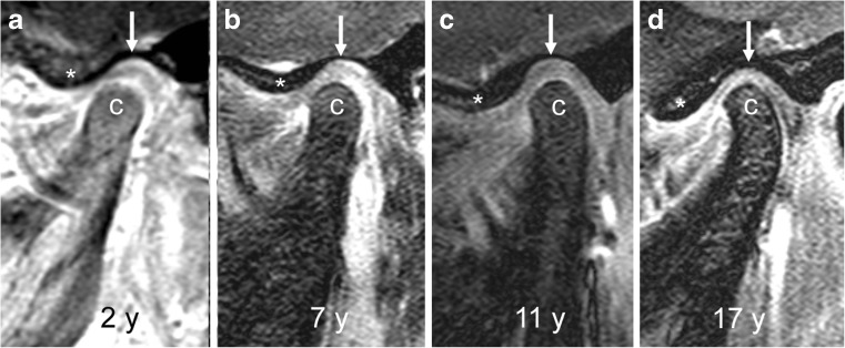
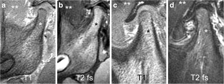
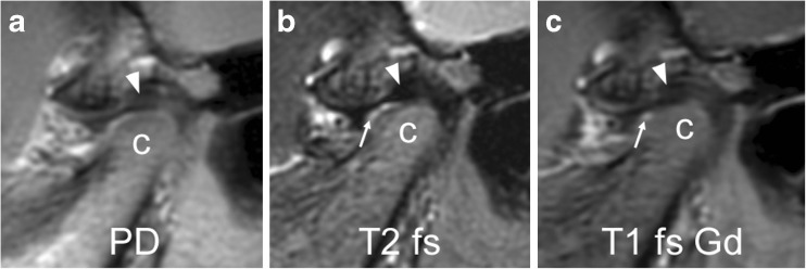
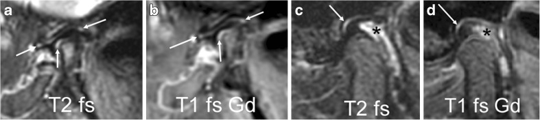
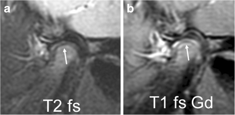
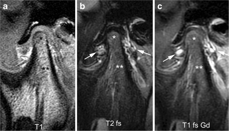
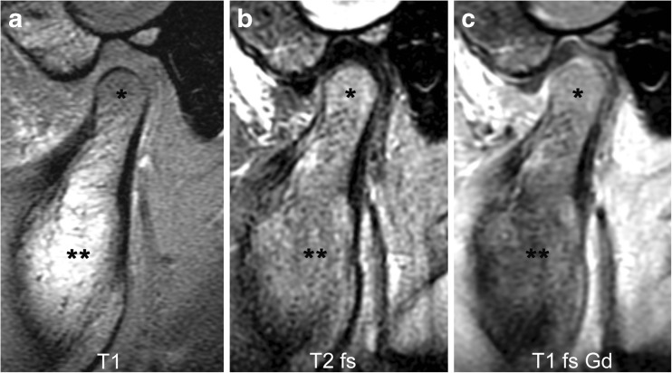
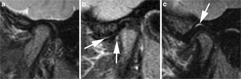
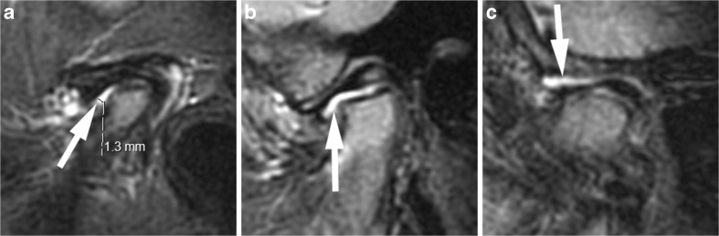
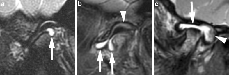
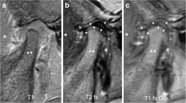
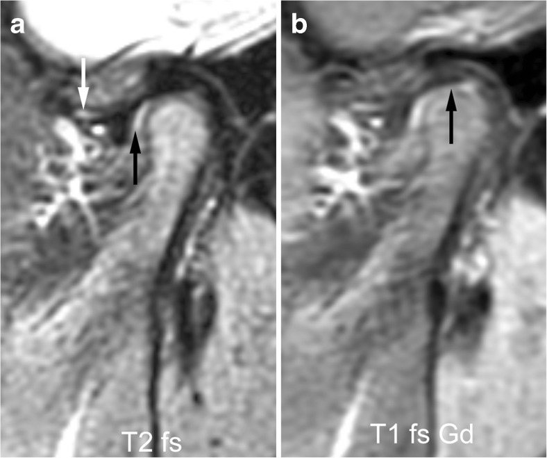
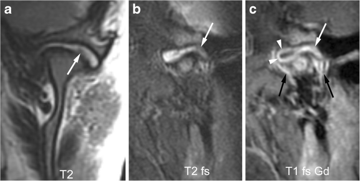
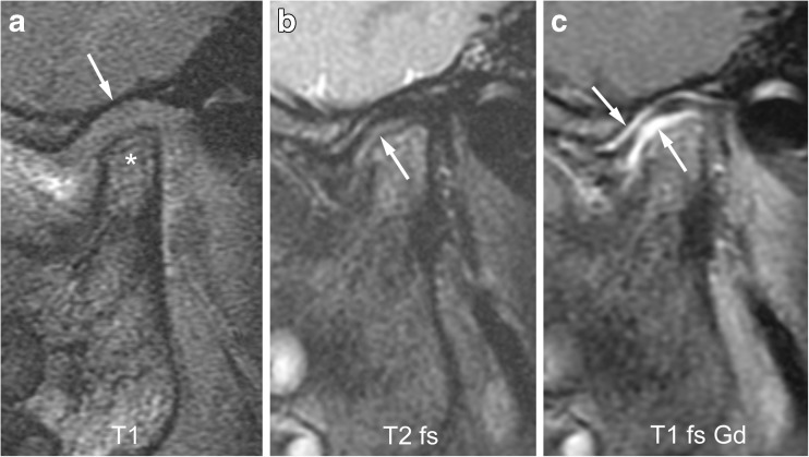
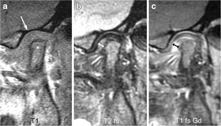
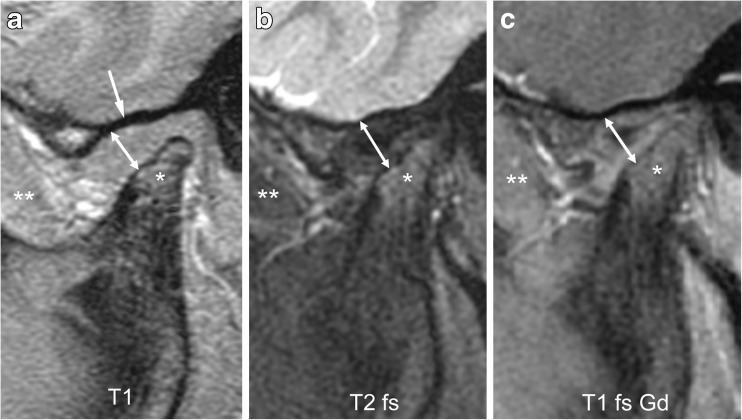
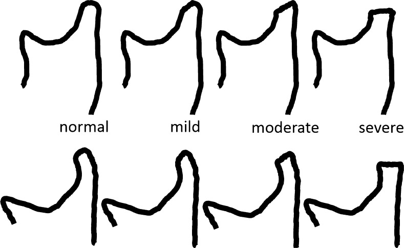
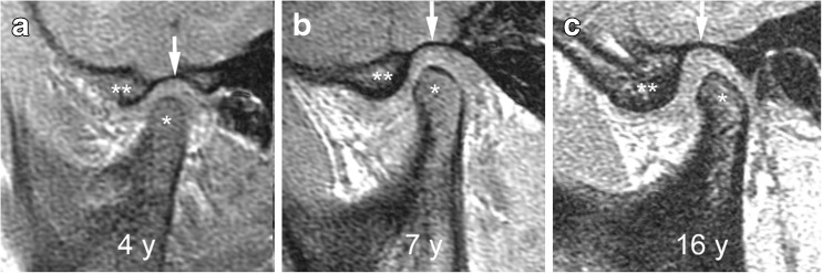
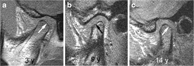
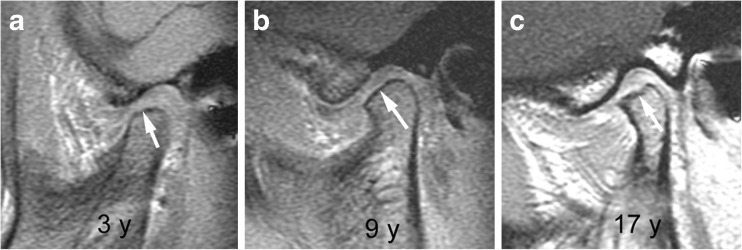
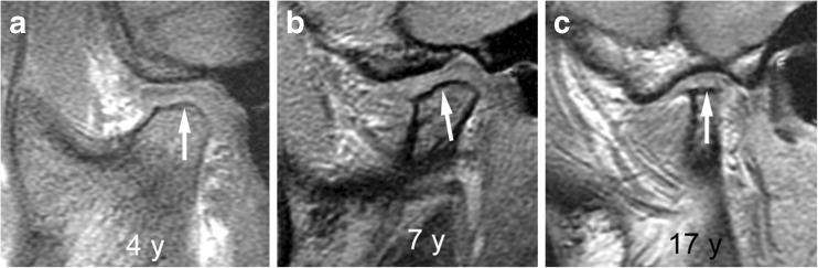
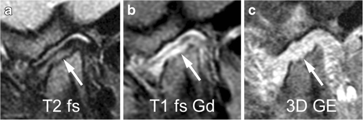
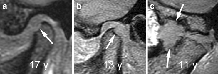
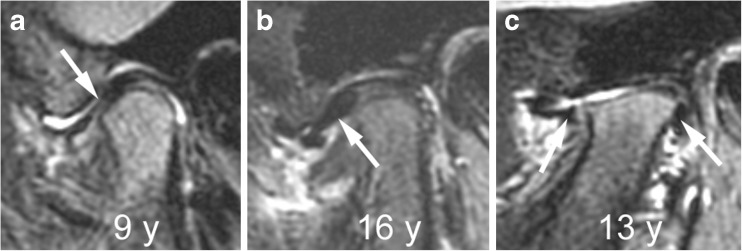
Contrast-enhanced magnetic resonance imaging (MRI) is considered the diagnostic standard for identifying involvement of the temporomandibular joint by juvenile idiopathic arthritis. Early or active arthritis is shown as bone marrow oedema, joint effusion, synovial thickening and increased joint enhancement. Subsequent joint damage includes characteristic deformity of the mandibular condyle, bone erosion, disk abnormalities and short mandibular ramus due to impaired growth. In this pictorial essay, we illustrate normal MRI findings and growth-related changes of the temporomandibular joint in children. The rationale and practical application of semiquantitative MRI assessment of joint inflammation and damage are discussed and presented. This atlas can serve as a reference for grading temporomandibular joint arthritis according to the scoring systems proposed by working groups of OMERACT (Outcome Measures in Rheumatology and Clinical Trials) and the EuroTMjoint research network. Systematic assessment of the level of inflammation, degree of osteochondral deformation, and growth of the mandibular ramus by MRI may aid in monitoring the course of temporomandibular joint arthritis and evaluating treatment options.

Keywords: Children; Juvenile idiopathic arthritis; Magnetic resonance imaging; Synovitis; Temporomandibular joint.


Conflict of interest statement

None

Figures

Fig. 1
Fig. 1
Age-dependent bony configuration of the temporomandibular joint. a-d Sagittal oblique gradient echo images (TR/TE 10/4.2 ms, flip angle 20°) from different children, obtained, at ages 2 years (a), 7 years (b), 11 years (c) and 17 years (d) show changing configurations of the mandibular condyle (c). In the 2-year-old child (a), the superior articular contour is round with a straight condylar neck. With increasing age and growth (b-d), the condylar neck gains an anterior tilt and the head appears more angular with less rounding of the anterior-superior joint surface. Articular eminence (*) height increases and glenoid fossa (arrows) gets deeper with increasing age
Fig. 2
Fig. 2
Mandibular bone marrow signal. a-b Sagittal oblique images from a 2-year-old boy with predominantly red bone marrow in the mandible. Haematopoietic marrow (* ) shows low signal intensity on T1-weighted gradient echo image without fat saturation (TR/TE 300/4.2 ms, flip angle 80°, a), which is iso- or hypointense compared to muscle (**), and intermediate signal intensity on fluid-sensitive fast spin echo image with spectral fat saturation (TR/TE 5,400/77 ms), b), which is hyperintense compared to muscle (**). c-d Sagittal oblique images in a 12-year-old boy with predominantly yellow bone marrow. Fatty marrow (*) shows high signal intensity on T1-weighted gradient echo image without fat saturation (TR/TE 300/4.2 ms, flip angle 80°, c), which is hyperintense compared to muscle (**), and intermediate to low signal intensity on fluid-sensitive fast spin echo image with spectral fat saturation (TR/TE 5,400/77 ms, d), which is almost isointense to muscle (**). Note the normal shape of the articular disk resembling a bow tie with low signal intensity on the fluid-sensitive fast spin echo images. T1 T1-weighted gradient echo, T2 fs fat saturated T2-weighted
Fig. 3
Fig. 3
Normal articular disk in a 6-year-old girl. a-c Proton density weighted (TR/TE 3,000/13 ms, a), fat-saturated T2-weighted (TR/TE 5,400/77 ms, b), and postcontrast fat-saturated T1-weighted (TR/TE 670/10 ms, c) fast spin echo images all show a normal biconcave shape of the articular disk and the posterior band (arrowheads) located at the 11 o’clock position of the mandibular condyle (c). Note normal joint enhancement (arrow in c) confined to small amounts of fluid in the anterior lower joint space (arrow in b) and mild flattening of the anterior portion of condyle. PD proton density, T1 fs Gd contrast-enhanced fat saturated T1-weighted, T2 fs fat saturated T2-weighted
Fig. 4
Fig. 4
Normal joint fluid and enhancement. a-b Sagittal oblique images from a 3-year-old girl show normal amounts of fluid in joint recesses. Fluid-sensitive image (TR/TE 5,400/77 ms, fat-saturated, a) shows high signal intensity (arrows). On contrast enhanced fat saturated T1-weighted image (TR/TE 670/10 ms, b) there is corresponding enhancement (arrows). c-d Sagittal oblique fast spin echo images from a 13-year-old girl show normal amounts of fluid (arrow) in the joint recesses, with high signal intensity on fluid-sensitive images (c) and enhancement following contrast application shown as high signal intensity on post-contrast fat-saturated T1-weighted images (d). The slightly expanded venous plexus in the retrodiskal tissue shows high signal intensity on T2-weighted images (* in c) and enhancement (* in d). T1 fs Gd contrast-enhanced fat saturated T1-weighted, T2 fs fat saturated T2-weighted
Fig. 5
Fig. 5
Mandibular growth zone. Sagittal oblique fast spin echo images in a 3-year-old girl show the mandibular growth zone at the surface of the condyle. a Beneath the low signal intensity line of cartilage and subchondral bone, this is seen as a narrow zone of high signal intensity (arrow) on fluid-sensitive sequence (TR/TE 5,400/77 ms, fat-saturated). b On postcontrast fat-saturated T1-weighted images (TR/TE 670/10 ms) there is corresponding enhancement. These findings represent the well vascularised zone of endochondral ossification. Note enhancement of the whole lower joint compartment as well as anterior recess of upper compartment. T1 fs Gd contrast-enhanced fat saturated T1-weighted, T2 fs fat saturated T2-weighted
Fig. 6
Fig. 6
Normal bone marrow signal and enhancement (grade 0) in a 14-year-old boy. a-c Sagittal oblique T1-weighted (TR/TE 300/4.2 ms, flip angle 80°) gradient echo image without fat saturation (a), T2-weighted (TR/TE 5,400/77 ms) fat saturated image (b) and postcontrast fat-saturated T1-weighted (TR/TE 670/10 ms) fast spin echo images (c) show the marrow space of the mandibular condyle (*) with isointense signal compared to that of the mandibular ramus (**). Note prominent veins surrounding the joint (arrows in b,c) and slightly increased enhancement of the posterior upper joint compartment. For this age, the condyle appears rather round and lacks an anterior tilt of the mandibular neck. T1 T1-weighted, T1 fs Gd contrast-enhanced fat saturated T1-weighted, T2 fs fat saturated T2-weighted
Fig. 7
Fig. 7
Bone marrow oedema and enhancement (grade 1) of the mandibular condyle in a 9-year-old boy. a-c When compared to the mandibular ramus (**) there is lower signal from the mandibular condyle (*) on the T1-weighted gradient echo image without fat saturation (TR/TE 300/4.2 ms, flip angle 80°, a), and slightly increased signal on fat-saturated T2-weighted image (TR/TE 5,400/77 ms, b) and on postcontrast fat-saturated T1-weighted image (TR/TE 670/10 ms, c) images, Note mild joint enhancement with high signal intensity (hyperintense to muscle) in the posterior superior joint recess and lower joint compartment on postcontrast fat-saturated T1-weighted image (c)
Fig. 8
Fig. 8
No joint effusion (grade 0). a-c Sagittal oblique fat-saturated T2-weighted fast spin echo images (TR/TE 5,400/77 ms). There is no high signal intensity fluid within the joint compartments in a 3-year-old boy (a). There is a normal amount of fluid in the anterior recesses (arrows in b) in a 14-year-old boy and in the superior joint compartment (arrow in c) in a 14-year-old girl
Fig. 9
Fig. 9
Small joint effusion (grade 1). a-c Sagittal oblique fat-saturated T2-weighted fast spin echo images (TR/TE 5,400/77 ms) show high signal intensity (arrows) of >1-mm and ≤2-mm thickness within the joint spaces in the lower anterior recess in a 6-year-old girl (a), in the lower joint compartment (b) and upper joint compartment (c) in a 5-year-old boy. Note mild flattening of condyle in (a), and moderate/severe flattening of the condyle in (b) and (c)
Fig. 10
Fig. 10
Large joint effusion (grade 2) on sagittal oblique images. a-c Effusion exceeding 2-mm width (arrows) on fat-saturated T2-weighted fast spin echo images (TR/TE 5,400/77 ms) in the posterior recess in a 15-year-old girl (a), in the anterior recesses in a 13-year-old boy (b) and with bulging of the upper joint compartment in a 7-year-old girl (c). Note thickened synovium with intermediate signal intensity (arrowheads) on these posteriorly in (b) and (c)
Fig. 11
Fig. 11
No synovial thickening (grade 0) and no joint enhancement (grade 0) on sagittal oblique images in a 3-year-old boy. a There is a large proportion of haematopoietic marrow in the mandible, which is shown as low signal intensity (**) similar to that of muscle tissue (*) in an unenhanced T1-weighted gradient echo image without fat saturation (T1, TR/TE 300/4.2 ms, flip angle 80°). b-c The width of both joint compartments (including articular cartilage, synovium and joint space) between the bony articular surface and disk is smaller than 1 mm (small double arrows) on fat-saturated T2-weighted image (TR/TE 5,400/77 ms, b) and on contrast-enhanced fat-saturated T1-weighted image (TR/TE 670/10 ms, c). Postcontrast signal intensity of the joint spaces is similar to that muscle (*). There is higher signal intensity compared to that of muscle (*) in the fluid-sensitive image (b) and postcontrast fat-saturated T1-weighted image (c). T1 T1-weighted, T1 fs Gd contrast-enhanced fat saturated T1-weighted, T2 fs fat saturated T2-weighted
Fig. 12
Fig. 12
No synovial thickening (grade 0) but mild increase of joint enhancement (grade 1) on sagittal oblique images in a 6-year-old boy. a Fat saturated T2-weighted image (TR/TE 5,400/77 ms) shows normal amount of joint fluid in upper and lower anterior recesses (arrows). b Postcontrast T1-weighted fat-saturated image (TR/TE 670/10 ms) shows high signal intensity, isointense to that of veins, in both joint compartments. At the midportion of the lower joint compartment (arrow) this exceeds the extent of high signal intensity seen in (a). Note mild bone marrow oedema and enhancement of the normally shaped mandibular condyle. T1 fs Gd contrast-enhanced fat saturated T1-weighted, T2 fs fat saturated T2-weighted
Fig. 13
Fig. 13
Mild synovial thickening (grade 1) and severe joint enhancement (grade 2). a-b Coronal T2-weighted image (TR/TE 2,600/110 ms, a) and sagittal oblique fat-saturated T2-weighted image (TR/TE 5,400/77 ms, b) show a large effusion with high signal intensity and some nodular areas of synovial thickening with intermediate signal intensity in posterior portion of the upper joint compartment (arrows). c Corresponding early postcontrast fat-saturated T1-weighted (TR/TE 670/10 ms) image demonstrates enhancement of the synovium (white arrow) and peripheral joint fluid (arrowheads) involving the entire upper compartment, with same signal intensity as surrounding veins (black arrows). T1 fs Gd contrast-enhanced fat saturated T1-weighted, T2 T2-weighted, T2 fs fat saturated T2-weighted
Fig. 14
Fig. 14
Mild synovial thickening (grade 1) and severe joint enhancement (grade 2) on sagittal oblique images in a 5-year-old boy. a There moderate flattening of condyle (*) and mild flattening of temporal bone (arrow) on T1-weighted image (TR/TE 300/4.2 ms, flip angle 80°, without fat saturation). b Fluid-sensitive, fat-saturated T2-weighted fast spin echo image (TR/TE 5,400/77 ms) shows tissue with intermediate signal intensity exceeding 1-mm width in lower joint space (arrow). c Postcontrast fat-saturated T1-weighted image (TR/TE 670/10 ms) shows high signal intensity (arrows) involving both the upper and lower joint compartments. T1-weighted, T2 T2-weighted, T2 fs fat saturated T2-weighted
Fig. 15
Fig. 15
Severe synovial thickening (grade 2) and severe joint enhancement (grade 2) on sagittal oblique images in a 11-year-old girl. a There is moderate flattening of the condyle (*) and mild flattening of the temporal bone (arrow) on T1-weighted image (TR/TE 300/4.2 ms, flip angle 80°). b Thickened synovium (arrow) is shown in the anterior inferior portion of the joint space as intermediate signal intensity tissue exceeding 2-mm width on fat saturated T2-weighted fast spin echo (TR/TE 5,400/77 ms). c There is strong enhancement of the thickened synovium (arrow) but also the entire lower joint space and the posterior half of the upper joint space are shown as high signal intensity on postcontrast fat-saturated T1-weighted image (TR/TE 670/10 ms). T1 T1-weighted, T1 fs Gd contrast-enhanced fat saturated T1-weighted, T2 fs fat saturated T2-weighted
Fig. 16
Fig. 16
Severe synovial thickening (grade 2) but no increased joint enhancement (grade 0) on sagittal oblique images in an 8-year-old girl. a-c There is moderate flattening of the mandibular condyle (*) and mandibular fossa (arrow in a), and joint space expansion (double arrow) on T1-weighted image (TR/TE 300/4.2 ms, flip angle 80°, a), fat-saturated T2-weighted fast spin echo image (TR/TE 5,400/77 ms, b) and on postcontrast fat-saturated T1-weighted image (TR/TE 670/10 ms, c). The synovium is isointense to muscle (**) on all images. T1 T1-weighted, T1 fs Gd contrast-enhanced fat saturated T1-weighted, T2 fs fat saturated T2-weighted
Fig. 17
Fig. 17
Grades of condylar flattening. Schematic drawing showing different degrees of condylar flattening viewed in the sagittal oblique plane, in a young child (upper row) up to about 5 years of age and in an older child (lower row)
Fig. 18
Fig. 18
Normal shape of mandibular condyle (grade 0) and temporal bone on sagittal oblique T1-weighted gradient echo images (TR/TE 300/4.2 ms, flip angle 80°, without fat saturation). a-c Images from three different children show the changing configuration of the mandibular condyle (*), the articular eminence (**) and the glenoid fossa (arrows) among a 4-year-old (a), a 7-year-old (b) and a 16-year-old (c). In a 4-year-old child (a) the superior articular contour is round with a straight condylar neck. With increasing age and growth, the condylar neck gains an anterior tilt and the head appears more angular, with less rounding of the anterior-superior joint surface. The articular eminence gets larger resulting in a deeper glenoid fossa
Fig. 19
Fig. 19
Mild condylar flattening (grade 1). a-c Sagittal oblique T1-weighted gradient echo images (TR/TE 300/4.2 ms, flip angle 80°, without fat saturation) from different children obtained at 3 years (a), 9 years (b) and 14 years (c) show flattening involving part of the condylar surface (arrows)
Fig. 20
Fig. 20
Moderate condylar flattening (grade 2). a-c. Sagittal oblique T1-weighted gradient echo images (TR/TE 300/4.2 ms, flip angle 80°, without fat saturation) from different children obtained at 3 years (a), 9 years (b) and 17 years (c) show flattening involving the entire surface of the condyle with preserved anterior angulation (arrows)
Fig. 21
Fig. 21
Severe condylar flattening (grade 2). a-c Sagittal oblique T1-weighted gradient echo images (TR/TE 300/4.2 ms, flip angle 80°, without fat saturation) from different children obtained at 4 years (a), 7 years (b) and 17 years (c) show flattening involving the entire surface of the condyle and horizontally oriented joint surface (arrows)
Fig. 22
Fig. 22
Small erosions in a 7-year-old girl on sagittal oblique images. a-c Irregularity of the bony articular surface (arrows) with small breaks of the subchondral bone is evident on fat saturated fast spin echo image (TR/TE 5,400/77 ms, a) and on fat-saturated postcontrast T1-weighted image (TR/TE 670/10 ms), but is best delineated on gradient echo image (TR/TE 10/4.2 ms, flip angle 20°, c). For assessing the extent of these erosions (part of condylar surface versus entire articular surface), all successive images covering the condyle from lateral to medial need to be viewed. Note bone marrow oedema, mild synovial thickening and severe joint enhancement. 3D GE volumetric T1-weighted gradient echo, T1 fs Gd contrast-enhanced fat saturated T1-weighted, T2 fs fat saturated T2-weighted
Fig. 23
Fig. 23
Large erosions. a-c Sagittal oblique gradient echo images (TR/TE 10/4.2 ms, flip angle 20°) from different children show large defect (arrow) of the anterior condylar surface in a 17-year-old (a), of the posterior condylar surface (arrow) in a 13-year-old (b) and involving both a severely deformed mandibular condyle and the articular eminence of the adjacent temporal bone (arrows) in an 11-year-old (c)
Fig. 24
Fig. 24
Abnormalities of articular disk. a-c Sagittal oblique fat-saturated T2-weighted fast spin echo images (TR/TE 5,400/77 ms) from different children show a flat disk (arrow) in a 9-year-old (a), an anteriorly dislocated disk (arrow) in a 16-year-old (b) and a perforated disk with peripheral remnants (arrows) seen in the anterior and posterior joint recesses in a 13-year-old (c)

References

    1. Larheim TA, Doria AS, Kirkhus E, et al. TMJ imaging in JIA patients—an overview. Semin Orthod. 2015;21:102–110. doi: 10.1053/j.sodo.2015.02.006. - DOI
    1. Kellenberger CJ, Arvidsson LZ, Larheim TA. Magnetic resonance imaging of temporomandibular joints in juvenile idiopathic arthritis. Semin Orthod. 2015;21:111–120. doi: 10.1053/j.sodo.2015.02.007. - DOI - PubMed
    1. Kuseler A, Pedersen TK, Herlin T, et al. Contrast enhanced magnetic resonance imaging as a method to diagnose early inflammatory changes in the temporomandibular joint in children with juvenile chronic arthritis. J Rheumatol. 1998;25:1406–1412. - PubMed
    1. Muller L, Kellenberger CJ, Cannizzaro E, et al. Early diagnosis of temporomandibular joint involvement in juvenile idiopathic arthritis: a pilot study comparing clinical examination and ultrasound to magnetic resonance imaging. Rheumatology (Oxford) 2009;48:680–685. doi: 10.1093/rheumatology/kep068. - DOI - PMC - PubMed
    1. Weiss PF, Arabshahi B, Johnson A, et al. High prevalence of temporomandibular joint arthritis at disease onset in children with juvenile idiopathic arthritis, as detected by magnetic resonance imaging but not by ultrasound. Arthritis Rheum. 2008;58:1189–1196. doi: 10.1002/art.23401. - DOI - PubMed

MeSH terms

LinkOut - more resources