. 2018 Feb;135(2):227-247.
doi: 10.1007/s00401-017-1785-8. Epub 2017 Nov 13.

Artificial intelligence in neurodegenerative disease research: use of IBM Watson to identify additional RNA-binding proteins altered in amyotrophic lateral sclerosis

Affiliations

Artificial intelligence in neurodegenerative disease research: use of IBM Watson to identify additional RNA-binding proteins altered in amyotrophic lateral sclerosis

Nadine Bakkar et al. Acta Neuropathol. 2018 Feb.

Abstract

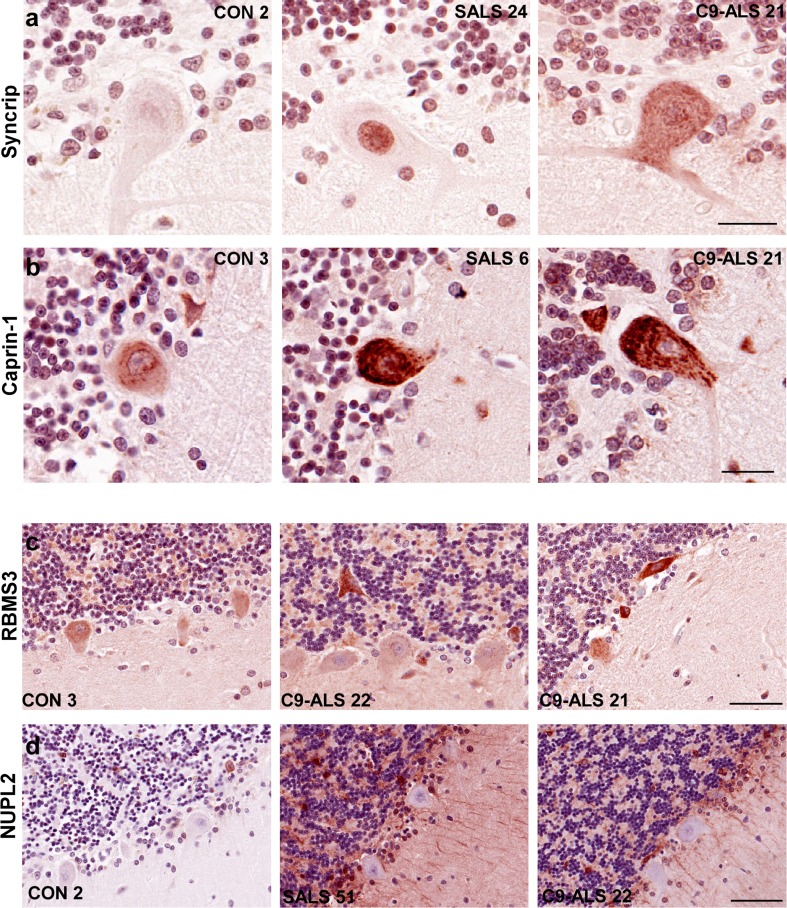
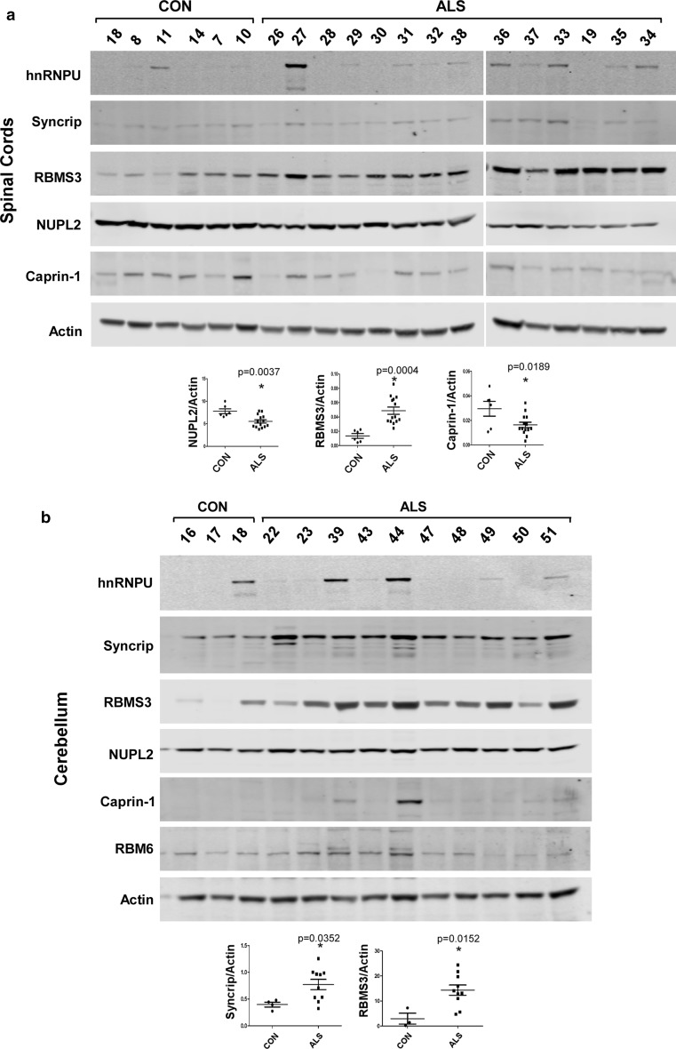
Amyotrophic lateral sclerosis (ALS) is a devastating neurodegenerative disease with no effective treatments. Numerous RNA-binding proteins (RBPs) have been shown to be altered in ALS, with mutations in 11 RBPs causing familial forms of the disease, and 6 more RBPs showing abnormal expression/distribution in ALS albeit without any known mutations. RBP dysregulation is widely accepted as a contributing factor in ALS pathobiology. There are at least 1542 RBPs in the human genome; therefore, other unidentified RBPs may also be linked to the pathogenesis of ALS. We used IBM Watson® to sieve through all RBPs in the genome and identify new RBPs linked to ALS (ALS-RBPs). IBM Watson extracted features from published literature to create semantic similarities and identify new connections between entities of interest. IBM Watson analyzed all published abstracts of previously known ALS-RBPs, and applied that text-based knowledge to all RBPs in the genome, ranking them by semantic similarity to the known set. We then validated the Watson top-ten-ranked RBPs at the protein and RNA levels in tissues from ALS and non-neurological disease controls, as well as in patient-derived induced pluripotent stem cells. 5 RBPs previously unlinked to ALS, hnRNPU, Syncrip, RBMS3, Caprin-1 and NUPL2, showed significant alterations in ALS compared to controls. Overall, we successfully used IBM Watson to help identify additional RBPs altered in ALS, highlighting the use of artificial intelligence tools to accelerate scientific discovery in ALS and possibly other complex neurological disorders.

Keywords: Amyotrophic lateral sclerosis; Artificial intelligence; Motor neuron; Protein aggregation; RNA-binding protein.


Conflict of interest statement

RB is a founder of Iron Horse Diagnostics, Inc., a biotechnology company focused on diagnostic and prognostic biomarkers for ALS and other neurologic disorders.

Figures

Fig. 1
Fig. 1
Immunolocalization of IBM Watson top-ranked RBPs in lumbar spinal cord. IHC for hnRNPU, SC-35, Caprin-1 and RBM6 in the lumbar spinal cord of 4 C9-ALS, 4 non-neurological disease controls and 6–14 SALS cases. Representative images of motor neurons are shown, counterstained with hematoxylin. a Control motor neurons stained with hnRNPU show weak nuclear staining, while ALS motor neurons exhibit either strong nuclear staining, or cytoplasmic thread-like inclusions. b SRSF2/SC-35 labeled nuclear speckles in control motor neurons. ALS neurons exhibit a variety of phenotypes ranging from large dark speckles, to strong nuclear staining, and rare cytoplasmic inclusions and neuropil staining in one ALS case (SALS 49). c Caprin-1 labels cytoplasmic granules in control motor neurons, with larger granules and strong immunostaining in ALS neurons. In addition, most SALS cases but no C9-ALS cases exhibit Caprin-1 staining in the nucleolus. d RBM6 is negative or weak in control motor neurons, while ALS cases exhibit nucleolar staining. All images were taken at ×40 magnification. Scale bar: 50 μm
Fig. 2
Fig. 2
Immunolocalization of IBM Watson top-ranked RBPs in the cerebellum. IHC for Syncrip, Caprin-1, RBMS3 and NUPL2 in 4 C9-ALS, 3–5 non-neurological disease controls and 8 SALS cases. Representative images are shown, counterstained with hematoxylin. a Weak Syncrip-labeled nuclei in control Purkinje cells, as well as granule cell nuclei in one out of five control cases. C9-ALS Purkinje cells exhibited medium-to-strong diffuse cytoplasmic Syncrip staining (four out of four), while SALS displayed weak granule cell and strong Purkinje cell nuclear staining. b Weak Caprin-1 IHC in four out of five control cerebellum; while Purkinje cells in C9-ALS (four out of four cases) and SALS (four out of seven) exhibited medium-to-strong cytoplasmic staining. c Negative-to-weak RBMS3 IHC in control cerebellum with three out of five cases showing no interneuron staining, while three out of five had some granular layer interstitial immunostaining. Interneurons in the Purkinje, molecular and granule cell layers displayed strong RBMS3 IHC in all C9-ALS and SALS cases. In addition, six ALS case showed some strong inter-granule cell staining, while three cases had RBMS3 staining in Purkinje cells. d NUPL2 was negative to weak in all controls, but labeled astrocytes in all SALS and one out of four C9-ALS cases examined. Purkinje cells were negative for NUPL2 in all subject groups. All images were taken at ×40 magnification. Scale bar: 25 μm for a, b, 50 μm for c and 70 μm for d
Fig. 3
Fig. 3
Quantification of hnRNPU and Syncrip staining intensities. a hnRNPU motor neuron nuclear staining intensity ranges were measured using the Aperio ImageScope software in lumbar spinal cord sections (see “Materials and methods”). The four intensity ranges (negative, weak, medium and strong) were combined into two categories for ease of viewing, with negative/weak depicted by −/+ and medium/strong depicted by ++/+++ and the results were plotted for controls, C9-ALS and SALS. One-way ANOVA with Bonferroni’s multiple comparison testing demonstrated that for the medium/strong group (++/+++), SALS was significantly different from CON (*p < 0.01), and C9-ALS (**p < 0.01), while C9-ALS and CON were not different from each other. b Syncrip staining intensities of Purkinje cells, with categories pooled into negative/weak (−/+) and medium/strong (++/+++). One-way ANOVA shows that for the negative/weak group, CON was significantly different from SALS (*p < 0.01) and from C9-SALS (**p < 0.001); while for the medium/strong group, CON was statistically different from the C9-ALS group (**p < 0.01), and from the SALS group (*p < 0.001). Values depicted are means ± SEM (standard error of the mean)
Fig. 4
Fig. 4
Co-localization of top-ranked RBPs with TDP-43 or p62. Lumbar spinal cords sections from SALS sections were co-stained with a hnRNPU and TDP-43, b SC-35 and TDP-43, or c Caprin-1 and p62. Nuclei were co-stained with DAPI and images were captured on a confocal microscope at ×63 magnification. Scale bars represent 10 μm
Fig. 5
Fig. 5
Cell type determination of RBMS3 and NUPL2 positive cells in the cerebellum by confocal microscopy. a Cerebellar sections from SALS were co-labeled with RBMS3, parvalbumin or calretinin to identify interneurons. b Double-label confocal microscopy of NUPL2 and GFAP to identify astrocytes. Large arrowheads in a depict calretinin-positive, parvalbumin-negative interneurons (possibly unipolar brush cells), small arrowheads point to calretinin-positive, parvalbumin-positive (potentially Golgi cells), while small arrows point to calretinin-negative, parvalbumin-positive interneurons (possible basket or stellate cells). Scale bars represent 10 μm
Fig. 6
Fig. 6
Gene expression of IBM Watson RBPs in spinal cord and cerebellum. a RNA from 4–5 control and 8 SALS spinal cords were extracted, cDNA was made and real-time PCR was performed for the IBM Watson top and bottom-ranked RBPs. b Cerebellum tissue from 4 controls, 7–8 SALS, and 2 C9-ALS (shown in blue) were used for real-time PCR. Individual values depicted are average of three experimental replicates, and mean ± SEM are shown. Significance is indicated by stars and p values are listed in each plot
Fig. 7
Fig. 7
Gene expression of IBM Watson proteins in iPSC-MN and laser-captured Purkinje cells. a iPSC-derived motor neurons were differentiated for 45–60 days, RNA extracted and real-time PCR was performed for IBM Watson-ranked RBPs. 3 separate control iPSC lines, 5 different C9-ALS lines (2 independent differentiations of 4 lines C9-ALS 2–5, and one differentiation of line C9-ALS1), and two SALS lines were used. The different colors depict the various lines used, ran in experimental triplicates, and values shown are means ± SEM. Asterisks denote significance, with p values of 0.0495 for Caprin-1 (CON vs. C9-ALS), 0.0052 for RBMS3 (CON vs. SALS), and 0.0314 for Syncrip (CON vs. C9-ALS). b Frozen cerebellar sections were stained with methyl green pyronin, and at least 250 Purkinje cells were captured from each case, RNA extracted and real-time PCR was performed on each sample. 3 controls and 7 SALS cases were used. Bars represent individual data points calculated from experimental replicates
Fig. 8
Fig. 8
Immunoblot analysis of IBM Watson-ranked proteins in spinal cord and cerebellum. a Immunoblot of protein lysates prepared from 6 control and 14 SALS spinal cord sample. b Immunoblot of cerebellum protein lysates from 4 control, 8 SALS and 2 C9-ALS (#22 and 23) cases. Graphs and statistical analysis are shown for proteins that are significantly altered in ALS when compared to controls

References

    1. Al-Chalabi A, Jones A, Troakes C, King A, Al-Sarraj S, van den Berg LH. The genetics and neuropathology of amyotrophic lateral sclerosis. Acta Neuropathol. 2012;124:339–352. doi: 10.1007/s00401-012-1022-4. - DOI - PubMed
    1. Al-Sarraj S, King A, Troakes C, Smith B, Maekawa S, Bodi I, Rogelj B, Al-Chalabi A, Hortobagyi T, Shaw CE. p62 positive, TDP-43 negative, neuronal cytoplasmic and intranuclear inclusions in the cerebellum and hippocampus define the pathology of C9orf72-linked FTLD and MND/ALS. Acta Neuropathol. 2011;122:691–702. doi: 10.1007/s00401-011-0911-2. - DOI - PubMed
    1. Bain JM, Cho MT, Telegrafi A, Wilson A, Brooks S, Botti C, Gowans G, Autullo LA, Krishnamurthy V, Willing MC, Toler TL, Ben-Zev B, Elpeleg O, Shen Y, Retterer K, Monaghan KG, Chung WK. Variants in HNRNPH2 on the X chromosome are associated with a neurodevelopmental disorder in females. Am J Hum Genet. 2016;99:728–734. doi: 10.1016/j.ajhg.2016.06.028. - DOI - PMC - PubMed
    1. Blokhuis AM, Koppers M, Groen EJ, van den Heuvel DM, Dini Modigliani S, Anink JJ, Fumoto K, van Diggelen F, Snelting A, Sodaar P, Verheijen BM, Demmers JA, Veldink JH, Aronica E, Bozzoni I, den Hertog J, van den Berg LH, Pasterkamp RJ. Comparative interactomics analysis of different ALS-associated proteins identifies converging molecular pathways. Acta Neuropathol. 2016;132:175–196. doi: 10.1007/s00401-016-1575-8. - DOI - PMC - PubMed
    1. Boylan K. Familial amyotrophic lateral sclerosis. Neurol Clin. 2015;33:807–830. doi: 10.1016/j.ncl.2015.07.001. - DOI - PMC - PubMed

Publication types

MeSH terms

Substances