. 2018 Feb 15;526(3):480-495.
doi: 10.1002/cne.24346. Epub 2017 Nov 13.

Long-term, dynamic synaptic reorganization after GABAergic precursor cell transplantation into adult mouse spinal cord

Affiliations

Long-term, dynamic synaptic reorganization after GABAergic precursor cell transplantation into adult mouse spinal cord

Ida J Llewellyn-Smith et al. J Comp Neurol. .

Abstract

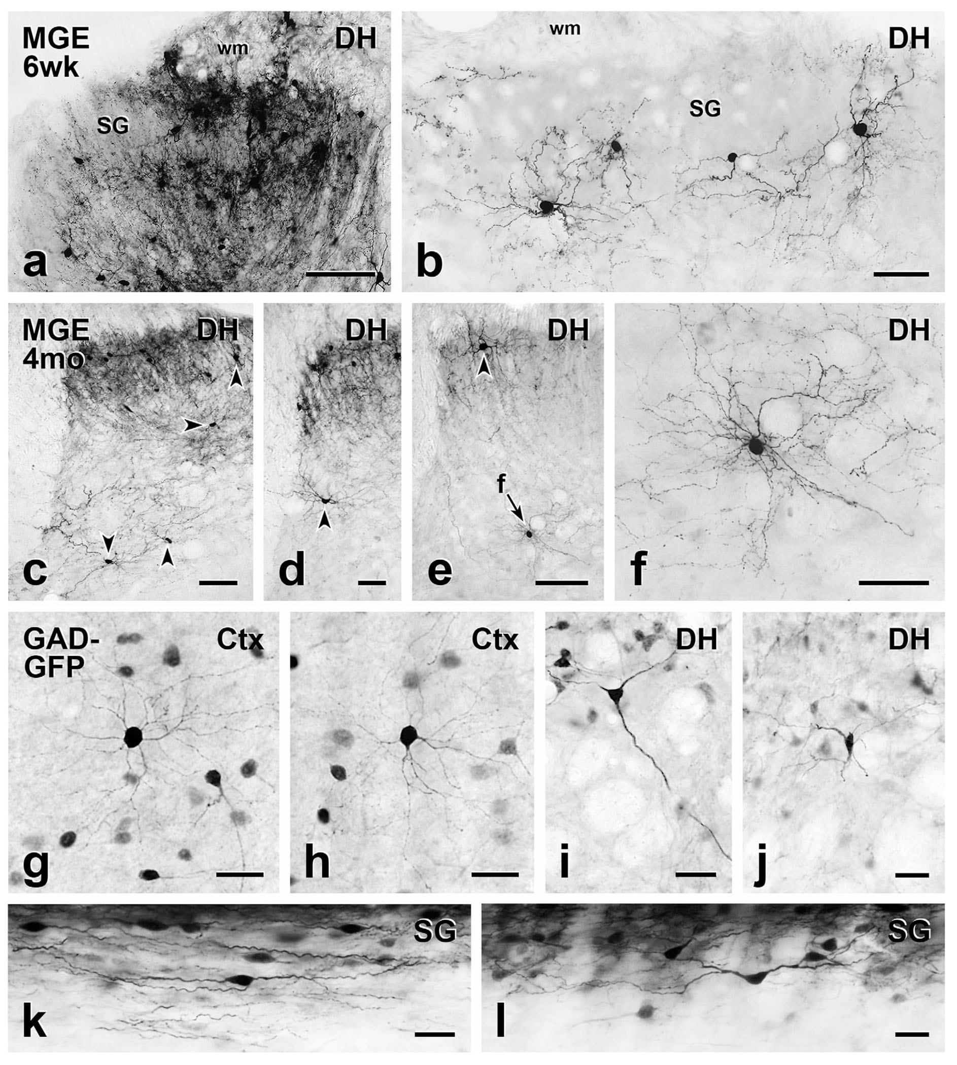
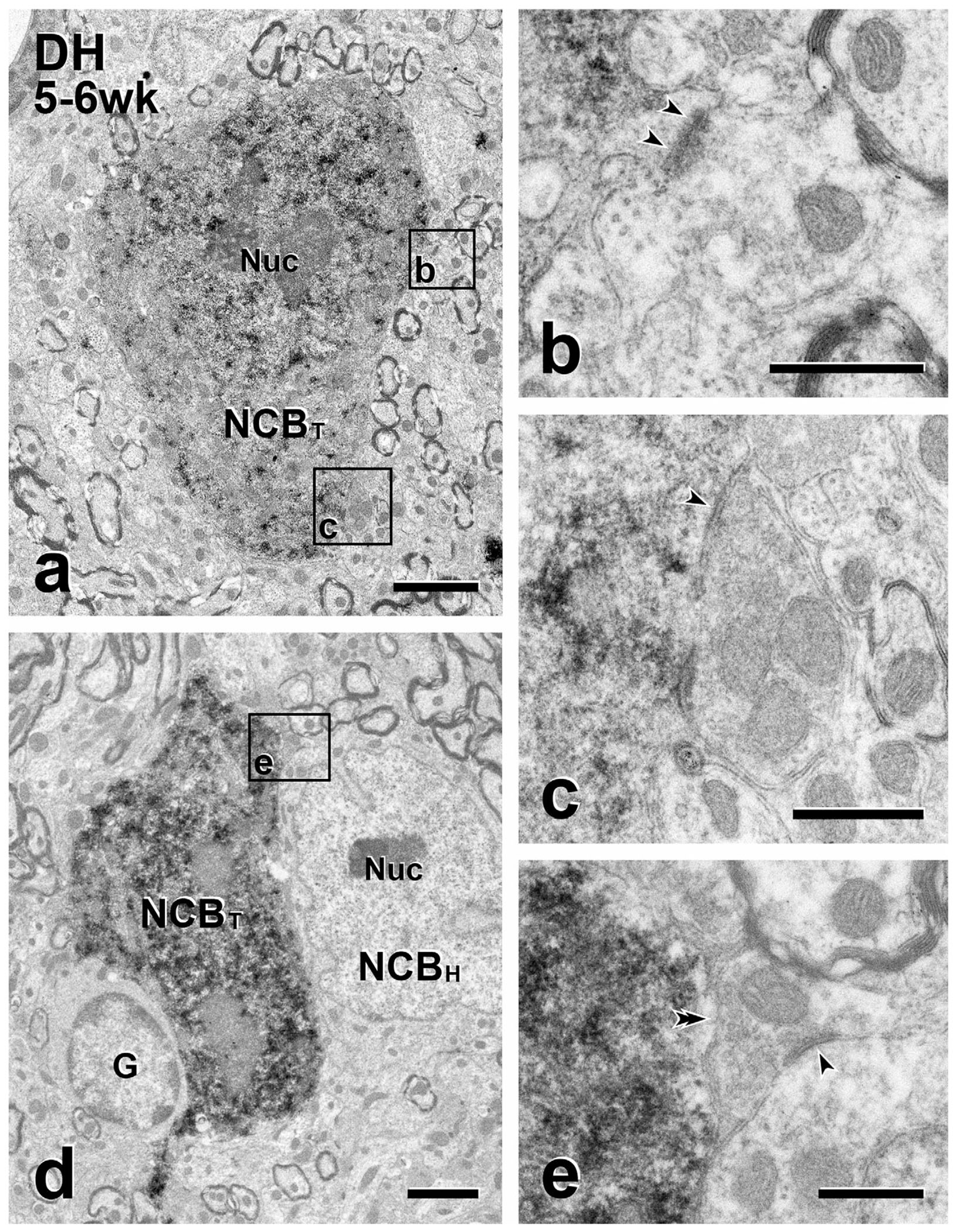
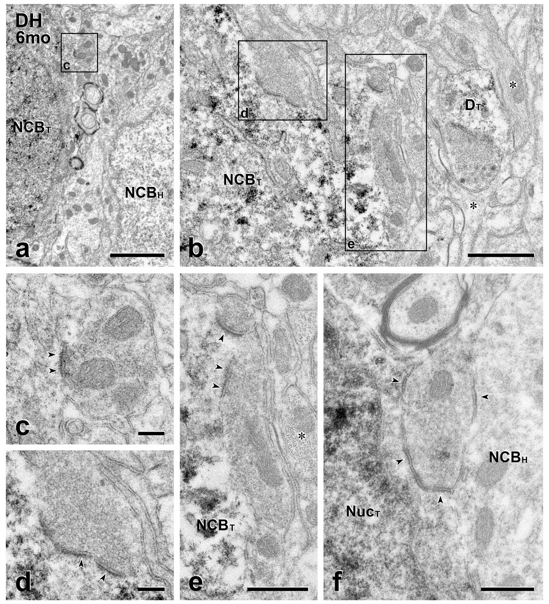
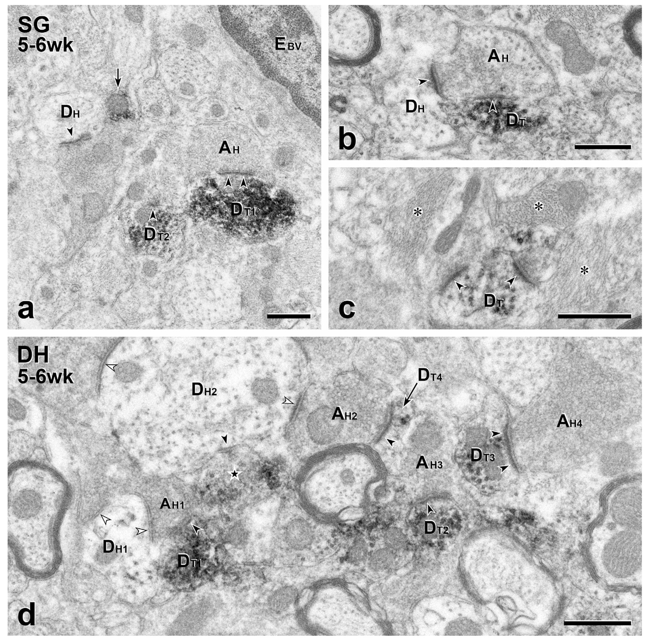
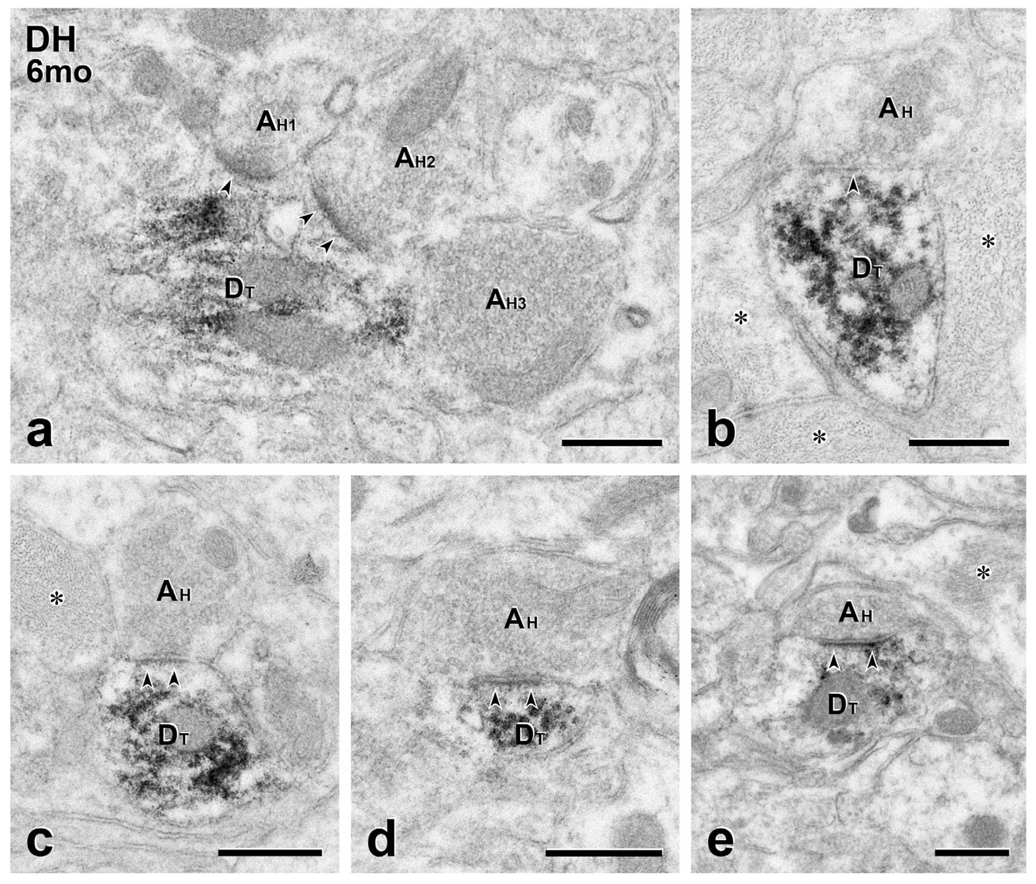
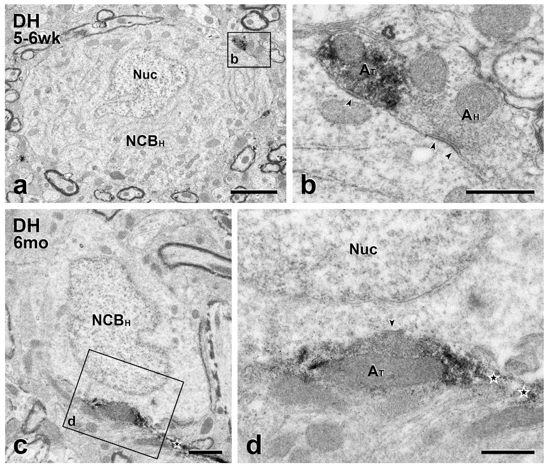
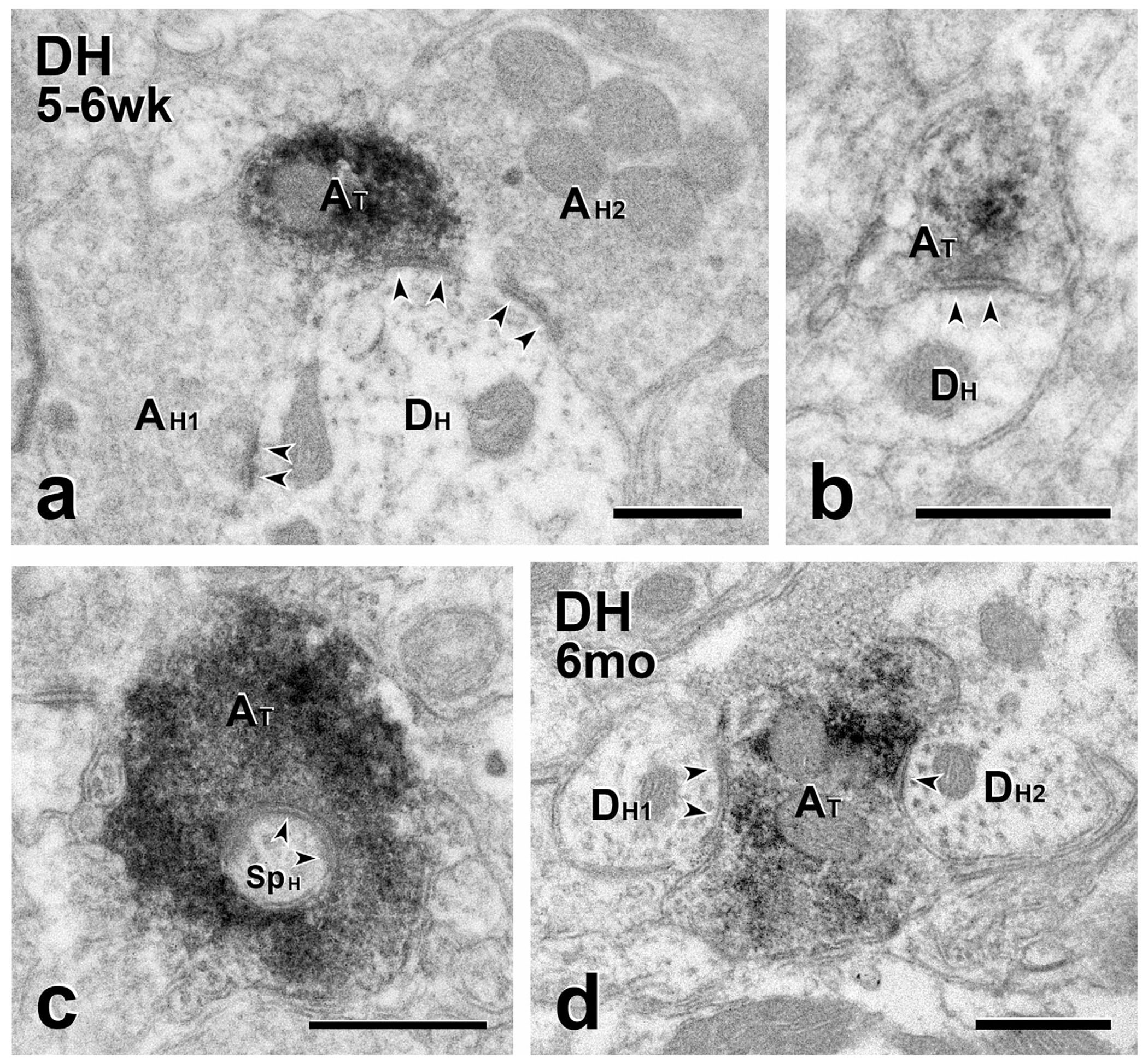
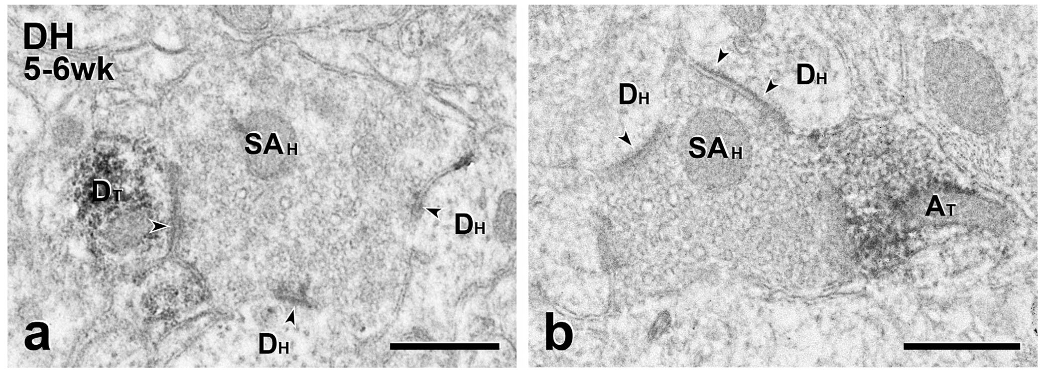
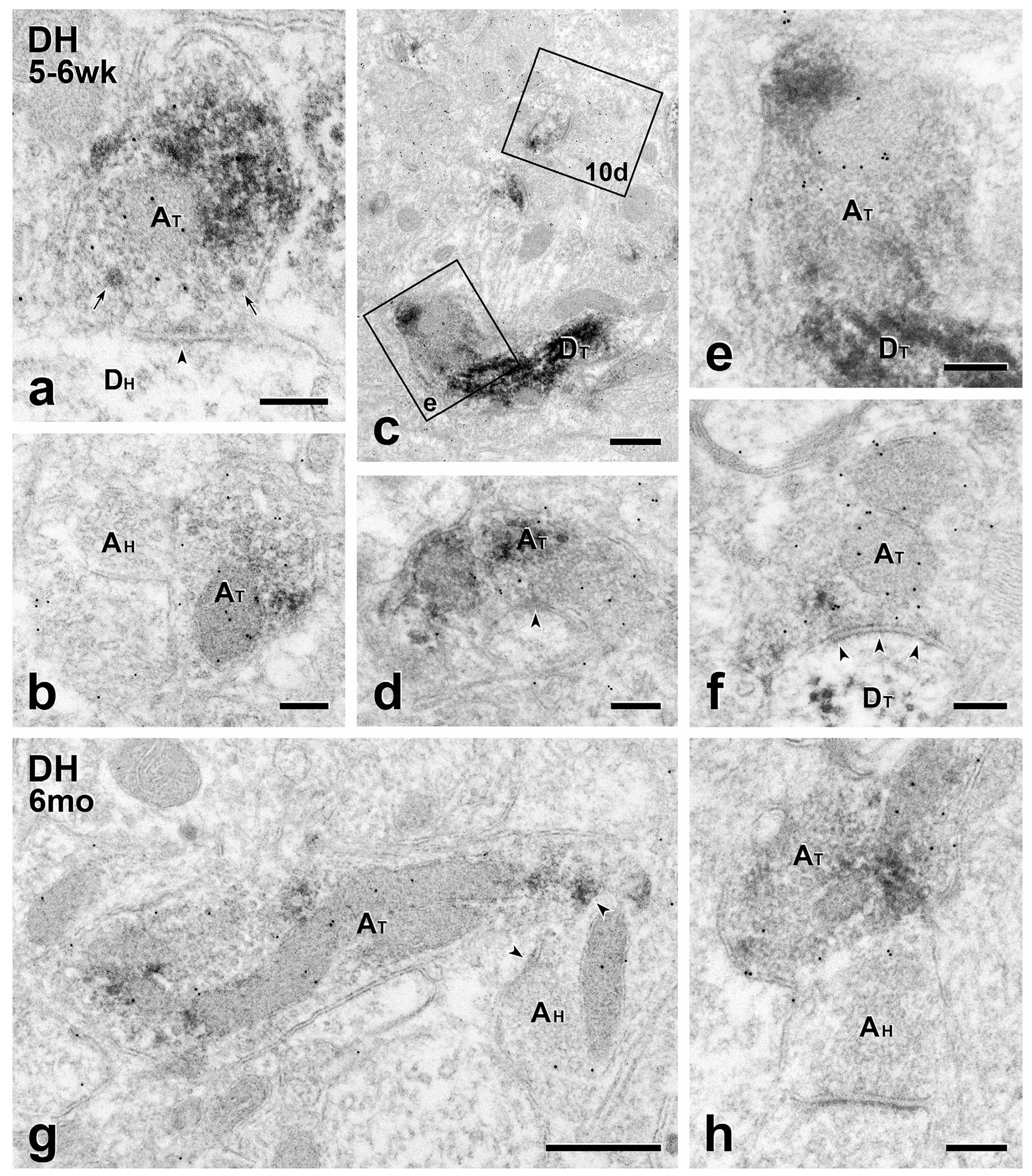
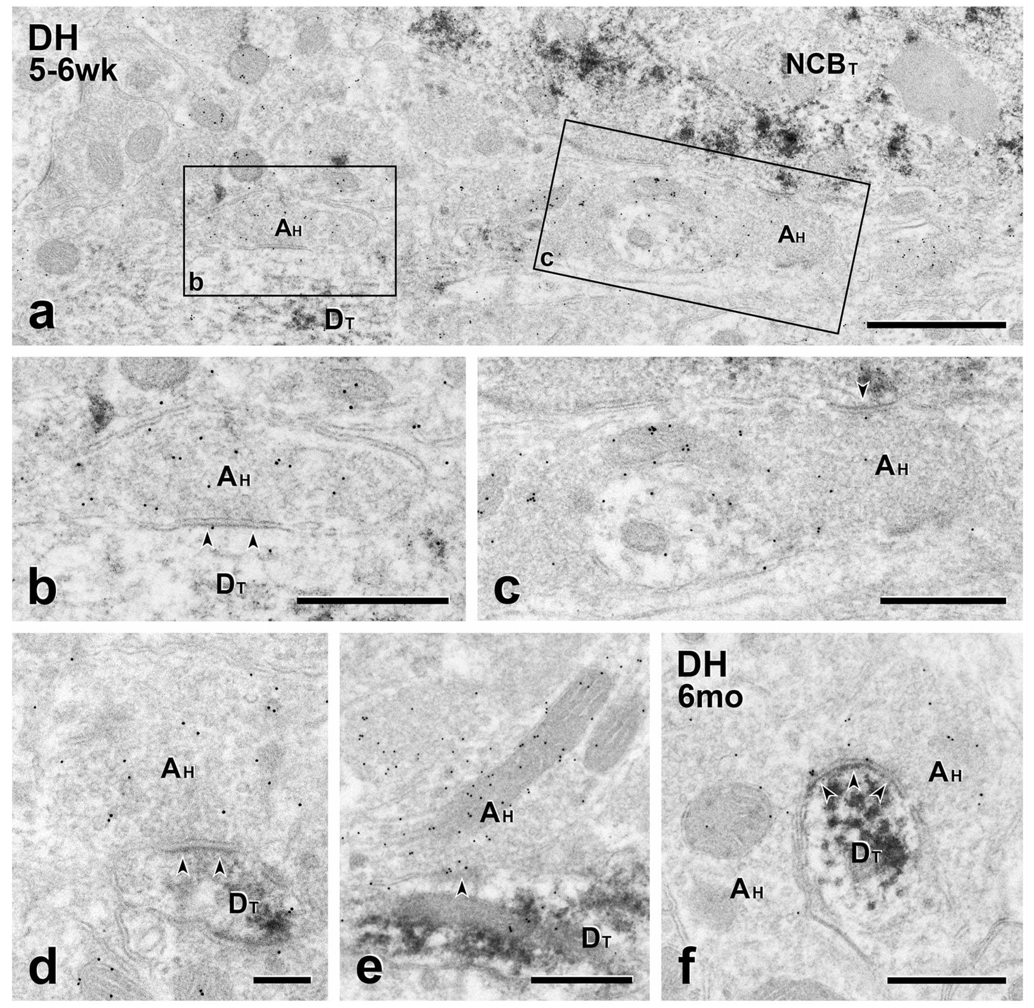
Transplanting embryonic precursors of GABAergic neurons from the medial ganglionic eminence (MGE) into adult mouse spinal cord ameliorates mechanical and thermal hypersensitivity in peripheral nerve injury models of neuropathic pain. Although Fos and transneuronal tracing studies strongly suggest that integration of MGE-derived neurons into host spinal cord circuits underlies recovery of function, the extent to which there is synaptic integration of the transplanted cells has not been established. Here, we used electron microscopic immunocytochemistry to assess directly integration of GFP-expressing MGE-derived neuronal precursors into dorsal horn circuitry in intact, adult mice with short- (5-6 weeks) or long-term (4-6 months) transplants. We detected GFP with pre-embedding avidin-biotin-peroxidase and GABA with post-embedding immunogold labeling. At short and long times post-transplant, we found host-derived synapses on GFP-immunoreactive MGE cells bodies and dendrites. The proportion of dendrites with synaptic input increased from 50% to 80% by 6 months. In all mice, MGE-derived terminals formed synapses with GFP-negative (host) cell bodies and dendrites and, unexpectedly, with some GFP-positive (i.e., MGE-derived) dendrites, possibly reflecting autoapses or cross talk among transplanted neurons. We also observed axoaxonic appositions between MGE and host terminals. Immunogold labeling for GABA confirmed that the transplanted cells were GABAergic and that some transplanted cells received an inhibitory GABAergic input. We conclude that transplanted MGE neurons retain their GABAergic phenotype and integrate dynamically into host-transplant synaptic circuits. Taken together with our previous electrophysiological analyses, we conclude that MGE cells are not GABA pumps, but alleviate pain and itch through synaptic release of GABA.

Keywords: GABA; RRID:AB_476667; RRID:SCR_003577; RRID:SCR_003970; RRID:SCR_007418; RRID:SCR_008487; spinal cord; structural plasticity; transplants; ultrastructure.


Conflict of interest statement

CONFLICT OF INTEREST

The authors have no conflicts of interest to declare.

Figures

FIGURE 1
FIGURE 1
Comparison of short- and long-term MGE cell morphology with cortical and spinal cord neurons from GAD-GFP transgenic mice. (a) At the level of the injection site, MGE-derived, GFP-immunoreactive cell bodies and processes are densely distributed throughout the dorsal horn (DH) 6 weeks post-transplantation. SG, substantia gelatinosa; wm, white matter. (b) Morphology of the MGE cells that migrated from the injection site is readily appreciated. They have small round or oval cell bodies with many fine dendrites, which are characteristics of cortical (Ctx) GABAergic interneurons (g & h). (c–f) Five months after transplantation, the MGE cells retain their cortical morphology. Arrowheads point to cell bodies of MGE neurons. Arrow F in E points to a cell shown at higher magnification in F. (g–l) Compared to cortical GAD-GFP neurons from transgenic GAD-GFP mice (g & h), those in deeper dorsal horn (i & j) are generally multipolar with a few primary dendrites that radiate circumferentially. Longitudinal sections (k & l) revealed that transgenic GAD-GFP neurons in the SG have a characteristic islet cell morphology, with fusiform cell bodies and rostrocaudally oriented dendrites. Scale bars: 100 μm in (a) and (c); 50 μm in (b), (d-f); 25 μm in (g-l)
FIGURE 2
FIGURE 2
Short-term transplants: presynaptic inputs from the host to transplant cell bodies. (a & d) Immunoperoxidase reaction product identifies GFP-immunoreactive transplant cell bodies (NCBT) in the dorsal horn (DH). Boxed areas in (a) (Boxes b, c) and in (d) (Box e) illustrate unlabeled (host) axon terminals presynaptic (arrowheads) to GFP-labeled transplant cell bodies. In (a), the transplant cell body receives both an asymmetric and a symmetric synapse. The density postsynaptic to the unlabeled terminal in (b) is thick (i.e., an asymmetric synapse) whereas the density postsynaptic to the unlabeled terminal in (c) is thin (i.e., a symmetric synapse). In (d), the cell body of a GFP-negative (host) neuron (NCBH) and a glial cell (G) abut the GFP-positive transplanted cell body. (e) The unlabeled axon terminal in (e) directly contacts (double arrowhead) the GFP-positive cell body in (d), without an intervening glia process and also forms a symmetric synapse (arrowheads) with an unlabeled host cell body. Nuc, neuronal nucleus. Scale bars: 2 μm in (a, d); 500 nm in (b, c, e)
FIGURE 3
FIGURE 3
Long-term transplants: Presynaptic inputs from the host to transplant cell bodies. (a & b) In (a), the GFP-positive cell body of a transplanted neuron (NCBT) lies close to the unlabeled cell body of a host neuron (NCBH). In (b), a transplant cell body (NCBT) is located near the center of the injection site, where there are many glial processes (*) that are full of 10 nm filaments. Boxed area (c) in (a) and boxed areas (d) and (e) in (b) are shown at higher magnification in panels (c–e), respectively. DT, dendrite of a transplanted neuron. (c–e) Unlabeled host axon terminals (c, d) and (e) synapse (arrowheads) on the GFP-positive cell bodies in (a) and (b). Note presynaptic clustering of vesicles and asymmetric synapses. (f) This unlabeled host axon terminal is presynaptic (arrowheads) to both an unlabeled host and a GFP-labeled transplant cell body. NucT, nucleus of a transplanted neuron. Scale bars: 2 μm in (a); 1 μm in (b); 500 nm in (c–f)
FIGURE 4
FIGURE 4
Short term transplants: Presynaptic inputs from the host to transplant dendrites. (a–c) In the substantia gelatinosa (SG), unlabeled host terminals (AH) are presynaptic (arrowheads) to GFP-immunoreactive transplant dendrites (DT). In (a), the unlabeled synapse on DT1 is asymmetric. A nearby unlabeled host dendrite (DH) also receives an asymmetric synapse (arrowhead). The arrow points to a likely intervaricose segment of a transplant axon, a GFP-positive profile common in the SG. EBV, endothelial cell of a blood vessel. (b) A GFP-immunoreactive dendrite (DT) receives a symmetric synapse (arrowheads) from an unlabeled host terminal (AH) that also forms an asymmetric synapse (arrowhead) with an unlabeled host dendrite (DH). (c) Two unlabeled host terminals form asymmetric synapses (arrowheads) on a GFP-positive MGE-derived dendrite (DT). The area shown contains many glial processes (asterisks). (d) This 10.45 μm2 field in deeper dorsal horn (DH) illustrates the density and complexity of MGE-derived synaptic connections. Four GFP-positive dendrites (DT1–4) receive synapses from unlabeled host axon terminals (AH1–4). The field also includes a GFP-immunoreactive transplant-derived axon terminal (star) that is presynaptic to an unlabeled host dendrite (DH2). DH2 receives convergent input from two unlabeled host terminals, one of which (AH2) is also presynaptic to a GFP-labeled transplant dendrite (DT4). Black arrowheads, synapses in which MGE neurons are pre- or postsynaptic; white arrowheads, synapses involving only host neurons. Scale bars: 500 nm in (a–d)
FIGURE 5
FIGURE 5
Long term transplants: presynaptic inputs from the host to transplant dendrites. (a) Two unlabeled host terminals (AH1, AH2) converge (arrowheads) on a large GFP-labeled transplant-derived dendrite (DT), forming symmetric synapses. There is also likely input from a third host terminal (AH3). (b–e) Both asymmetric (b) and asymmetric (c–e) synaptic specializations (arrowheads) are found between GFP-negative host axon terminals (AH) and GFP-positive transplant dendrites (DT). Scale bars: 500 nm in (a–e)
FIGURE 6
FIGURE 6
Short- and long-term transplants: MGE-derived axons are presynaptic to host cell bodies. GFP-immunoreactive transplant-derived terminals (AT) form synapses (arrowheads) on the unlabeled cell bodies of host neurons (NCBH). (a & c), Low magnification micrographs of host neuronal cell bodies after short (a) and long-term (c) transplantation. The transplant-derived (symmetric) synapses (boxed areas b and d in a and c) are magnified (AT; arrowheads) in panels (b) and (d), respectively. The terminal in (d) is continuous with one of its intervaricose segments (star). In (b), the adjacent host terminal (AH) synapses on the host cell body and directly contacts the transplant-derived terminal. Nuc, neuronal nucleus. Scale bars: 2 μm in (a); 1 μm in (c); 500 nm in (c) and (d)
FIGURE 7
FIGURE 7
Short- and long-term transplants: MGE-derived axons are presynaptic to host dendrites. (a–e) Short-term transplants. (a) A GFP–immunoreactive transplant-derived terminal (AT) makes a symmetric synapse (arrowheads) with an unlabeled host dendrite (DH) that is also postsynaptic to two unlabeled host-derived terminals (AH1 and AH2). (b) GFP-labeled transplant terminal (AT) forming a symmetric synapse on an unlabeled host dendrite (DH). (c) A GFP-labeled transplant terminal (AT) that is presynaptic to a spine arising from an unlabeled host neuron (SpH). (d) Long-term transplant. Two unlabeled host dendrites (DH1 and DH2) receive symmetric synapses from the same GFP-immunoreactive transplant-derived axon terminal (AT). Scale bars: 500 nm
FIGURE 8
FIGURE 8
Short-term transplants: MGE-derived neurons participate in synaptic glomeruli. (a) This unlabeled scalloped central terminal (SAH), likely of primary afferent origin, is presynaptic to a GFP-positive transplant-derived dendrite (DT) as well as to two unlabeled host dendrites (Dh). (b) This unlabeled scalloped terminal is also presynaptic to two unlabeled host dendrites (DH) and directly contacts a GFP-positive transplant-derived axon terminal (AT). Although a synaptic specialization is not apparent, there is no intervening glial process between the transplant-derived terminal and the scalloped terminal, suggesting that the MGE terminal is presynaptic to the host terminal. Scale bars: 500 nm
FIGURE 9
FIGURE 9
Short- and long-term transplants: MGE-derived axon terminals are GABA-immunoreactive. (a–f) Short term transplants. GABA immunolabeling of GFP-immunoreactive MGE-derived axon terminals (AT) with 10 nm gold illustrates the transmitter phenotype of transplant terminals and the breadth of the circuitry in which they engage. (a) A symmetric transplant-derived presynaptic input (arrowheads) to an unlabeled host dendrite (DH). Both small clear and large granular vesicles (arrows) occur in the transplant terminal. (b) Axoaxonic apposition between transplant (GFP- and GABA-positive; AT) and host-derived (GFP- and GABA-negative; AH) terminals. (c) 10nm gold-immunolabeled transplant-derived dendrites (DT). Boxed areas (e) and 10d are shown at higher magnification in Figures 9e and 10d, respectively. (d) Symmetric GABA-immunoreactive, transplant-derived (AT) presynaptic input (arrowhead) to a small, unlabeled host dendrite. (e) A GABA-gold labeled transplant-derived terminal (AT) directly contacts a transplant-derived dendrite (DT). (f) Transplant-derived (AT), GABA-immunoreactive symmetric input (arrowheads) to a transplant-derived dendrite (DT). (g & h) Long term transplants. Transplant-derived, GABA-immunoreactive terminals (AT) directly contact host axon terminals (AH). The latter are presynaptic to unlabeled host dendrites. Scale bars: 500 nm in (c) and (g); 200 nm in all others
FIGURE 10
FIGURE 10
Short- and long-term transplants: Host GABAergic neurons are presynaptic to transplant cell bodies and dendrites (a–e), Shortterm transplants. GFP-negative host axon terminals (AH) labeled with 10 nm GABA immunogold form symmetric synapses on a GFP-positive, transplant-derived cell body (NCBT in a) and dendrites (DT in b, d and e). Boxed areas (b) and (c) are shown at higher magnification in panels (b) and (c), respectively. (f) Long-term transplant. 10 nm GABA immunogold labeled GFP-negative (host) axon terminal (AH) forms a symmetric synapse on a GFP-positive transplant-derived dendrite. Scale bars: 1 μm in (a); 500 nm in (b–f)

References

    1. Baraban SC, Southwell DG, Estrada RC, Jones DL, Sebe JY, Alfaro-Cervello C, … Alvarez-Buylla A (2009). Reduction of seizures by transplantation of cortical GABAergic interneuron precursors into Kv1.1 mutant mice. Proceedings of the National Academy of Sciences of the United States of America, 106(36), 15472–15477. https://doi.org/10.1073/pnas.0900141106 - DOI - PMC - PubMed
    1. Basbaum AI, Glazer EJ, & Oertel W (1986). Immunoreactive glutamic acid decarboxylase in the trigeminal nucleus caudalis of the cat: A light- and electron-microscopic analysis. Somatosensory Research, 4(1), 77–94. - PubMed
    1. Blizzard CA, Chuckowree JA, King AE, Hosie KA, McCormack GH, Chapman JA, … Dickson TC (2011). Focal damage to the adult rat neocortex induces wound healing accompanied by axonal sprouting and dendritic structural plasticity. Cerebral Cortex, 21(2), 281–291. https://doi.org/10.1093/cercor/bhq091 - DOI - PubMed
    1. Braz J, Solorzano C, Wang X, & Basbaum AI (2014). Transmitting pain and itch messages: A contemporary view of the spinal cord circuits that generate gate control. Neuron, 82(3), 522–536. https://doi.org/10.1016/j.neuron.2014.01.018 - DOI - PMC - PubMed
    1. Braz JM, Juarez-Salinas D, Ross SE, & Basbaum AI (2014). Transplant restoration of spinal cord inhibitory controls ameliorates neuropathic itch. Journal of Clinical Investigation, 124(8), 3612–3616. https://doi.org/10.1172/JCI75214 - DOI - PMC - PubMed

MeSH terms

Substances