Review
. 2018 Feb;66(2):221-238.
doi: 10.1002/glia.23256. Epub 2017 Nov 14.

Origin and dynamics of oligodendrocytes in the developing brain: Implications for perinatal white matter injury

Affiliations
Review

Origin and dynamics of oligodendrocytes in the developing brain: Implications for perinatal white matter injury

Erik van Tilborg et al. Glia. 2018 Feb.

Abstract

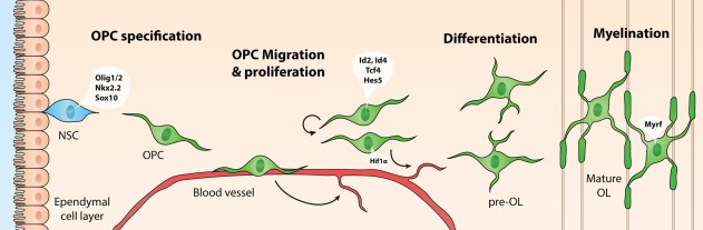
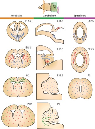
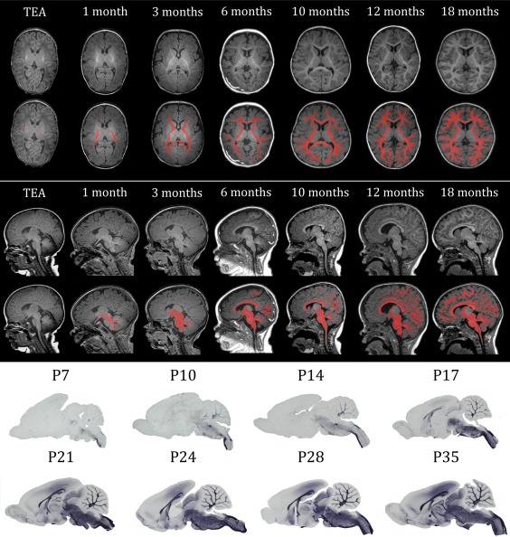
Infants born prematurely are at high risk to develop white matter injury (WMI), due to exposure to hypoxic and/or inflammatory insults. Such perinatal insults negatively impact the maturation of oligodendrocytes (OLs), thereby causing deficits in myelination. To elucidate the precise pathophysiology underlying perinatal WMI, it is essential to fully understand the cellular mechanisms contributing to healthy/normal white matter development. OLs are responsible for myelination of axons. During brain development, OLs are generally derived from neuroepithelial zones, where neural stem cells committed to the OL lineage differentiate into OL precursor cells (OPCs). OPCs, in turn, develop into premyelinating OLs and finally mature into myelinating OLs. Recent studies revealed that OPCs develop in multiple waves and form potentially heterogeneous populations. Furthermore, it has been shown that myelination is a dynamic and plastic process with an excess of OPCs being generated and then abolished if not integrated into neural circuits. Myelination patterns between rodents and humans show high spatial and temporal similarity. Therefore, experimental studies on OL biology may provide novel insights into the pathophysiology of WMI in the preterm infant and offers new perspectives on potential treatments for these patients.

Keywords: brain development; myelination; oligodendrocyte precursor cells; preterm birth; white matter injury.


Figures

Figure 1
Figure 1
The clinical problem of perinatal white matter injury (WMI) has evolved over time. Upper panels: photographs of postmortem brain slices of preterm infants with WMI (published with permission from http://neuropathology-web.org/). Lower panels: T2‐weighted MR images of preterm infants with WMI. Left part of the figure: in the 1980s, cystic periventricular leukomalacia (cPVL) was often observed in preterm infants. cPVL is associated with large cystic lesions in the white matter clearly present at macroscopic postmortem tissue and at (transverse) MRI scan (T2 sequence) as indicated by the green arrowheads. cPVL leads to severe disabilities such as cerebral palsy. Right part of the figure: at present, diffuse WMI is most often associated with atrophy of white matter causing loss of brain volume (middle (coronal) MRI scan) and punctate white matter lesions (PWML) (right (coronal) MRI scan: red arrowheads). Diffuse types of WMI are mainly associated with impaired cognitive functioning later in life
Figure 2
Figure 2
Schematic representation of oligodendrocyte (OL) development and transcription factors that contribute to OL lineage progression at different developmental stages. OL precursor cells (OPCs) originate from neuroepithelial zones surrounding the ventricles, where neural stem cells (NSCs) differentiate into (OPCs) under the influence of OL‐specific transcription factors Olig1/2, Nkx2.2, and Sox10. OPCs migrate toward an appropriate site via blood vessels, while at the same time promoting angiogenesis in a HIF1α‐dependent manner, in areas requiring more oxygen. At their final destination, OPCs proliferate to expand the pool of OPCs, under the regulation of transcription factors such as Id2, Id4, Tcf4, and Hes5. When proliferation is inhibited, OLs differentiate into premyelinating OLs (pre‐OLs), and finally into mature OLs that enwrap neuronal axons with myelin sheaths, under the influence of, for example, Myrf
Figure 3
Figure 3
Schematic representation of how different waves of OPC generation populate different regions of the CNS throughout development (panels: left/orange: forebrain; middle/green: cerebellum; right/purple: spinal cord). OPCs originating from different niches are represented by differently colored dots (blue, red, and green) (based on data by Fogarty, Richardson, & Kessaris, 2005; Grimaldi, Parras, Guillemot, Rossi, & Wassef, 2009; Hashimoto et al., 2016; Kessaris et al., 2006; Ravanelli & Appel, 2015; Vallstedt, Klos, & Ericson, 2005)
Figure 4
Figure 4
Overview of myelination throughout development from term‐equivalent age (TEA) to 18 months in human infants and from postnatal day (P)7 to P35 in rats. The upper panel shows transverse sections of T1‐weighted MRI scans at different ages, in the lower sections the myelinated white matter is manually colorized (red). The middle panel shows the sagittal sections of T1‐weighted MRI scans at different ages, in the lower sections the myelinated white matter is manually colorized (red). The lower panel shows sagittal sections of rat brains at different ages, stained for myelin basic protein (MBP), a myelin marker. The gross spatio‐temporal pattern of myelination in humans shows high resemblance with that of rodents
Figure 5
Figure 5
Developmental progression of OPCs, pre‐OLs, and myelination. The population of OPCs is expanded between 15 and 20 weeks gestational age (GA) (green line). Pre‐OLs peak between 30 and 40 weeks GA (orange line). Myelination starts before birth, but mostly occurs during the first year of life and continues for several decades (blue line). Children born prematurely are exposed to perinatal insults during the peak of pre‐OLs (red window), which hamper their ability to differentiate into myelinating OLs resulting in increased numbers of pre‐OLs and reduced myelination as observed in diffuse WMI (dashed orange and blue lines) (based on data by Back et al., 2001; Buser et al., 2012)

References

    1. Alexandrou, G. , Martensson, G. , Skiold, B. , Blennow, M. , Aden, U. , & Vollmer, B. (2014). White matter microstructure is influenced by extremely preterm birth and neonatal respiratory factors. Acta Paediatrics, 103(1), 48–56. https://doi.org/10.1111/apa.12445 - DOI - PubMed
    1. Ancel, P. Y. , Goffinet, F. , Epipage, 2., Writing Group, Kuhn, P. , Langer, B. , Matis, J. , … Kaminski, M. (2015). Survival and morbidity of preterm children born at 22 through 34 weeks' gestation in France in 2011: Results of the EPIPAGE‐2 cohort study. JAMA Pediatrics, 169(3), 230–238. https://doi.org/10.1001/jamapediatrics.2014.3351 - DOI - PubMed
    1. Aubert‐Broche, B. , Fonov, V. , Leppert, I. , Pike, G. B. , & Collins, D. L. (2008). Human brain myelination from birth to 4.5 years. Medical Image Computing and Computer‐Assisted Intervention, 11(Pt 2), 180–187. - PubMed
    1. Back, S. A. (2006). Perinatal white matter injury: The changing spectrum of pathology and emerging insights into pathogenetic mechanisms. Mental Retardation and Developmental Disabilities Research Reviews, 12(2), 129–140. https://doi.org/10.1002/mrdd.20107 - DOI - PubMed
    1. Back, S. A. (2017). White matter injury in the preterm infant: Pathology and mechanisms. Acta Neuropathology, https://doi.org/10.1007/s00401-017-1718-6 - DOI - PMC - PubMed

Publication types