. 2018 Feb 1;14(2):345-360.
doi: 10.1080/21645515.2017.1403703. Epub 2017 Dec 21.

Generation and testing anti-influenza human monoclonal antibodies in a new humanized mouse model (DRAGA: HLA-A2. HLA-DR4. Rag1 KO. IL-2Rγc KO. NOD)

Affiliations

Generation and testing anti-influenza human monoclonal antibodies in a new humanized mouse model (DRAGA: HLA-A2. HLA-DR4. Rag1 KO. IL-2Rγc KO. NOD)

Mirian Mendoza et al. Hum Vaccin Immunother. .

Abstract

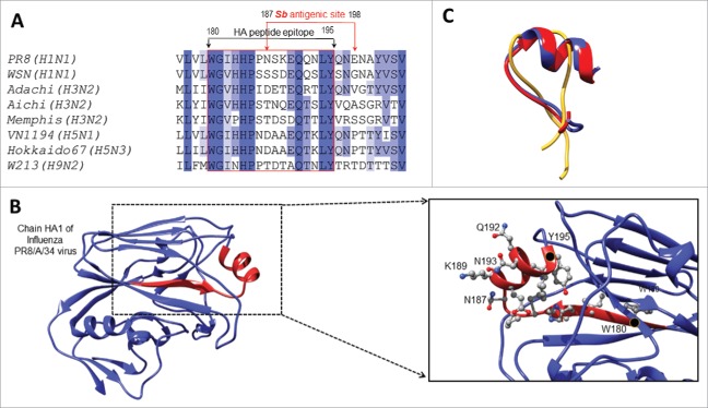
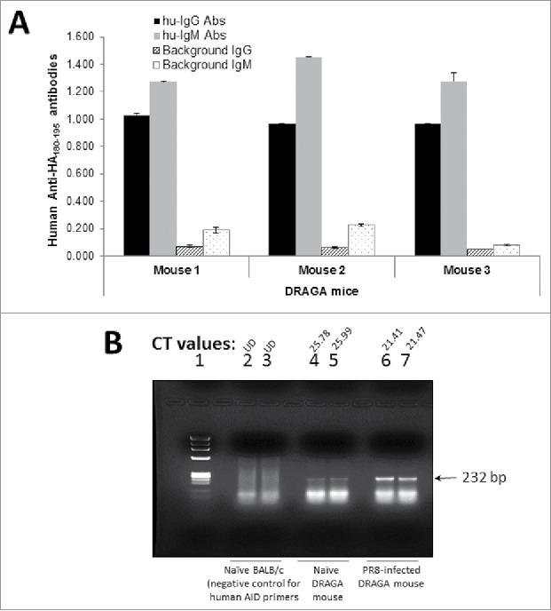
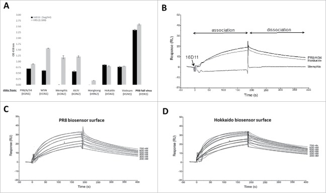
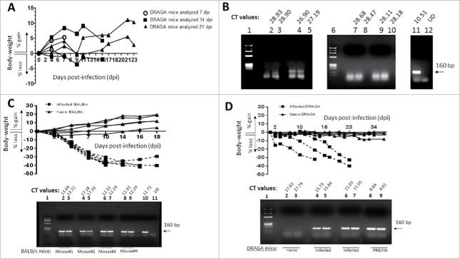
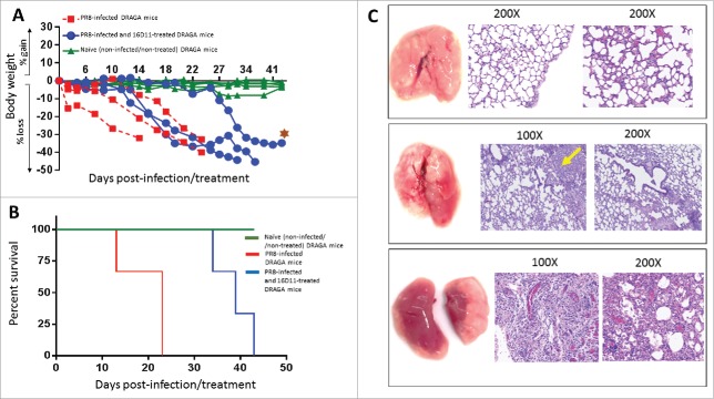
Pandemic outbreaks of influenza type A viruses have resulted in numerous fatalities around the globe. Since the conventional influenza vaccines (CIV) provide less than 20% protection for individuals with weak immune system, it has been considered that broadly cross-neutralizing antibodies may provide a better protection. Herein, we showed that a recently generated humanized mouse (DRAGA mouse; HLA-A2. HLA-DR4. Rag1KO. IL-2Rgc KO. NOD) that lacks the murine immune system and expresses a functional human immune system can be used to generate cross-reactive, human anti-influenza monoclonal antibodies (hu-mAb). DRAGA mouse was also found to be suitable for influenza virus infection, as it can clear a sub-lethal infection and sustain a lethal infection with PR8/A/34 influenza virus. The hu-mAbs were designed for targeting a human B-cell epitope (180WGIHHPPNSKEQ QNLY195) of hemagglutinin (HA) envelope protein of PR8/A/34 (H1N1) virus with high homology among seven influenza type A viruses. A single administration of HA180-195 specific hu-mAb in PR8-infected DRAGA mice significantly delayed the lethality by reducing the lung damage. The results demonstrated that DRAGA mouse is a suitable tool to (i) generate heterotype cross-reactive, anti-influenza human monoclonal antibodies, (ii) serve as a humanized mouse model for influenza infection, and (iii) assess the efficacy of anti-influenza antibody-based therapeutics for human use.

Keywords: Anti-flu antibody therapy; Human influenza infection model; Human monoclonal antibodies; Humanized mice.


Figures

Figure 1.
Figure 1.
HA180-195 epitope homology among different influenza virus heterotypes. (A) Sequence alignment of the HA proteins from several influenza virus strains and homologous residues 180–195 comprising part of the Sb antigenic site. (B) Ribbon representation of the HA protein (PDB ID 1ru7:A, residues 54–270) revealing the Sb antigenic site in red. Shown is in detailed view of the Sb antigenic site, the 180–195 residues side chains in ball and stick representation together with their carbon atoms colored in grey, nitrogen in blue and oxygen in red. The first and last residues of the Sb site are indicated with a black dot and labeled, and the highly exposed residues of the HA180-195 epitope located within the a-helix are labeled. (C) Overlapped conformations of soluble HA180-195 epitope from PR8/A/34 (red ribbon), Memphis (yellow ribbon) and Hokkaido (blue ribbon) viruses according to “de novo” modeling approach and PEP-FOLD server (described in materials and methods section).
Figure 2.
Figure 2.
HA180-195 specific hu-Ab responses and human AID expression in DRAGA mice. (A) DRAGA mice were immunized and boosted 2 weeks later with KLH-HA180-195 conjugate. Two weeks after the boost, sera of immunized mice was assessed in ELISA plates coated with rHA of PR8 virus (2 μg/well) for titers of human anti-HA180-195 IgM and IgG antibodies. Shown are the specific antibody titers for 3 representative mice and their corresponding signal-to-noise background of secondary antibody. (B) DRAGA mice (n = 3) were infected or not by the intranasal route with a sub-lethal dose of PR8 /A/34 virus (LD50) and 7 days later the splenic RNA was extracted and analyzed by RT-qPCR using our designed primers for human AID (described in the materials and methods section). One representative DRAGA infected mouse (lanes 6 and 7) and naive DRAGA mouse (lanes 4 and 5) are shown. Control negative for human AID primers specificity was splenic RNA extracted from naive BALB/c mice (lanes 2 and 3). The CT values for duplicate samples are shown for each mouse. Arrow indicates the size of human AID amplicon.
Figure 3.
Figure 3.
Western Blot isotyping of HA180-195 specific hu-mAbs. Shown are the HA180-195 specific hu-mAbs selected for this study as assessed by Western blot for the presence of IgG and IgM heavy and light chains. Affinity purified hu-mAbs were SDS-denatured and 2ME-reduced, applied at 5 μg/lane in 8–16% gradient gels of polyacrylamide, separated by SDS-PAGE, gels electro-transferred onto PVDF membranes, and the membranes were probed with specific antibodies for the μ, λ, κ, and λ chains followed by incubation with specie-specific secondary Abs-HRP conjugates, as indicated in each panel.
Figure 4.
Figure 4.
Structural analyses of anti-HA180-195 hu-mAbs. (A) Silver stain of 8–16% gradient SDS-PAGE gels ran under denaturing and reducing condition for four affinity purified, HA180-195 specific hu-mAbs at 1 μg/lane. (B) Immunoelectrophoresis of 16D11 hu-mAb showing the monoclonal bands of human μ heavy chain and human l light chain as compared with the human polyclonal m heavy chain and l light chain. (C) Agarose Titan gel analysis showing the monoclonality and difference in the electrophoretic mobility of HA180-195 specific hu-mAbs. (D) Histograms of FPLC analysis showing intact pentameric molecules of 16D11 hu-mAb. Arrows in each histogram indicates the earlier elution time for 16D11 IgM and control human IgM pentameric molecules than for human control IgG monomeric molecules as detected at 280 nm.
Figure 5.
Figure 5.
Amino acid sequences of CDR3 VH and VL regions of hu-mAbs and their isotype controls. (A) CDR3 VH sequences of four HA180-195 specific IgM hu-mAbs (16D11, 10B2, 8D12, and 13C10) and two non-specific, isotype control IgM/lambda hu-mAbs (8A4 and 25-3 hu-mAbs). (B) CDR3 VL sequences of the same hu-mAbs like in panel A. Shown are the signal peptides for both the Heavy and Light chains, the flanking frame regions FR1 to FR4, the CDR3s, and 10 amino acids adjacent to constant regions CH1 and CL1. Similar amino acid residues among all hu-mAbs are highlighted in green, those with more than 80% similarity in light green, those between 60 to 80% similarity in yellow, and those with less than 60% similarity are left uncolored. Blue arrows indicate the position where amino acid differences occurred for 10B2, 8D12, and 13C10 hu-mAbs.
Figure 6.
Figure 6.
Germline identification and 3D homology model for 16D11 hu-mAb. (A) Sequence alignment of the VH and VL chain of 16D11 hu-mAb and their corresponding germlines. Sequence corresponding to the CDRs are colored, and those corresponding to the V, D and J genes are indicated at the top of alignment. Protein sequence corresponding to the junction is highlighted in gray. Dots indicate sequence conservation, and dashes indicate gaps. (B) Colliers de Perles representation of the VH and VL protein chains. Residues corresponding to CDR loops are colored as in A, and square boxes represent the boundary residues between the framework and the CDR loops. Positions with red and bold letters indicate the five-conserved position of a V domain. (C) Ribbon and surface representation of the homology model of 16D11 Fab showing the VH and VL chains in dark, and respectively light grey. CDR loops are labeled and colored as in B. (D) Coulomb surface coloring of 16D11 homology model. (E) Showed are the areas with no potential charges in red and positive charges in blue.
Figure 7.
Figure 7.
HA cross-reactivity and binding affinity of 16D11 hu-mAb. (A) Binding of 16D11 hu-mAb to rHA proteins from influenza virus heterotypes in ELISA. Shown are duplicate rHAs-coated wells and+/- SD for 99% confidence. Signal-to-noise background of the anti-human IgM-HRP secondary antibody (0D 450 nm = 0.045 average) has been subtracted from each sample. PR8 virus-coated wells and repository human sera (HRS) were used as controls. (B) SPR comparative binding of 16D11 hu-mAb at 200 nanoMoles to rHA proteins of PR8/a/34 virus (black), Hokkaido virus (grey) and Memphis virus (red). 16D11 injection point and association and dissociation phases are indicated. Representative Sensograms of different concentrations of 16D11 hu-mAbs across biosensor surfaces coupled to rHA protein of PR8 virus (C) and Hokkaido virus (D) at 30 ml/min and 25°C. Sensograms were analyzed using a simultaneous fit algorithm (BIAevaluation 3.1) to calculate the kinetic parameters and binding affinities (as shown in Table 1). SPR sensograms for each response are shown as gray lines whilst fit analyses are shown as black lines.
Figure 8.
Figure 8.
Body-weights of PR8-infected DRAGA mice vs. titers of viral HA expression in the lungs. (A) Sub-lethal infection of DRAGA mice with PR8 virus. Body-weights of PR8 sub-lethally infected DRAGA mice (n = 6) were monitored every other 3rd or 4th day post-infection (dpi). (B) Agarose gel electrophoresis of the HA amplicons obtained by RT-qPCR and corresponding CT values in the lungs of DRAGA mice from panel A (sub-lethal infection), as measured at 7, 14, and 21 dpi. Shown are duplicate samples from a representative naïve (not infected) DRAGA mouse (lanes 2 and 3), and sublethally-infected 7 dpi (4–5) and 21 dpi (lanes 7–8 and 9–10). Arrow indicates the size (160 bp) amplicon of HA of PR8 virus (positive control, lane 11). Lane 1, molecular markers; Lane 12 distilled water (primers control). (C) Body-weights of PR8 lethally-infected (LD100) BALB/c mice and naïve BALB/c mice (n = 4 mice/group) monitored every other 3rd or 4th day post-infection (dpi). Lower panel, the agarose gel electrophoresis of HA amplicons in the lungs measured by RT-qPCR, and CT corresponding values in duplicate samples of lethally-infected BALB/c mice (lanes 2 to 9) at 18 dpi; Lane 1, molecular markers; Lane 10, the 160 bp amplicon of HA of PR8 virus (positive control); Lane 11, a representative naïve (non-infected) BALB/c mouse. (D) Body-weights of PR8 lethally-infected (LD100) DRAGA mice. Mice were monitored every other 3rd or 4th day. Lower panel, agarose gel electrophoresis of HA amplicons in the lungs measured by RT-qPCR, and CT corresponding values in duplicate samples. Lane 1, molecular markers; lanes 2-3, naïve DRAGA mouse; Lanes 4–5 and 6–7, two representative DRAGA mice lethally infected, as measured at 14 dpi and 18 dpi; Lanes 8–9, 160 bp amplicon of HA of PR8 virus (positive control).
Figure 9.
Figure 9.
The effect of 16D11 hu-mAb in PR8 lethally-infected DRAGA mice. (A) Body-weights of naïve (non-infected) DRAGA mice and PR8 lethally-infected (LD100) DRAGA mice with and without 16D11 hu-mAb treatment. A single i.p. injection of 600 μg per mouse (n = 3-4 mice /group) was administered at the time of infection. Mice were monitored every other 3rd or 4th dpi. Brown star indicates the most resilient mouse in the treatment group. (B) Survival rates for groups of DRAGA mice described in panel A. Of note, the most resilient DRAGA mouse in the PR8-infected/treated group showed 40% less loss in body-weight at day 43 post-infection when was sacrificed for analysis, which is a significantly when compared with the average loss in body-weight for the control infection group (p = 0.026 according to Mantel-Cox test, and p = 0.016 according to pairwise curves comparisons of Gehan-Breslow-Wilcoxon test). (C) Lung analysis of DRAGA mice described in panel A. Upper panels, lungs and Hematoxilin-Eosin (HE) staining of lung sections from a representative naïve DRAGA mouse; Middle panels, lungs and HE staining of lung sections from the most resilient DRAGA mouse to PR8 lethal infection upon 16D11 hu-mAb treatment as analyzed 40 days post-infection. Shown is a mild lung damage in the lower lobe of the right lung (diffuse grayish area) with slightly distorted alveolar architecture and scattered lymphocyte infiltrates (yellow arrow); Lower panels, lungs and HE staining of lung sections from a representative PR8-lethally DRAGA mouse left untreated, and analyzed 20 days post-infection. Shown is massive pneumonia in both lungs (dark reddish areas) with heavily distorted alveolar architecture, and interstitial and intra-alveolar lymphocyte infiltration.

Similar articles

Cited by

References

    1. Kilbourne ED. Influenza. New York, NY: Plenum Medical Book Co.; 1987
    1. Li OT, Poon LL. One step closer to universal influenza epitopes. Expert Rev Anti Infect Ther. 2009;7(6):687–690 doi:10.1586/eri.09.48. PMID:19681695 - DOI - PubMed
    1. Velkov T, Ong C, Baker MA, Kim H, Li J, Nation RL, Huang JX, Cooper MA, Rockman S. The antigenic architecture of the hemagglutinin of influenza H5N1 viruses. Mol Immunol. 2013;56(4):705–719 doi:10.1016/j.molimm.2013.07.010. PMID:23933511 - DOI - PubMed
    1. Osterholm MT. Preparing for the next pandemic. N Engl J Med. 2005;352:1839–1842 doi:10.1056/NEJMp058068. PMID:15872196 - DOI - PubMed
    1. McMurry JA, Johanson BE, De Groot AS. A call to cellular and humoral arms. Human Vaccines. 2008;4(2):148–157 doi:10.4161/hv.4.2.5169. PMID:18382131 - DOI - PubMed

Publication types

Substances

LinkOut - more resources