

The Apoptogenic Toxin AIP56 Is Secreted by the Type II Secretion System of Photobacterium damselae subsp. piscicida
- PMID: 29135911
- PMCID: PMC5705983
- DOI: 10.3390/toxins9110368
The Apoptogenic Toxin AIP56 Is Secreted by the Type II Secretion System of Photobacterium damselae subsp. piscicida
Abstract
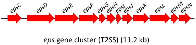
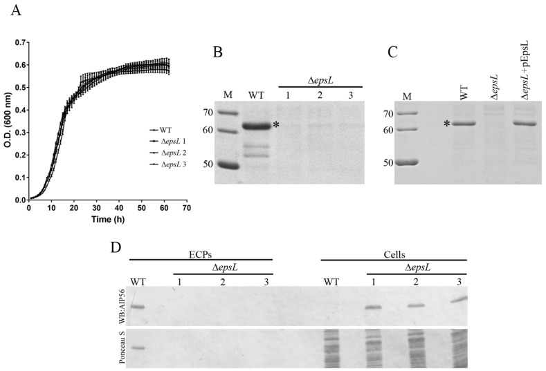
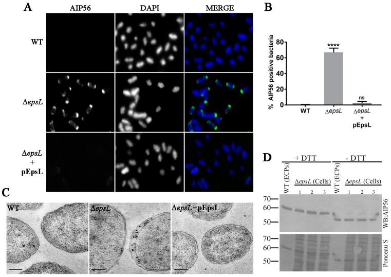
AIP56 (apoptosis-inducing protein of 56 kDa) is a key virulence factor of Photobacterium damselae subsp. piscicida (Phdp), the causative agent of a septicaemia affecting warm water marine fish species. Phdp-associated pathology is triggered by AIP56, a short trip AB toxin with a metalloprotease A domain that cleaves the p65 subunit of NF-κB, an evolutionarily conserved transcription factor that regulates the expression of inflammatory and anti-apoptotic genes and plays a central role in host responses to infection. During infection by Phdp, AIP56 is systemically disseminated and induces apoptosis of macrophages and neutrophils, compromising the host phagocytic defence and contributing to the genesis of pathology. Although it is well established that the secretion of AIP56 is crucial for Phdp pathogenicity, the protein secretion systems operating in Phdp and the mechanism responsible for the extracellular release of the toxin remain unknown. Here, we report that Phdp encodes a type II secretion system (T2SS) and show that mutation of the EpsL component of this system impairs AIP56 secretion. This work demonstrates that Phdp has a functional T2SS that mediates secretion of its key virulence factor AIP56.
Keywords: T2SS; exotoxin; polar localisation; secretion.
Conflict of interest statement
The authors declare no conflict of interest.
Figures
Similar articles
-
Involvement of Hsp90 and cyclophilins in intoxication by AIP56, a metalloprotease toxin from Photobacterium damselae subsp. piscicida.Sci Rep. 2019 Jun 21;9(1):9019. doi: 10.1038/s41598-019-45240-w. Sci Rep. 2019. PMID: 31227743 Free PMC article.
-
Susceptibility of Sea Bream (Sparus aurata) to AIP56, an AB-Type Toxin Secreted by Photobacterium damselae subsp. piscicida.Toxins (Basel). 2022 Feb 5;14(2):119. doi: 10.3390/toxins14020119. Toxins (Basel). 2022. PMID: 35202146 Free PMC article.
-
AIP56: a novel bacterial apoptogenic toxin.Toxins (Basel). 2010 Apr;2(4):905-18. doi: 10.3390/toxins2040905. Epub 2010 Apr 26. Toxins (Basel). 2010. PMID: 22069616 Free PMC article. Review.
-
AIP56, a novel plasmid-encoded virulence factor of Photobacterium damselae subsp. piscicida with apoptogenic activity against sea bass macrophages and neutrophils.Mol Microbiol. 2005 Nov;58(4):1025-38. doi: 10.1111/j.1365-2958.2005.04893.x. Mol Microbiol. 2005. PMID: 16262788
-
Photobacterium damselae subsp. piscicida: an integrated view of a bacterial fish pathogen.Int Microbiol. 2002 Mar;5(1):3-9. doi: 10.1007/s10123-002-0051-6. Int Microbiol. 2002. PMID: 12102234 Review.
Cited by
-
Unconventional structure and mechanisms for membrane interaction and translocation of the NF-κB-targeting toxin AIP56.Nat Commun. 2023 Nov 16;14(1):7431. doi: 10.1038/s41467-023-43054-z. Nat Commun. 2023. PMID: 37973928 Free PMC article.
-
Involvement of Hsp90 and cyclophilins in intoxication by AIP56, a metalloprotease toxin from Photobacterium damselae subsp. piscicida.Sci Rep. 2019 Jun 21;9(1):9019. doi: 10.1038/s41598-019-45240-w. Sci Rep. 2019. PMID: 31227743 Free PMC article.
-
Horizontal Transfer of Microbial Toxin Genes to Gall Midge Genomes.Genome Biol Evol. 2021 Sep 1;13(9):evab202. doi: 10.1093/gbe/evab202. Genome Biol Evol. 2021. PMID: 34450656 Free PMC article.
-
Assessing the impact, genomics and evolution of type II secretion across a large, medically important genus: the Legionella type II secretion paradigm.Microb Genom. 2019 Jun;5(6):e000273. doi: 10.1099/mgen.0.000273. Epub 2019 Jun 5. Microb Genom. 2019. PMID: 31166887 Free PMC article.
-
A Highly Unstable and Elusive Plasmid That Encodes the Type III Secretion System Is Necessary for Full Virulence in the Marine Fish Pathogen Photobacterium damselae subsp. piscicida.Int J Mol Sci. 2022 Apr 25;23(9):4729. doi: 10.3390/ijms23094729. Int J Mol Sci. 2022. PMID: 35563122 Free PMC article.
References
-
- Barnes A.C., dos Santos N.M., Ellis A.E. Update on bacterial vaccines: Photobacterium damselae subsp. piscicida. In: Midtlyng P.J., editor. Progress in Fish Vaccinology. Volume 121. Karger; Basel, Switzerland: 2005. pp. 75–84. - PubMed
-
- Do Vale A., Costa-Ramos C., Silva A., Silva D.S., Gartner F., dos Santos N.M., Silva M.T. Systemic macrophage and neutrophil destruction by secondary necrosis induced by a bacterial exotoxin in a gram-negative septicaemia. Cell. Microbiol. 2007;9:988–1003. doi: 10.1111/j.1462-5822.2006.00846.x. - DOI - PubMed
-
- Do Vale A., Silva M.T., dos Santos N.M.S., Nascimento D.S., Reis-Rodrigues P., Costa-Ramos C., Ellis A.E., Azevedo J.E. AIP56, a novel plasmid-encoded virulence factor of Photobacterium damselae subsp. piscicida with apoptogenic activity against sea bass macrophages and neutrophils. Mol. Microbiol. 2005;58:1025–1038. doi: 10.1111/j.1365-2958.2005.04893.x. - DOI - PubMed
Publication types
MeSH terms
Substances
LinkOut - more resources
Full Text Sources
Other Literature Sources
