. 2017 Nov 14;18(11):2414.
doi: 10.3390/ijms18112414.

A Role of Sp1 Binding Motifs in Basal and Large T-Antigen-Induced Promoter Activities of Human Polyomavirus HPyV9 and Its Variant UF-1

Affiliations

A Role of Sp1 Binding Motifs in Basal and Large T-Antigen-Induced Promoter Activities of Human Polyomavirus HPyV9 and Its Variant UF-1

Ugo Moens et al. Int J Mol Sci. .

Abstract

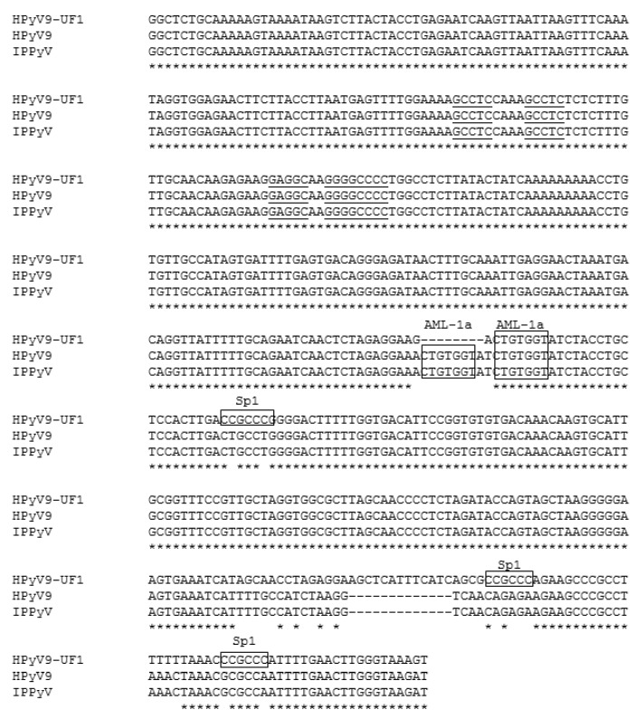
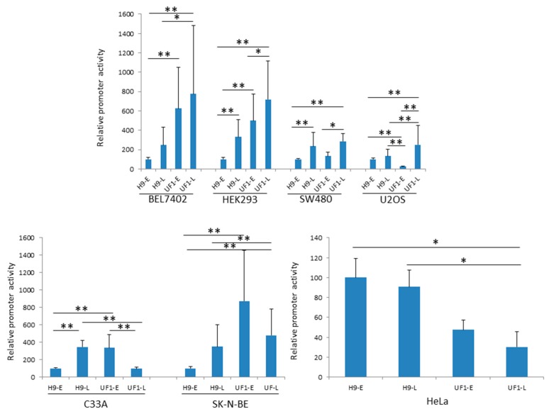
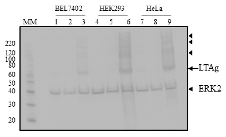
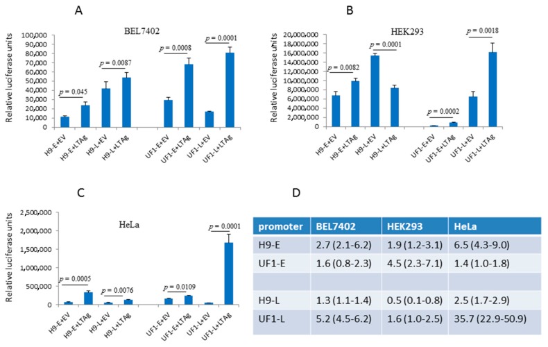
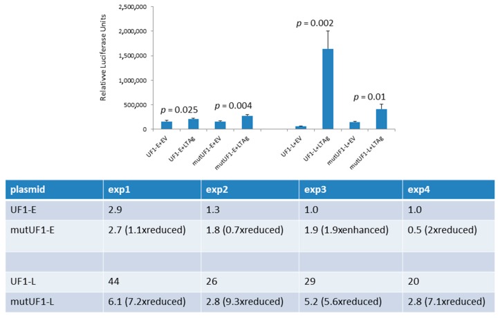
Human polyomavirus 9 (HPyV9) was originally detected in the serum of a renal transplant patient. Seroepidemiological studies showed that ~20-50% of the human population have antibodies against this virus. HPyV9 has not yet been associated with any disease and little is known about the route of infection, transmission, host cell tropism, and genomic variability in circulating strains. Recently, the HPyV9 variant UF-1 with an eight base-pair deletion, a thirteen base-pair insertion and with point mutations, creating three putative Sp1 binding sites in the late promoter was isolated from an AIDS patient. Transient transfection studies with a luciferase reporter plasmid driven by HPyV9 or UF1 promoter demonstrated that UF1 early and late promoters were stronger than HPyV9 promoters in most cell lines, and that the UF1 late promoter was more potently activated by HPyV9 large T-antigen (LTAg). Mutation of two Sp1 motifs strongly reduced trans-activation of the late UF1 promoter by HPyV9 LTAg in HeLa cells. In conclusion, the mutations in the UF1 late promoter seem to strengthen its activity and its response to stimulation by HPyV9 LTAg in certain cells. It remains to be investigated whether these promoter changes have an influence on virus replication and affect the possible pathogenic properties of the virus.

Keywords: Sp1; large T antigen; luciferase; mutation; non-coding control region.


Conflict of interest statement

The authors declare no conflict of interest.

Figures

Figure 1
Figure 1
Alignment of the non-coding control region (NCCR) of human polyomavirus 9 (HPyV9), IPPyV and UF-1. IPPyV was the name given to the HPyV9 variant described by Sauvage et al. [14]. Indels are indicated by dashes, asterisk show identical nucleotides. The putative Sp1 and AML-1a motifs are shown in a frame, while the putative HPyV9 LTAg-binding motifs are underlined. The NCCR is depicted in an early-to-late direction.
Figure 2
Figure 2
Comparison of the early and late promoter activity of HPyV9 and UF-1 in different human cell lines. Cells that were ~70% confluent were transfected with 400 ng of a reporter plasmid containing the luciferase gene driven by either the early or the late promoter of HPyV9 (respectively UF-1). Cells were harvest the next day and luciferase activity was measured and corrected for protein concentration in the sample. Each experiment was repeated three to five times, each time with three independent parallels and the results are the average of 9–15 values ± standard deviation. The activity of the HPyV9 early promoter (H9-E) was arbitrary set as 100. * p < 0.05; ** p < 0.01.
Figure 3
Figure 3
Detection of ectopic expressed HPyV9 LTAg in BEL7402, HEK293, and HeLa cells. Lanes 1–3: lysates of BEL7402 cells, lanes 4–6: lysates of HEK293 cells; lanes 7–9: lysates of HeLa cells. Lanes 1, 4, and 7: mock transfected cells; lanes 2, 5, and 8: cells transfected with empty vector pcDNA3.1(+); lanes 3, 6, and 9: cells transfected with expression plasmid for hemagglutinin (HA) tagged LTAg of HPyV9. Western blots were performed with anti-HA and anti-ERK2 antibodies. The molecular mass (in kDa) of the markers (lane MM) is indicated. The arrows indicate the bands corresponding to HPyV9, LTAg, and ERK2, respectively, while the bands marked with arrowheads may represent post-translationally modified HPyV9 LTAg.
Figure 4
Figure 4
Effect of HPyV9 LTAg on the early and late promoters of HPyV9 and UF1. BEL7402 (panel A), HEK293 (panel B), and HeLa cells (panel C) were co-transfected with the luciferase reporter plasmid containing the H9-E, H9-L, UF1-E, or UF1-L promoter, and empty vector pcDNA3.1(+) or expression plasmid for HPyV9 LTAg. Luciferase values were measured and corrected for the protein concentration. Each experiment was repeated three to five times (Appendix A). A representative experiment for each promoter and each cell line is shown. Each bar represents the average of three independent parallels ± standard deviation. (Panel D) Fold induction of the promoter by HPyV9 LTAg. The average (range) of three to four independent experiments is given.
Figure 5
Figure 5
Effect of mutation of putative Sp1 motifs on HPyV9 LTAg-induced UF1-E and UF1-L promoter activity. HeLa cells were co-transfected with the luciferase reporter plasmid with wild-type UF1-E (respectively UF1-L) promoter or with mutated UF1-E (respectively mutated UF1-L) promoter and empty expression vector pcDNA3.1(+) (EV) or HPyV9 LTAg expression vector. Luciferase activity was measured and corrected for protein concentration. Top panel: a representative experiment is shown. The bars show the average of three parallels ± SD. Each experiment was performed 4 times (Appendix A). The table summarizes the promoter activity in the presence of HPyV9 LTAg (shown as fold induction) of the four experiments.

Similar articles

Cited by

References

    1. Moens U., Krumbholz A., Ehlers B., Zell R., Johne R., Calvignac-Spencer S., Lauber C. Biology, evolution, and medical importance of polyomaviruses: An update. Infect. Genet. Evol. 2017;54:18–38. doi: 10.1016/j.meegid.2017.06.011. - DOI - PubMed
    1. Buck C.B., Van Doorslaer K., Peretti A., Geoghegan E.M., Tisza M.J., An P., Katz J.P., Pipas J.M., McBride A.A., Camus A.C., et al. The Ancient Evolutionary History of Polyomaviruses. PLoS Pathog. 2016;12:e1005574. doi: 10.1371/journal.ppat.1005574. - DOI - PMC - PubMed
    1. Moens U., Calvignac-Spencer S., Lauber C., Ramqvist T., Feltkamp M.C.W., Daugherty M.D., Verschoor E.J., Ehlers B. ICTV Report Consortium. ICTV Virus Taxonomy Profile: Polyomaviridae. J. Gen. Virol. 2017;98:1159–1160. - PMC - PubMed
    1. Gardner S.D., Field A.M., Coleman D.V., Hulme B. New human papovavirus (B.K.) isolated from urine after renal transplantation. Lancet. 1971;1:1253–1257. doi: 10.1016/S0140-6736(71)91776-4. - DOI - PubMed
    1. Padgett B.L., Walker D.L., ZuRhein G.M., Eckroade R.J., Dessel B.H. Cultivation of papova-like virus from human brain with progressive multifocal leucoencephalopathy. Lancet. 1971;1:1257–1260. doi: 10.1016/S0140-6736(71)91777-6. - DOI - PubMed

Substances

LinkOut - more resources