Review
. 2017 Nov 14;18(11):2416.
doi: 10.3390/ijms18112416.

Relax, Cool Down and Scaffold: How to Restore Surface Expression of Folding-Deficient Mutant GPCRs and SLC6 Transporters

Affiliations
Review

Relax, Cool Down and Scaffold: How to Restore Surface Expression of Folding-Deficient Mutant GPCRs and SLC6 Transporters

H M Mazhar Asjad et al. Int J Mol Sci. .

Abstract

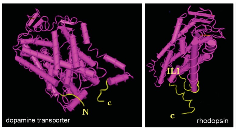
Many diseases arise from mutations, which impair protein folding. The study of folding-deficient variants of G protein-coupled receptors and solute carrier 6 (SLC6) transporters has shed light on the folding trajectory, how it is monitored and how misfolding can be remedied. Reducing the temperature lowers the energy barrier between folding intermediates and thereby eliminates stalling along the folding trajectory. For obvious reasons, cooling down is not a therapeutic option. One approach to rescue misfolded variants is to use membrane-permeable orthosteric ligands. Antagonists of GPCRs are-in many instances-effective pharmacochaperones: they restore cell surface expression provided that they enter cells and bind to folding intermediates. Pharmacochaperoning of SLC6 transporters is less readily achieved because the ionic conditions in the endoplasmic reticulum (ER) are not conducive to binding of typical inhibitors. The second approach is to target the heat-shock protein (HSP) relay, which monitors the folding trajectory on the cytosolic side. Importantly, orthosteric ligands and HSP-inhibitors are not mutually exclusive. In fact, pharmacochaperones and HSP-inhibitors can act in an additive or synergistic manner. This was exemplified by rescuing disease-causing, folding-deficient variants of the human dopamine transporters with the HSP70 inhibitor pifithrin-μ and the pharmacochaperone noribogaine in Drosophila melanogaster.

Keywords: G protein coupled receptors/GPCRs; heat-shock protein inhibitors; heat-shock protein relay; misfolding; pharmacochaperoning; solute carrier 6/SLC6.


Conflict of interest statement

The authors declare no conflict of interest.

Figures

Figure 1
Figure 1
Cumulative number of point mutations in the coding sequence of mutations, which result in folding-deficient solute carriers (SLC) transporters. The publications were identified in PubMed (www.ncbi.nlm.nih.gov). The numbers are a conservative estimate: only coding variants were counted, where the experimental evidence indicated a loss of function due to misfolding. Truncations due to premature stop codons were ignored, as were mutations, which resulted in a disrupted binding site for substrate and co-substrate ions. The pertinent references are for the norepinephrine transport (NET/SLC6A2 [17], for the creatine transporter-1 (CT1/SLC6A8 [18,19,20,21,22,23,24,25,26,27,28]), for the glycine transporter-2 (GlyT2/SLC6A5 [29,30]), for the dopamine transporter (DAT/SLC6A3 [31,32,33]) and for the GABA-transporter-1 (GAT1 [34]).
Figure 2
Figure 2
Structures of an SLC transporter (dopamine transporter) and a GPCR (rhodopsin) highlighting the circular arrangement of the helices in hydrophobic core. In spite of the truncations of the N- and C-terminal peptide segments, the structures indicate that juxtamembrane N- and C-terminal portions (highlighted in yellow) meet. In the transporter, the structure suggests an end-to-end contact of the cytoplasmic segments. In the receptor, helix 8 of the cytoplasmic carboxyterminus approaches α-helix 1/cytoplasmic loop 1 (IL1). Either arrangement likely serves as latch to stabilize the circular structure. It is also evident that several helices do not run perpendicular to the plane of the membrane; tilting is a reflection of the hydrophobic mismatch, which imposes an energy barrier during the conformational search associated with folding. View from an intracellular (cytosolic) perspective of the tilted dopamine transporter. Extracellular and lateral view of rhodopsin. Structure models (dopamine transporter 4XP4; rhodopsin 2I36) taken from www.ncbi.nlm.nih.gov.
Figure 3
Figure 3
Extended chaperone/COPII-exchange model. Upon release from the SEC61 translocon channel (not shown), the nascent transmembrane protein (GPCR or SLC6 transporter symbolised by blue TM helices) is glycosylated and engaged by calnexin; subsequently a heat-shock protein relay is recruited to the C-terminus with sequential binding of HSP40 and HSP70, (which can be inhibited by pifithin-μ, YM01, YM08, etc.) followed by transfer to HSP90 (which can be inhited by DMAG = 17-(dimethylaminoethylamino)-17-demethoxygeldanamycin = alvespimycin, radiciol and related compounds). If the client protein reaches its stable fold, the heat-shock proteins are released. This licences the C-terminus for an interaction with the cognate SEC24-isoform. The SEC23/SEC24-dimer (bow tie-shaped red and blue triangles) incorporates the protein cargo at ER exit sites (ERES) into the nascent COPII (coatomer protein complex II) coated vesicle, the curvature of which is induced by the bow tie-shape and stabilized by the outer layer COPII components SEC13/SEC31. For the sake of simplicity, the additional co-chaperones of the heat-shock protein relay (HOP, AHA1, etc.) and the additional components of the COPII machinery (the guanine nucleotide exchange factor SEC12 and the G protein SAR1) are not shown. If a stable fold cannot be reached, the protein is eventually marked for ER-associated degradation (ERAD) by recruitment of an E3 ubiquitin ligase. This can be triggered by the cytosolic heat-shock protein relay (shown in the upper part) or by lumenal chaperones (not shown). Initiation of ERAD is also contingent on a kifunensine-inhibited lumenal mannisodase (ER degradation-enhancing alpha-mannosidase-like protein 1—EDEM1). After retrotanslocation, the protein is degraded by the proteasome, which is susceptible to inhibition by MG132, bortezomib and related compounds. Note that for the sake of clarity the series of events have been depicted in two separate schematic representations, but they occur in the same plane of the membrane.

Similar articles

Cited by

References

    1. Morello J.P., Petäjä-Repo U.E., Bichet D.G., Bouvier M. Pharmacological chaperones: A new twist on receptor folding. Trends Pharmacol. Sci. 2000;21:466–469. doi: 10.1016/S0165-6147(00)01575-3. - DOI - PubMed
    1. Leidenheimer N.J., Ryder K.G. Pharmacological chaperoning: A primer on mechanism and pharmacology. Pharmacol. Res. 2014;83:10–19. doi: 10.1016/j.phrs.2014.01.005. - DOI - PMC - PubMed
    1. Germain D.P., Hughes D.A., Nicholls K., Bichet D.G., Giugliani R., Wilcox W.R., Feliciani C., Shankar S.P., Ezgu F., Amartino H., et al. Treatment of Fabry’s disease with the pharmacologic chaperone migalastat. N. Engl. J. Med. 2016;375:545–555. doi: 10.1056/NEJMoa1510198. - DOI - PubMed
    1. Van Goor F., Hadida S., Grootenhuis P.D., Burton B., Stack J.H., Straley K.S., Decker C.J., Miller M., McCartney J., Olson E.R., et al. Correction of the F508del-CFTR protein processing defect in vitro by the investigational drug VX-809. Proc. Natl. Acad. Sci. USA. 2011;108:18843–18848. doi: 10.1073/pnas.1105787108. - DOI - PMC - PubMed
    1. Alderman E.L., Coltart D.J., Wettach G.E., Harrison D.C. Coronary artery syndromes after sudden propranolol withdrawal. Ann. Intern. Med. 1974;81:625–627. doi: 10.7326/0003-4819-81-5-625. - DOI - PubMed

MeSH terms

Substances

LinkOut - more resources