

doi: 10.1038/ismej.2017.184.
Epub 2017 Nov 14.
Build your own soil: exploring microfluidics to create microbial habitat structures
Affiliations
- PMID: 29135971
- PMCID: PMC5776464
- DOI: 10.1038/ismej.2017.184
Item in Clipboard
Build your own soil: exploring microfluidics to create microbial habitat structures
ISME J.
2018 Feb.
Abstract
Soil is likely the most complex ecosystem on earth. Despite the global importance and extraordinary diversity of soils, they have been notoriously challenging to study. We show how pioneering microfluidic techniques provide new ways of studying soil microbial ecology by allowing simulation and manipulation of chemical conditions and physical structures at the microscale in soil model habitats.
Conflict of interest statement
The authors declare no conflict of interest.
Figures
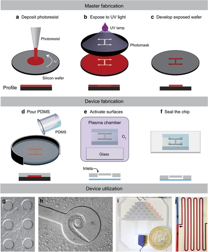
Fabrication of microfluidic devices. A common method to make microfluidic devices is to make a master by photolithography, which is then used to mold PDMS silicone. (a) Deposition of photoresist on a silicon or glass wafer. The thickness of the photoresist is defined by spinning the wafer at a certain rotational speed for a certain time. (b) UV light exposure through mask. UV light illuminates the desired pattern through a photomask and catalyses photoresist crosslinking. (c) Development of the exposed wafer. Non-crosslinked photoresist is removed using a solvent bath. The pattern is now visible on the surface of the master. (d) PDMS molding. PDMS is poured on the developed master and allowed to polymerize in an oven, forming a flexible polymer block. (e) Surface activation. Holes for desired inlets are punched into the PDMS slab, and both PDMS and the glass slide are activated in a plasma chamber. Other materials such as membranes or other PDMS layers can also be used to seal the chip. (f) Sealing the chip by placing the surfaces in contact, which form covalent bonds between the PDMS and the glass surface. (g) Hyphae of Mycetinis scorodonius growing in a pillar system with 100 μm wide pillars. (h) Nematode that migrated into a chip channel from a natural soil inoculum. (i) Microfluidic chip where a dye gradient is generated by sequential mixing and introduced into a culture chamber. (j) Zoom-in on the gradient generator showing and dye diffusion.
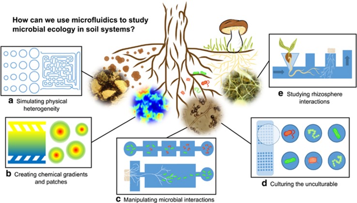
Five aspects of how microfluidics can be used to mimic the soil-environment and study microbial behavior in a small-structured environment. (a) Simulating physical heterogeneity. Pillars and walls of different sizes and shapes can be used to simulate differences in soil structure and porosity to study how variation in physical heterogeneity affects, for example, microbial establishment, behavior (Held et al., 2010; Deng et al., 2015), and feedback interactions with their environment. (b) Creating chemical gradients and patches. Chemical gradients or plume-like injections can be created inside the chips to mimic spatial heterogeneity of nutrients or other soluble compounds and study, for example, chemotaxis (Stocker et al., 2008). (c) Manipulating microbial interactions. Arenas for the study of microbial physiology, behavior and interactions can be fabricated, allowing minute control over when and where microbes enter the system, with the possibility to restrict encounters to few individual cells or hyphae (Stanley et al., 2014; Hol et al., 2016). (d) Culturing the unculturable. With the development of the Ichip (Nichols et al., 2010), new possibilities have opened up for culturing soil bacteria that have not previously been possible in solid medium cultures. The main design factor thought to facilitate this is the micro-confinement of individual cells in diffusion chambers sealed off with membranes, still allowing for metabolic transfer to and from the surrounding environment. This strongly expands the species pool for laboratory studies, and facilitates identification of their special requirements for pure culture isolation. (e) Studying rhizosphere interactions. Plant roots can be grown from seeds, for example, through pipette tips, into channels of microfluidic devices (Grossmann et al., 2011), permitting close monitoring of root morphology, and giving us the ability to control nutrient supply as well as microbial exposure within the root system. This will open up possibilities to study, for example, the colonization success and succession of root symbiosis such as those involving rhizobia and mycorrhizas, or monitoring of pathogens under differentiated nutrient conditions.
References
-
- Antwis RE, Griffiths SM, Harrison XA, Aranega-Bou P, Arce A, Bettridge AS et al. (2017). Fifty important research questions in microbial ecology. FEMS Microbiol Ecol 93: 1–10. - PubMed
-
- Bhatia SN, Ingber DE. (2014). Microfluidic organs-on-chips. Nat Biotechnol 32: 760–772. - PubMed
-
- Boswell G. (2003). Growth and function of fungal mycelia in heterogeneous environments. Bull Math Biol 65: 447–477. - PubMed
-
- Deng J, Orner EP, Chau JF, Anderson EM, Kadilak AL, Rubinstein RL et al. (2015). Synergistic effects of soil microstructure and bacterial EPS on drying rate in emulated soil micromodels. Soil Biol Biochem 83: 116–124.
