. 2017 Nov 14;12(11):e0187186.
doi: 10.1371/journal.pone.0187186. eCollection 2017.

Arginase expression modulates nitric oxide production in Leishmania (Leishmania) amazonensis

Affiliations

Arginase expression modulates nitric oxide production in Leishmania (Leishmania) amazonensis

Stephanie Maia Acuña et al. PLoS One. .

Abstract

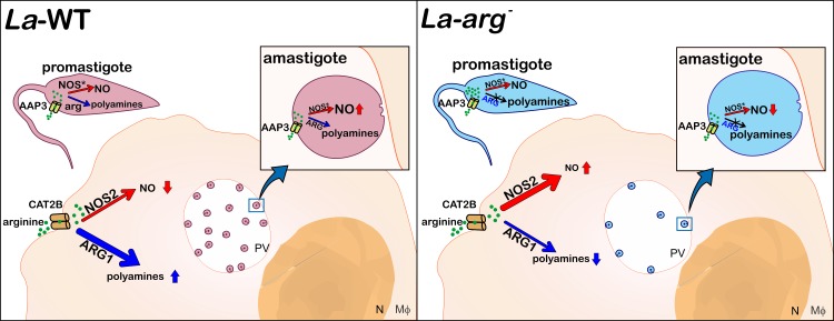
Background: Arginase is an enzyme that converts L-arginine to urea and L-ornithine, an essential substrate for the polyamine pathway supporting Leishmania (Leishmania) amazonensis replication and its survival in the mammalian host. L-arginine is also the substrate of macrophage nitric oxide synthase 2 (NOS2) to produce nitric oxide (NO) that kills the parasite. This competition can define the fate of Leishmania infection.

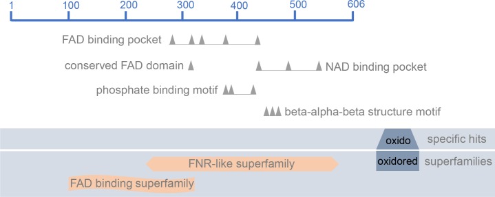
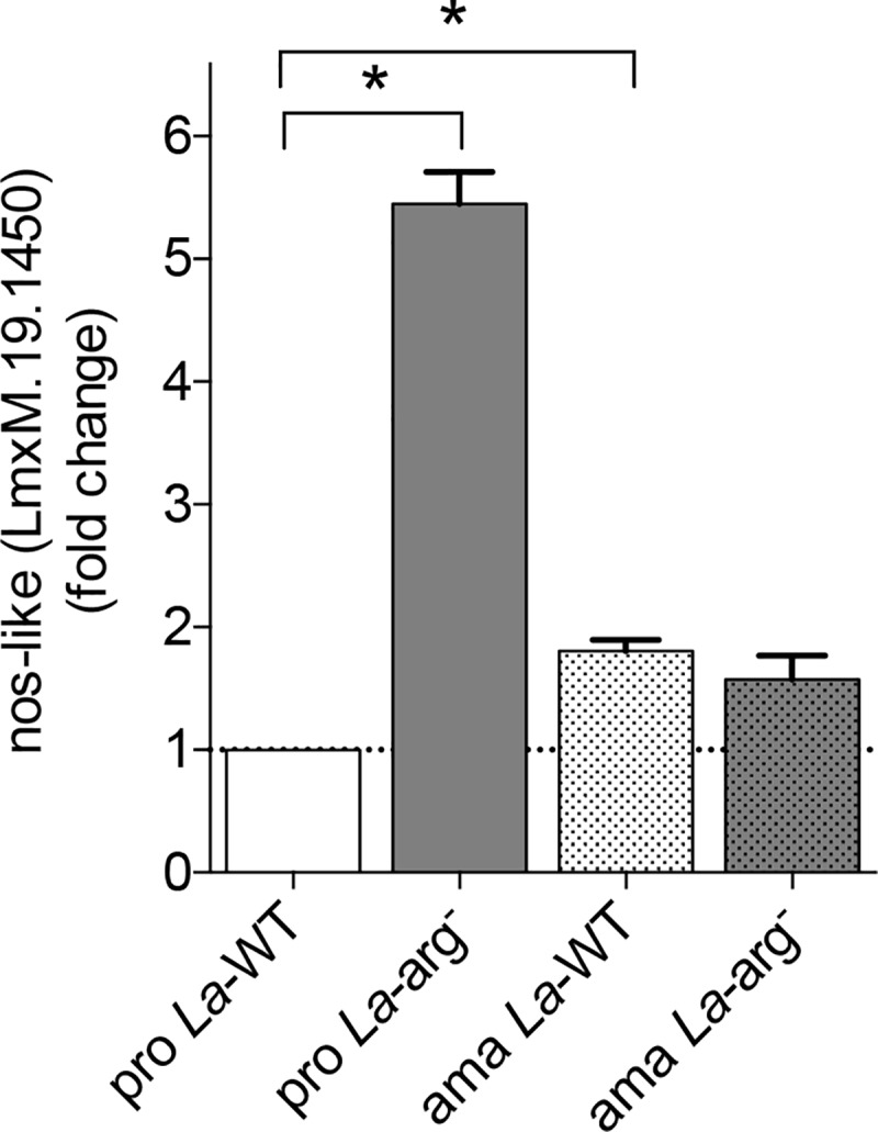
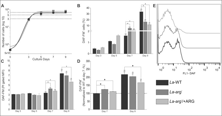
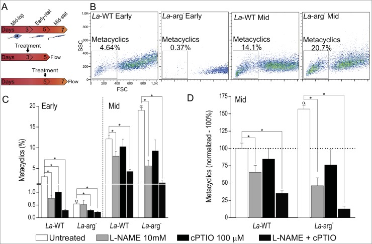
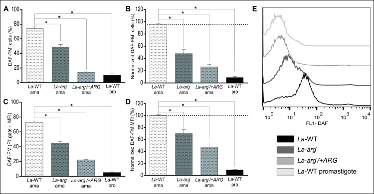
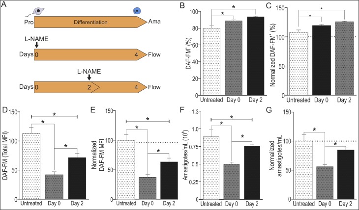
Methodology/principal findings: The transcriptomic profiling identified a family of oxidoreductases in L. (L.) amazonensis wild-type (La-WT) and L. (L.) amazonensis arginase knockout (La-arg-) promastigotes and axenic amastigotes. We highlighted the identification of an oxidoreductase that could act as nitric oxide synthase-like (NOS-like), due to the following evidences: conserved domain composition, the participation of NO production during the time course of promastigotes growth and during the axenic amastigotes differentiation, regulation dependence on arginase activity, as well as reduction of NO amount through the NOS activity inhibition. NO quantification was measured by DAF-FM labeling analysis in a flow cytometry.

Conclusions/significance: We described an arginase-dependent NOS-like activity in L. (L.) amazonensis and its role in the parasite growth. The increased detection of NO production in the mid-stationary and late-stationary growth phases of La-WT promastigotes could suggest that this production is an important factor to metacyclogenesis triggering. On the other hand, La-arg- showed an earlier increase in NO production compared to La-WT, suggesting that NO production can be arginase-dependent. Interestingly, La-WT and La-arg- axenic amastigotes produced higher levels of NO than those observed in promastigotes. As a conclusion, our work suggested that NOS-like is expressed in Leishmania in the stationary growth phase promastigotes and amastigotes, and could be correlated to metacyclogenesis and amastigotes growth in a dependent way to the internal pool of L-arginine and arginase activity.


Conflict of interest statement

Competing Interests: The authors have declared that no competing interests exist.

Figures

Fig 1
Fig 1. NOS-like graphical summary of conserved domains.
Structure of the oxidoreductase synthase-like protein (LmxM.1950/EC 1.14.13.39) with the following conserved domains hits: ferredoxin reductase (FNR) binding domain, flavin adenine dinucleotide (FAD) binding pocket, conserved FAD binding motif, nicotinamide adenine dinucleotide (NAD) binding pocket, phosphate binding motif and beta-alpha-beta structure motif. The representation was based on in silico search of conserved domain of NCBI database.
Fig 2
Fig 2. Nos-like mRNA expression by RT-qPCR.
The nos-like mRNA levels were based on quantification of the target and were normalized by gapdh expression in promastigotes (pro) and amastigotes (ama) of La-WT and La-arg-. The values are the mean ± SEM of three independent biological replicates (n4-6). (*) p < 0.05.
Fig 3
Fig 3. Growth curve and NO production in La-WT, La-arg- and La-arg-/+ARG on days 3, 5, 7 and 9.
L. (L.) amazonensis promastigotes wild-type (La-WT—black); arginase null knockout (La-arg-—dark gray) and the arginase addback (La-arg-/+ARG–light gray) growth curve and the NO production by DAF-FM labeling in flow cytometry. A total of 2x10⁶ cells was labeled with 1 ng/mL propidium iodide (PI) and was analyzed using FACSCalibur (Becton Dickinson). The viable cells (PI-) were used for the analysis of the frequency of DAF-FM-labeled cells and the mean fluorescence (MFI). (A) Growth curves and the establishment of the logarithmic growth phase (day 3); early-stationary (day 5), mid-stationary (day 7) and late-stationary phase (day 9). (B) Frequency of DAF-FM+ cells. Each bar represents the mean of the percent ±SEM of DAF-FM+ cells (n = 3–9). (C) MFI of DAF-FM+ cells. Each bar represents the mean of the fluorescence intensity ±SEM of viable cells (n = 3–9). (D) Normalization of MFI values in relation to day 7 of La-WT (100%). (E) Histogram of MFI of the PI- gate at the 9th day of culture. These data were analyzed by Student’s t-test, and (*) p < 0.05, (α) p < 0.05 compared the day 7 with the days 3 and 5, (β) p < 0.05 compared the day 9 with the days 3, 5 and 7. The data are representative of 3 independent experiments.
Fig 4
Fig 4. Inhibition of NOS activity or NO amount and metaclyclogenesis in La-WT and La-arg- promastigote.
(A) L. (L.) amazonensis wild-type (La-WT) and arginase null knockout (La-arg-) at logarithmic (log—day 3) and early-stat (early-stationary—day 5) phase promastigotes were untreated (white) or treated with 10 mM of L-NAME (light gray), 100 μM of cPTIO (dark gray) and L-NAME plus cPTIO (dark). After 2 days of culture metacyclic promastigotes forms were quantified by flow cytometry. (B) Dot plot of SSC and FSC features representative of metacyclics promastigote forms viable cells analyzed using FACSCalibur (Becton Dickinson). (C) Frequency of metacyclics promastigotes. Each bar represents the mean of the percent ±SEM of metaclyclic cells (n = 3–9). (D) Normalization of the percentage of metacyclic forms in relation to log phase of La-WT (100%). These data were analyzed by Student’s t-test, and (*) p < 0.05, (α) p < 0.05 compared the La-WT to La-arg-. The data are representative of 3 independent experiments (n = 6–9).
Fig 5
Fig 5. L. (L.) amazonensis axenic amastigotes produced NO in a higher amount than promastigotes.
La-WT, La-arg- and La-arg-/+ARG promastigotes at the early-stationary phase (day 5 – 5x10⁶ cells/10 mL) were differentiated into amastigotes by changing the M199 medium to pH 5.5 and maintaining the culture at 34°C during 4 days. Next, the cells were labeled with 1 μM DAF-FM and 1 ng/mL PI and were analyzed by flow cytometry. La-WT promastigotes in the late-stationary phase were used as a positive control. The viable cells (PI-) were used to analyze the frequency of DAF-FM+ cells and MFI. (A) Frequency of DAF-FM+ cells. These are representative of 1 independent experiment (n = 3–4). (B) Normalization of the percentage of DAF-FM+ La-arg- and La-arg-/+ARG in relation to La-WT (100%). These are representative of 3 independent experiments (n = 9). (C) MFI of DAF-FM+ cells. These are representative of 1 independent experiment (n = 3–4). (D) Normalization of the MFI values of La-arg- and La-arg-/+ARG in relation to La-WT (100%). These data are representative of 3 independent experiments (n = 9). These data were analyzed were analyzed by one-way ANOVA and Tukey’s comparison post-test. Significance p-value is represented by *, p<0.05. (E) Histogram of MFI of DAF-FM.
Fig 6
Fig 6. Inhibition of NOS activity during La-WT and La-arg- amastigotes differentiation.
(A) L. (L.) amazonensis La-WT at early-stationary phase promastigotes (pro) were differentiated into amastigotes (ama), by changing the M199 medium to pH 5.5 and maintaining the culture at 34°C during 4 days, untreated (white) or treated with 10 mM of L-NAME (light gray) at day 0 or day of culture. Next, a total of 2x10⁶ cells was labeled with 1 μM DAF-FM and 1 ng/mL propidium iodide (PI) and analyzed using FACSCalibur (Becton Dickinson). B) Frequency of of DAF-FM+ cells. (C) Normalization of the percentage of DAF-FM+ cells in relation to La-WT-untreated (100%). (D) MFI of DAF-FM+ cells. (E) Normalization of the MFI of DAF-FM+ cells in relation La-WT-untreated (100%). (F) Number of amastigotes forms. (G) Normalization of number of amastigote forms in relation La-WT-untreated (100%). Each bar represents the mean of the values ±SEM (n = 3–9). These data were analyzed by Student’s t-test, and (*) p < 0.05. The data are representative of 3 independent experiments.
Fig 7
Fig 7. Schematic representation of L. (L.) amazonensis promastigote and amastigote NO production in the infection context.
In L. (L.) amazonensis, the AAP3 transporter takes up L-arginine, and this amino acid is metabolized by arginase into ornithine to produce polyamines or via NOS-like to produce NO. La-WT-infected macrophages increase the activity of the polyamine pathway, increasing the expression of the L-arginine transporter from the host (CAT2B) and parasite (AAP3), arginase1 (host) and parasite-arginase to metabolizing L-arginine to produce polyamines, consequently sustaining the basal level of NOS2 expression and NO production. Indeed, the La-WT amastigote differentiation induces NOS-like expression and NO production, higher levels than promastigotes, but lower than those produced by macrophages. The lack of arginase activity in promastigotes increases the L-arginine levels, even that the reduction in AAP3 expression, inducing the expression of NOS-like, NO and citrulline production. The La-arginfected macrophages present lower levels of CAT2B and AAP3, which could reduce L-arginine uptake, and lower the levels of arginase 1 and polyamine production compared with La-WT infection allowing the increase in NOS2 expression and NO production, reducing infectivity. In addition, the La-arg- amastigote differentiation induces NOS-like expression but not AAP3 expression and L-arginine uptake sufficient to increase NO production compared with La-WT promastigotes. PV: parasitophorous vacuole.

Similar articles

Cited by

References

    1. Bogdan C (2008) Mechanisms and consequences of persistence of intracellular pathogens: leishmaniasis as an example. Cell Microbiol 10: 1221–1234. doi: 10.1111/j.1462-5822.2008.01146.x - DOI - PubMed
    1. Liese J, Schleicher U, Bogdan C (2008) The innate immune response against Leishmania parasites. Immunobiology 213: 377–387. doi: 10.1016/j.imbio.2007.12.005 - DOI - PubMed
    1. Sacks D, Kamhawi S (2001) Molecular aspects of parasite-vector and vector-host interactions in leishmaniasis. Annu Rev Microbiol 55: 453–483. doi: 10.1146/annurev.micro.55.1.453 - DOI - PubMed
    1. Teixeira DE, Benchimol M, Rodrigues JC, Crepaldi PH, Pimenta PF, de Souza W (2013) The cell biology of Leishmania: how to teach using animations. PLoS Pathog 9: e1003594 doi: 10.1371/journal.ppat.1003594 - DOI - PMC - PubMed
    1. Séguin O, Descoteaux A (2016) Leishmania, the phagosome, and host responses: The journey of a parasite. Cell Immunol 309: 1–6. doi: 10.1016/j.cellimm.2016.08.004 - DOI - PubMed