

Identification of topological features in renal tumor microenvironment associated with patient survival
- PMID: 29136101
- PMCID: PMC7263397
- DOI: 10.1093/bioinformatics/btx723
Identification of topological features in renal tumor microenvironment associated with patient survival
Abstract
Motivation: As a highly heterogeneous disease, the progression of tumor is not only achieved by unlimited growth of the tumor cells, but also supported, stimulated, and nurtured by the microenvironment around it. However, traditional qualitative and/or semi-quantitative parameters obtained by pathologist's visual examination have very limited capability to capture this interaction between tumor and its microenvironment. With the advent of digital pathology, computerized image analysis may provide a better tumor characterization and give new insights into this problem.
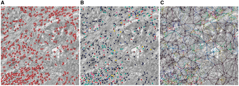
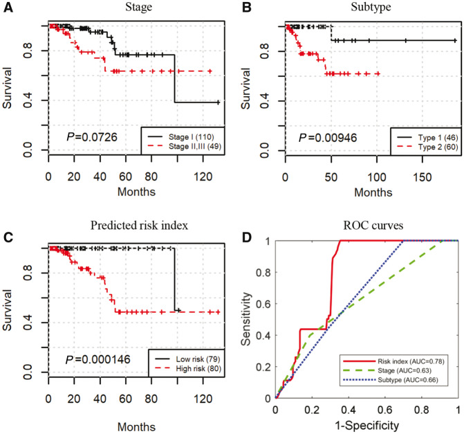
Results: We propose a novel bioimage informatics pipeline for automatically characterizing the topological organization of different cell patterns in the tumor microenvironment. We apply this pipeline to the only publicly available large histopathology image dataset for a cohort of 190 patients with papillary renal cell carcinoma obtained from The Cancer Genome Atlas project. Experimental results show that the proposed topological features can successfully stratify early- and middle-stage patients with distinct survival, and show superior performance to traditional clinical features and cellular morphological and intensity features. The proposed features not only provide new insights into the topological organizations of cancers, but also can be integrated with genomic data in future studies to develop new integrative biomarkers.
Availability and implementation: https://github.com/chengjun583/KIRP-topological-features.
Contact: 1271992826@qq.com or kunhuang@iu.edu.
Supplementary information: Supplementary data are available at Bioinformatics online.
Figures


References
-
- Phoulady H.A. et al. (2016) Nucleus segmentation in histology images with hierarchical multilevel thresholding. InProceedings SPIE 9791, Medical Imaging 2016 Digital Pathology. Vol. 9791,San Diego, California, United States (23 March 2016), p.979111. doi: 10.1117/12.2216940. - DOI
-
- Al-Kofahi Y. et al. (2010) Improved automatic detection and segmentation of cell nuclei in histopathology images.IEEE Trans. Biomed. Eng.,57,841–852. - PubMed
-
- Albarqouni S. et al. (2016) AggNet: deep learning from crowds for mitosis detection in breast cancer histology images.IEEE Trans. Med. Imaging,35,1313–1321. - PubMed
-
- Beck A.H. et al. (2011) Systematic analysis of breast cancer morphology uncovers stromal features associated with survival.Sci. Transl. Med.,3,108ra113–108ra113. - PubMed
Publication types
MeSH terms
Grants and funding
LinkOut - more resources
Full Text Sources
Other Literature Sources
Medical
Research Materials
