Review
. 2018 Jul 21;39(28):2618-2624.
doi: 10.1093/eurheartj/ehx653.

Calcific aortic valve stenosis: hard disease in the heart: A biomolecular approach towards diagnosis and treatment

Affiliations
Review

Calcific aortic valve stenosis: hard disease in the heart: A biomolecular approach towards diagnosis and treatment

Frederique E C M Peeters et al. Eur Heart J. .

Abstract

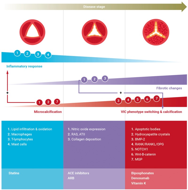
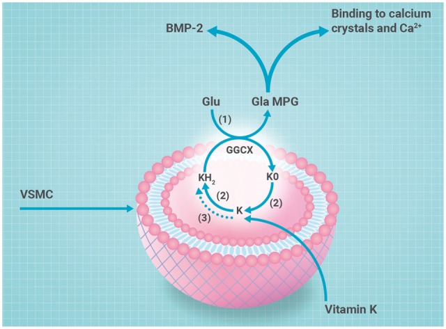
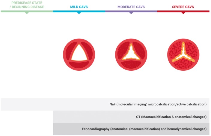
Calcific aortic valve stenosis (CAVS) is common in the ageing population and set to become an increasing economic and health burden. Once present, it inevitably progresses and has a poor prognosis in symptomatic patients. No medical therapies are proven to be effective in holding or reducing disease progression. Therefore, aortic valve replacement remains the only available treatment option. Improved knowledge of the mechanisms underlying disease progression has provided us with insights that CAVS is not a passive disease. Rather, CAVS is regulated by numerous mechanisms with a key role for calcification. Aortic valve calcification (AVC) is actively regulated involving cellular and humoral factors that may offer targets for diagnosis and intervention. The discovery that the vitamin K-dependent proteins are involved in the inhibition of AVC has boosted our mechanistic understanding of this process and has opened up novel avenues in disease exploration. This review discusses processes involved in CAVS progression, with an emphasis on recent insights into calcification, methods for imaging calcification activity, and potential therapeutic options.


Figures

Figure 1
Figure 1
Aortic valve. Left panel: 3D reconstruction (from bottom to top): aortic valve with three cusps and proximal ascending aorta. Middle panel: 2D view. Right panel: valvular histology (bottom to top): ventricularis, spongiosa, and fibrosa.
Figure 2
Figure 2
Pathophysiology and potential treatment targets (schematic overview). Upper panel: Progressive calcific aortic valve stenosis stages from non-stenotic to severe stenosis (left–right). Progressive thickening and calcification result in valvular dysfunction, characterized by decreased cusp mobility and opening, altered haemodynamics and stress. Middle panel: Cellular involvement in calcific aortic valve stenosis. Endothelial damage triggers lipid infiltration and upon oxidation an inflammatory response involving macrophages, T-lymphocytes, and mast cells. Inflammation triggers phenotypic switching of valvular interstitial cells resulting in increased extracellular vesicle release, providing a nidus for calcification. Microcalcification provokes an inflammatory response, resulting in increased apoptosis and/or delayed phagocytosis thereby expanding calcium deposition. Upon propagation, pro-fibrotic and pro-calcific processes dominate. Pro-fibrotic changes leading to collagen deposition and facilitating progressive calcification are mediated by reduced nitric oxide expression and up-regulation of renin–angiotensin system. Calcification is the dominant process driving disease progression. valvular interstitial cell phenotype switching to an osteoblast phenotype is thought to play a role in the progression phase by multiple regulatory pathways including Notch, receptor activator of nuclear factor kappa B/receptor activator of nuclear factor kappa B ligand/osteoprotegerin, Wnt/b-catenin, and bone morphogenetic protein-2. Lower panel: Potential pharmacological interventions.
Figure 3
Figure 3
Synthesis of active matrix-Gla protein: schematic overview of vitamin K metabolism. The vitamin K cycle has a central role in the posttranslational carboxylation of glutamate (Glu) to γ-carboxyglutamate (Gla). Reduction of vitamin K to vitamin K hydroxyquinone (KH2) in presence of vitamin K epoxide reductase (VKOR) (2) or DDT-diaphorase (3). Vitamin K hydroxyquinone is oxidized during γ-glutamyl carboxylation by gamma-glutamyl carboxylase (GGCX) (1) into vitamin K epoxide (KO). Vitamin K epoxide is reduced to vitamin K by vitamin K epoxide reductase (2). Carboxylated matrix-Gla protein is active matrix-Gla protein that is secreted in the extracellular environment and inhibits calcification via binding to bone morphogenetic protein-2 or direct inhibition of calcium crystal formation.
Figure 4
Figure 4
Role of imaging techniques in displaying calcification in stages of disease progression. Molecular imaging using 18F-sodium fluoride enables to visualize microcalcification and active calcification from beginning stages onwards. Computed tomography displays macrocalcification and anatomical changes in latter phases. Echocardiography visualizes anatomical changes and haemodynamic changes (2).

References

    1. Bonow RO, Greenland P.. Population-wide trends in aortic stenosis incidence and outcomes. Circulation 2015;131:969–971. - PubMed
    1. Baumgartner H, Falk V, Bax JJ, De Bonis M, Hamm C, Holm PJ, Iung B, Lancellotti P, Lansac E, Munoz DR, Rosenhek R, Sjogren J, Tornos Mas P, Vahanian A, Walther T, Wendler O, Windecker S, Zamorano JL.. 2017 ESC/EACTS Guidelines for the management of valvular heart disease: the Task Force for the Management of Valvular Heart Disease of the European Society of Cardiology (ESC) and the European Association for Cardio-Thoracic Surgery (EACTS). Eur Heart J 2017;38:2739–2791. - PubMed
    1. Dweck MR, Khaw HJ, Sng GK, Luo EL, Baird A, Williams MC, Makiello P, Mirsadraee S, Joshi NV, van Beek EJ, Boon NA, Rudd JH, Newby DE.. Aortic stenosis, atherosclerosis, and skeletal bone: is there a common link with calcification and inflammation? Eur Heart J 2013;34:1567–1574. - PubMed
    1. Aikawa E, Libby P.. A rock and a hard place: chiseling away at the multiple mechanisms of aortic stenosis. Circulation 2017;135:1951–1955. - PMC - PubMed
    1. Hutcheson JD, Aikawa E, Merryman WD.. Potential drug targets for calcific aortic valve disease. Nat Rev Cardiol 2014;11:218–231. - PMC - PubMed

Publication types

MeSH terms

Supplementary concepts