. 2018 Jun 6;44(4):854-864.
doi: 10.1093/schbul/sbx148.

Genetic Overlap Between Schizophrenia and Volumes of Hippocampus, Putamen, and Intracranial Volume Indicates Shared Molecular Genetic Mechanisms

Affiliations

Genetic Overlap Between Schizophrenia and Volumes of Hippocampus, Putamen, and Intracranial Volume Indicates Shared Molecular Genetic Mechanisms

Olav B Smeland et al. Schizophr Bull. .

Abstract

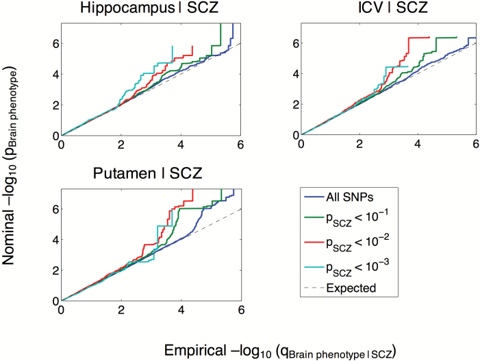
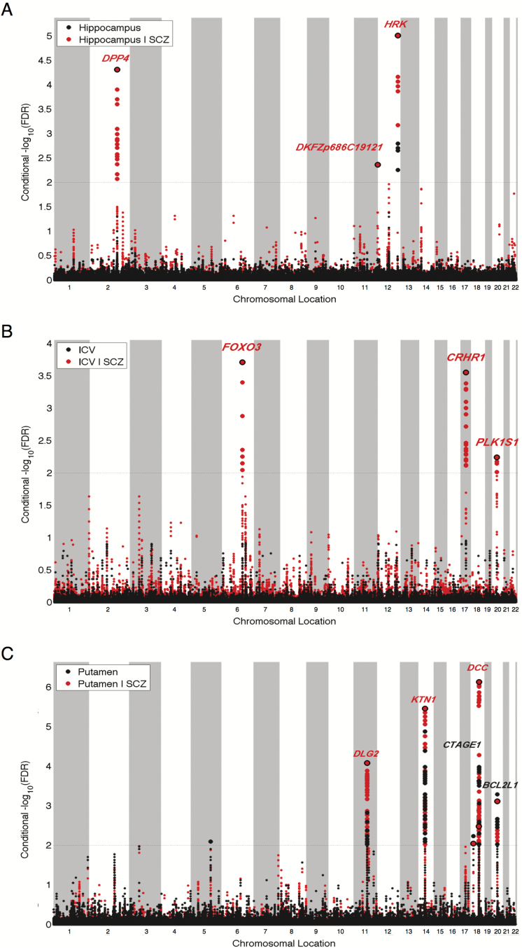
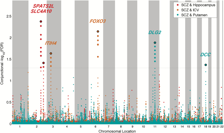
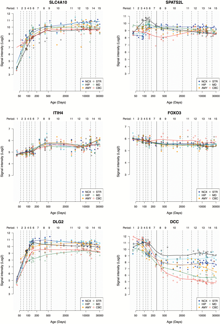
Schizophrenia (SCZ) is associated with differences in subcortical brain volumes and intracranial volume (ICV). However, little is known about the underlying etiology of these brain alterations. Here, we explored whether brain structure volumes and SCZ share genetic risk factors. Using conditional false discovery rate (FDR) analysis, we integrated genome-wide association study (GWAS) data on SCZ (n = 82315) and GWAS data on 7 subcortical brain volumes and ICV (n = 11840). By conditioning the FDR on overlapping associations, this statistical approach increases power to discover genetic loci. To assess the credibility of our approach, we studied the identified loci in larger GWAS samples on ICV (n = 26577) and hippocampal volume (n = 26814). We observed polygenic overlap between SCZ and volumes of hippocampus, putamen, and ICV. Based on conjunctional FDR < 0.05, we identified 2 loci shared between SCZ and ICV implicating genes FOXO3 (rs10457180) and ITIH4 (rs4687658), 2 loci shared between SCZ and hippocampal volume implicating SLC4A10 (rs4664442) and SPATS2L (rs1653290), and 2 loci shared between SCZ and volume of putamen implicating DCC (rs4632195) and DLG2 (rs11233632). The loci shared between SCZ and hippocampal volume or ICV had not reached significance in the primary GWAS on brain phenotypes. Proving our point of increased power, 2 loci did reach genome-wide significance with ICV (rs10457180) and hippocampal volume (rs4664442) in the larger GWAS. Three of the 6 identified loci are novel for SCZ. Altogether, the findings provide new insights into the relationship between SCZ and brain structure volumes, suggesting that their genetic architectures are not independent.


Figures

Fig. 1.
Fig. 1.
Conditional Q-Q plots of nominal vs empirical −log10P-values (corrected for inflation) in volumes of hippocampus, putamen, and intracranial volume (ICV) below the standard GWAS threshold of P < 5 × 10⁻⁸ as a function of significance of association with schizophrenia (SCZ) at the level of −log10(P) ≥ 1, −log10(P) ≥ 2, −log10(P) ≥ 3 corresponding to P ≤ .1, P ≤ .01, P ≤ .001, respectively. The blue lines indicate all SNPs. The dashed lines indicate the null hypothesis.
Fig. 2.
Fig. 2.
“Conditional FDR (condFDR) Manhattan plots” of conditional −log10 (FDR) values for (A) volume of hippocampus, (B) intracranial volume (ICV), and (C) volume of putamen conditioned on schizophrenia (SCZ). Unconditioned FDR values are shown in black, condFDR values in red. SNPs with conditional −log10 FDR > 2.0 (ie, condFDR < 0.01) are shown with large points. A black line around the large points indicates the most significant SNP in each LD block. This SNP is annotated with the closest gene.
Fig. 3.
Fig. 3.
“ConjFDR Manhattan plot” of conjunctional −log10 (FDR) values for schizophrenia (SCZ) and hippocampal volume, intracranial volume (ICV), and volume of putamen; the conjunctions are denoted as SCZ & hippocampus, SCZ & ICV, and SCZ & putamen, respectively. SNPs with conjFDR < 0.05 (ie, conjunctional −log10(FDR) > 1.3) are shown with enlarged data points. A black circle around the enlarged data points indicates the most significant SNP in each LD block and this SNP was annotated with the closest gene which is listed above the symbols in each locus. The figure shows the localization of the “conjunctional loci,” and further details are provided in table 1.
Fig. 4.
Fig. 4.
Expression trajectories of SLC4A10, SPATS2L, ITIH4, FOXO3, DLG2, and DCC in the developing and adult human brain, provided by the Human Brain Transcriptome project. Line plots show the log2-transformed gene exon array signal intensity from the early fetal period to late adulthood in 6 brain regions. The solid line between periods 7 and 8 (approximately post-conception day 280) separates prenatal from postnatal periods. NCX, neocortex; HIP, hippocampus; AMY, amygdala; STR, striatum; MD, mediodorsal nucleus of the thalamus; CBC, cerebellar cortex.

References

    1. van Erp TG, Hibar DP, Rasmussen JM, et al. Subcortical brain volume abnormalities in 2028 individuals with schizophrenia and 2540 healthy controls via the ENIGMA consortium. Mol Psychiatry. Apr 2016;21(4):547–553. - PMC - PubMed
    1. Okada N, Fukunaga M, Yamashita F, et al. Abnormal asymmetries in subcortical brain volume in schizophrenia. Mol Psychiatry. Oct 2016;21(10):1460–1466. - PMC - PubMed
    1. Haijma SV, Van Haren N, Cahn W, Koolschijn PC, Hulshoff Pol HE, Kahn RS. Brain volumes in schizophrenia: a meta-analysis in over 18 000 subjects. Schizophr Bull. 2013;39:1129–1138. - PMC - PubMed
    1. Lichtenstein P, Yip BH, Björk C, et al. Common genetic determinants of schizophrenia and bipolar disorder in Swedish families: a population-based study. Lancet. 2009;373:234–239. - PMC - PubMed
    1. Hibar DP, Stein JL, Renteria ME, et al. Common genetic variants influence human subcortical brain structures. Nature. Apr 09 2015;520(7546):224–229. - PMC - PubMed

Publication types

LinkOut - more resources