

Toll-Like Receptor 4 Reduces Occludin and Zonula Occludens 1 to Increase Retinal Permeability Both in vitro and in vivo
- PMID: 29136627
- PMCID: PMC5896294
- DOI: 10.1159/000480455
Toll-Like Receptor 4 Reduces Occludin and Zonula Occludens 1 to Increase Retinal Permeability Both in vitro and in vivo
Abstract
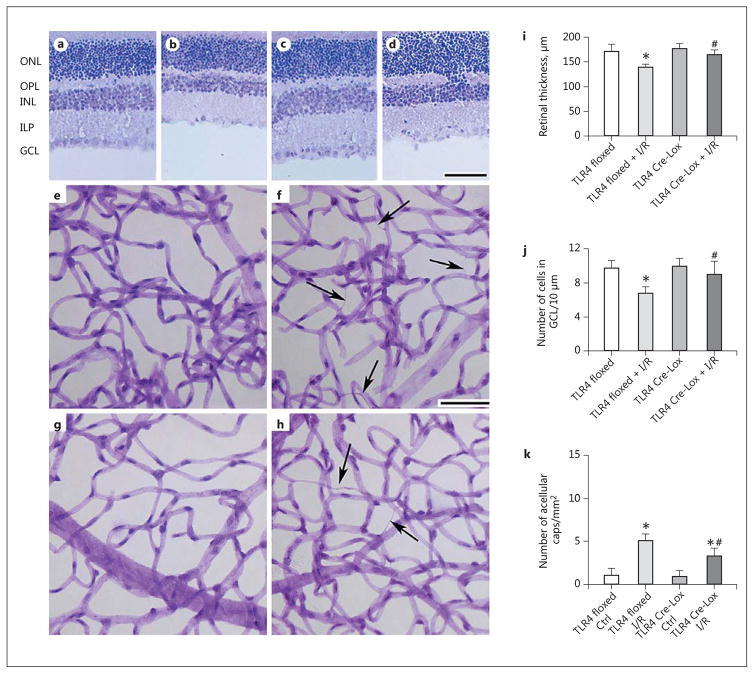
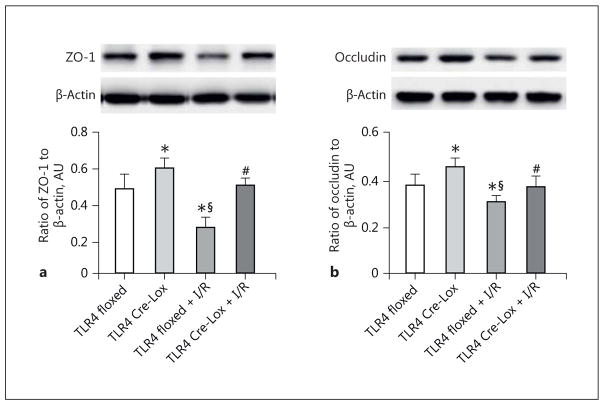
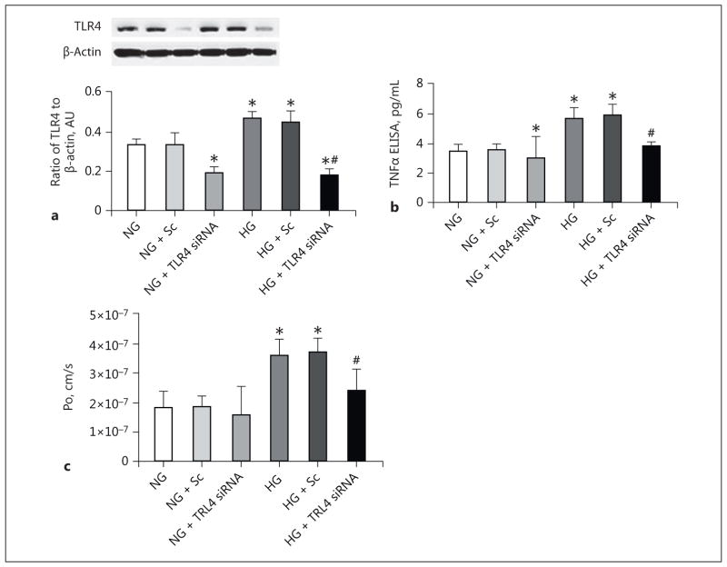
We reported that β-adrenergic receptors regulate toll-like receptor 4 (TLR4) signaling in the retina of diabetic mice and in retinal endothelial cells (REC) and Müller cells. We hypothesized that TLR4 regulates retinal permeability both in vitro and in vivo in the retinal vasculature. We used REC cultured in normal and high-glucose conditions and TLR4 siRNA treatments for cell culture studies of permeability and protein analyses of tumor necrosis factor α, occludin, and zonula occludens 1 (ZO-1). We used endothelial cell (EC)-specific Cre-Lox TLR4 knockout mice to study retinal permeability and neuronal and vascular changes following exposure to ocular ischemia/reperfusion (I/R) used as a retinal stressor. We found that the loss of TLR4 in the EC led to the reduced permeability following I/R and fewer degenerate capillaries. Retinal permeability was increased in REC grown in high-glucose conditions but was inhibited by TLR4 siRNA treatment. High-glucose culture conditions significantly reduced occludin and ZO-1 levels in REC, and TLR4 siRNA treatment restored levels to baseline. In conclusion, these studies demonstrate that TLR4 in EC strongly regulates retinal permeability and neuronal and vascular changes following exposure to stressors such as I/R.
Keywords: Endothelial cell; Inflammation; Ischemia/reperfusion; Permeability; Retina; Toll-like receptor 4.
© 2017 S. Karger AG, Basel.
Conflict of interest statement
No authors have any conflicts of interest with these studies.
Figures

Similar articles
-
Compound 49b Regulates ZO-1 and Occludin Levels in Human Retinal Endothelial Cells and in Mouse Retinal Vasculature.Invest Ophthalmol Vis Sci. 2017 Jan 1;58(1):185-189. doi: 10.1167/iovs.16-20412. Invest Ophthalmol Vis Sci. 2017. PMID: 28114578 Free PMC article.
-
Forskolin regulates retinal endothelial cell permeability through TLR4 actions in vitro.Mol Cell Biochem. 2021 Dec;476(12):4487-4492. doi: 10.1007/s11010-021-04252-9. Epub 2021 Aug 28. Mol Cell Biochem. 2021. PMID: 34499321 Free PMC article.
-
Epac1 regulates TLR4 signaling in the diabetic retinal vasculature.Cytokine. 2021 Aug;144:155576. doi: 10.1016/j.cyto.2021.155576. Epub 2021 May 18. Cytokine. 2021. PMID: 34020266 Free PMC article.
-
Prothymosin α Plays Role as a Brain Guardian through Ecto-F1 ATPase-P2Y12 Complex and TLR4/MD2.Cells. 2023 Feb 2;12(3):496. doi: 10.3390/cells12030496. Cells. 2023. PMID: 36766838 Free PMC article. Review.
-
Retinal endothelial cell apoptosis.Apoptosis. 2012 Dec;17(12):1258-60. doi: 10.1007/s10495-012-0777-3. Apoptosis. 2012. PMID: 23143137 Free PMC article. Review.
Cited by
-
Loss of TLR4 in mouse Müller cells inhibits both MyD88-dependent and -independent signaling.PLoS One. 2017 Dec 29;12(12):e0190253. doi: 10.1371/journal.pone.0190253. eCollection 2017. PLoS One. 2017. PMID: 29287085 Free PMC article.
-
Epac1 and PKA agonists inhibit ROS to reduce NLRP3 inflammasome proteins in retinal endothelial cells.Mol Vis. 2022 Dec 31;28:500-506. eCollection 2022. Mol Vis. 2022. PMID: 37089701 Free PMC article.
-
Commensal Escherichia coli Aggravates Acute Necrotizing Pancreatitis through Targeting of Intestinal Epithelial Cells.Appl Environ Microbiol. 2019 May 30;85(12):e00059-19. doi: 10.1128/AEM.00059-19. Print 2019 Jun 15. Appl Environ Microbiol. 2019. PMID: 30979838 Free PMC article.
-
Toll-Like Receptor Signalling Pathways and the Pathogenesis of Retinal Diseases.Front Ophthalmol (Lausanne). 2022 Mar 31;2:850394. doi: 10.3389/fopht.2022.850394. eCollection 2022. Front Ophthalmol (Lausanne). 2022. PMID: 38983565 Free PMC article. Review.
-
MicroRNA-based engineering of mesenchymal stem cell extracellular vesicles for treatment of retinal ischemic disorders: Engineered extracellular vesiclesand retinal ischemia.Acta Biomater. 2023 Mar 1;158:782-797. doi: 10.1016/j.actbio.2023.01.014. Epub 2023 Jan 11. Acta Biomater. 2023. PMID: 36638942 Free PMC article.
References
-
- Joussen AM, Poulaki V, Le ML, et al. A central role for inflammation in the pathogenesis of diabetic retinopathy. FASEB J. 2004;18:1450–1452. - PubMed
-
- Kern TS, Miller CM, Du Y, et al. Topical administration of nepafenac inhibits diabetes-induced retinal microvascular disease and underlying abnormalities of retinal metabolism and physiology. Diabetes. 2007;56:373–379. - PubMed
Publication types
MeSH terms
Substances
Grants and funding
LinkOut - more resources
Full Text Sources
Other Literature Sources
Molecular Biology Databases
Miscellaneous
