. 2017 Jul 26;8(47):82991-83008.
doi: 10.18632/oncotarget.19635. eCollection 2017 Oct 10.

miR-27a-3p targeting RXRα promotes colorectal cancer progression by activating Wnt/β-catenin pathway

Affiliations

miR-27a-3p targeting RXRα promotes colorectal cancer progression by activating Wnt/β-catenin pathway

Jiangtao Liang et al. Oncotarget. .

Abstract

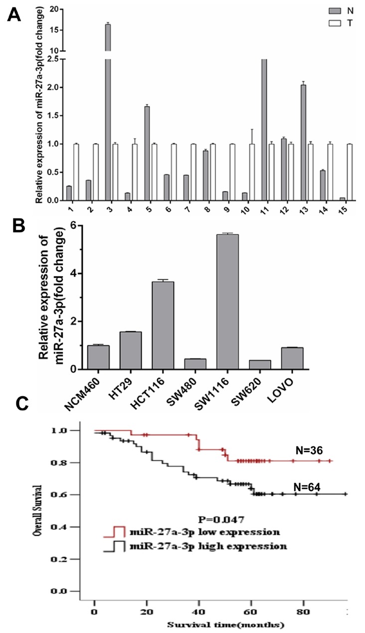
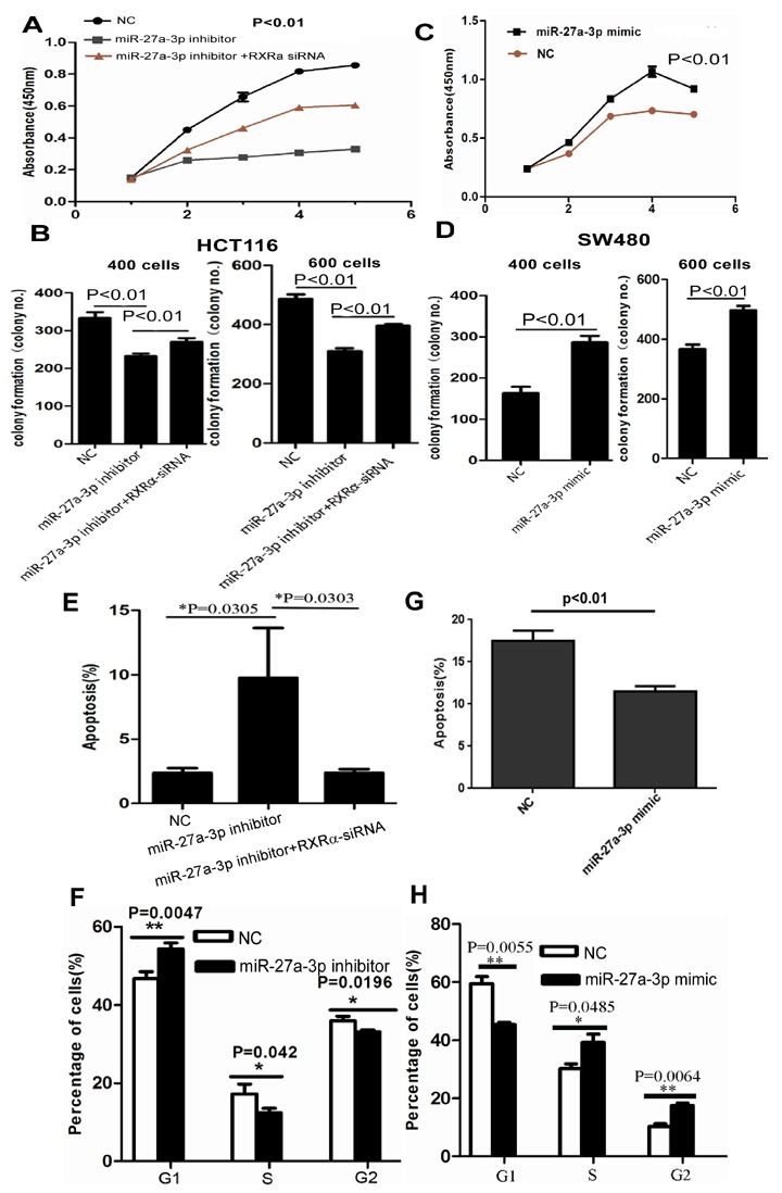
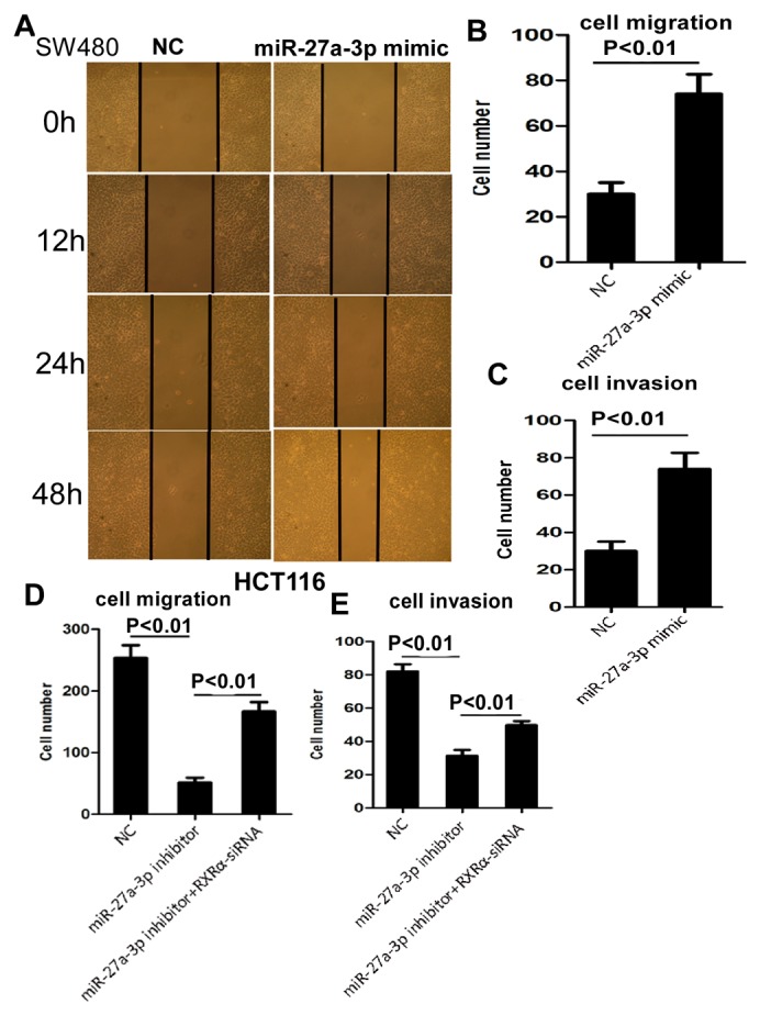
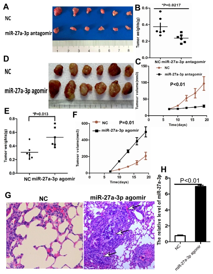
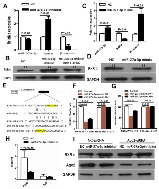
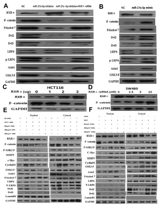
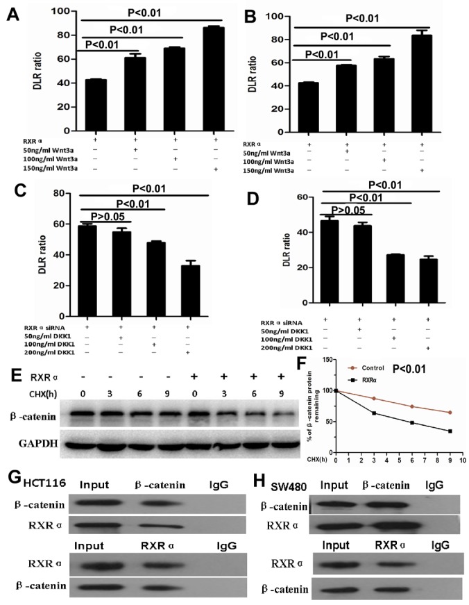
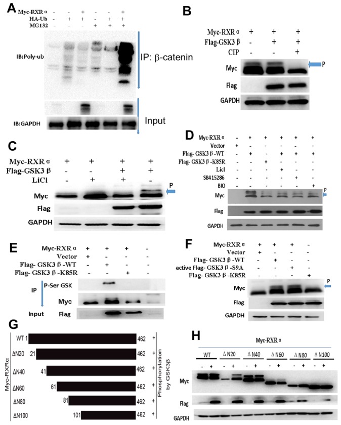
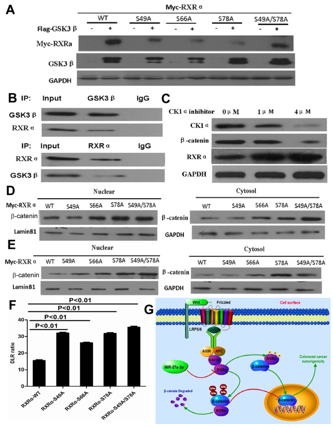
This study aimed to elucidate how miR-27a-3p modulates the Wnt/β-catenin signaling pathway to promote colorectal cancer (CRC) progression. Our results showed that the expression of miR-27a-3p was up-regulated in CRC and closely associated with histological differentiation, clinical stage, distant metastasis and CRC patients' survival. miR-27a-3p mimic suppressed apoptosis and promoted proliferation, migration, invasion of CRC cells in vitro and in vivo. Whereas miR-27a-3p inhibitor promoted apoptosis and suppressed proliferation, migration, invasion of CRC cells in vitro and in vivo. Furthermore, RXRα was the target gene of miR-27a-3p in CRC. miR-27a-3p expression negatively correlated with RXRα expression in CRC tissues. The underlining mechanism study showed that miR-27a-3p/RXRα/Wnt/β-catenin signaling pathway is involved in CRC progression. In conclusion, our findings first demonstrate that miR-27a-3p is a prognostic and/or potential therapeutic biomarker for CRC patients and RXRα as miR-27a-3p targeting gene plays an important role in activation of the Wnt/β-catenin pathway during CRC progression.

Keywords: RXRα; Wnt/β-catenin pathway; colorectal cancer; miR-27a-3p.


Conflict of interest statement

CONFLICTS OF INTEREST The authors declare no potential conflicts of interest.

Figures

Figure 1
Figure 1
(A) miR-27a-3p expression level in 15 paired of fresh CRC samples (T) and adjacent non-tumor colorectal mucosa tissues (N) by quantitative real-time PCR analysis. (B) miR-27a-3p expression level in CRC cell lines. Data were normalized against the miR-27a-3p expression level in NCM460 cells. (C) Overall survival of CRC patients with different levels of miR-27a-3p expression by Kaplan-Meier analysis.
Figure 2
Figure 2
(A-B) miR-27a-3p inhibitor significantly suppressed cell proliferation (A) and clone formation (B) in HCT116. However, RXRα knockdown reversed the suppression of cell proliferation and clone formation in HCT116 induced by miR-27a-3p inhibitor. (C-D) miR-27a-3p mimic significantly increased cell proliferation (C) and clone formation (D) in SW480 compared with the control group. (E-F) miR-27a-3p inhibitor significantly induced cell apoptosis (E) and cell cycle G-S phase arrest (F) in HCT116. However, RXRα knockdown reversed elevated cell apoptosis in HCT116 induced by miR-27a-3p inhibitor. (G-H) miR-27a-3p mimic significantly suppressed cell apoptosis (G) and cell cycle G-S phase arrest (H) in SW480 compared with the control group.
Figure 3
Figure 3
(A-B) miR-27a-3p mimic significantly increased SW480 cell migration by scratch wound healing assay (A) and cell migration assay (B), respectively. (C) miR-27a-3p mimic significantly increased SW480 cell invasion compared with the control group. (D-E) miR-27a-3p inhibitor significantly suppressed HCT116 cell migration (D) and invasion (E). However, RXRα knockdown reversed suppression of cell migration (D) and invasion (E) in HCT116 induced by miR-27a-3p inhibitor.
Figure 4
Figure 4
(A-C) miR-27a-3p antagomir significantly suppressed tumor growth (A), tumor weight (B), and tumor volume (C) of HCT116 cells implanted subcutaneously in BALB/c-nu mice compared with the control group, respectively. (D-F) miR-27a-3p agomir significantly increased tumor growth (D), tumor weight (E), and tumor volume (F) of SW480 cells implanted subcutaneously in BALB/c-nu mice compared with the control group, respectively. (G) Histological staining showed the lung metastatic carcinoma (arrow indicated) of tumor xenografts generated by HT29 cells transfected with miR-27a-3p agomir compared with the control group (NC), haematoxylin and eosin staining ×200. (H) miR-27-3p expression was significantly higher in tumor xenografts generated by HT29 cells transfected with miR-27a-3p agomir compared with the control group (NC) by quantitative real-time PCR.
Figure 5
Figure 5
(A) miR-27a-3p inhibitor increased RXRα mRNA and suppressed β-catenin mRNA expression in HCT116 by real-time PCR. (B) miR-27a-3p inhibitor increased RXRα protein expression in HCT116, however, RXRα knockdown reversed elevated RXRα protein expression which was induced by miR-27a-3p inhibitor in HCT116 by western blot analysis. The relative quantification of bands in Western blots was a ratio neutralized to GAPDH. (C) miR-27a-3p mimic suppressed RXRα mRNA and increased β-catenin mRNA expression in SW480 by real-time PCR. (D) miR-27a-3p mimic suppressed RXRα protein expression in SW480 by western blot analysis. The relative quantification of bands in Western blots was a ratio neutralized to GAPDH. (E) Predicted binding sites of miR-27a-3p in the wild type 3’-UTR of RXRα. Mutations in the 3’-UTR of RXRα and miR-27a-3p mimic were highlighted in yellow. (F-G) miR-27a-3p significantly inhibited the luciferase activities of RXRα-WT 3’UTR reporter in HCT116 (F) and SW480 (G) cells. However, miR-27a-3p had no effect on the luciferase activities of RXRα-Mut 3’UTR reporter in HCT116 (F) and SW480 (G) cells. (H) RNA binding protein immunoprecipitation assay showed Ago2 was associated with miR-27a-3p. (I) Ago2 knockdown dramatically decreased the effect of miR-27a-3p on RXRα expression in HCT116 cells.
Figure 6
Figure 6
(A) miR-27a-3p inhibitor increased RXRα and suppressed β-catenin, Frizzled-7, Dvl2, Dvl3, p-LRP6, Axin1, and GSK3β expression in HCT116 cells. However, RXRα knockdown reversed the suppression of β-catenin, Frizzled-7, Dvl2, Dvl3, p-LRP6, Axin1, and GSK3β expression in HCT116 cells by miR-27a-3p inhibitor. (B) miR-27a-3p mimic suppressed RXRα expression and increased β-catenin, Frizzled-7, Dvl2, Dvl3, p-LRP6, Axin1, and GSK3β expression in SW480 cells. (C) Ectopic RXRα suppressed β-catenin expression in HCT116 cells. (D) RXRα knockdown resulted in increased β-catenin protein level in SW480. (E) DKK1 dramatically reversed increased β-catenin, p-GSK3β, MMP9, c-Myc, cyclinD1, Axin1, Frizzled-7, p-LRP6, Dvl2, and Dvl3 in HCT116 with RXRα knockdown. (F) Wnt3a dramatically reversed suppressed β-catenin, p-GSK3β, MMP9, c-Myc, cyclinD1, Axin1, Frizzled-7, p-LRP6, Dvl2, and Dvl3 in SW480 transfected with RXRα expression plasmid.
Figure 7
Figure 7
(A-B) TCF/LEF Luciferase reporter assay showed Wnt3a significantly reversed suppressed β-catenin transcriptional activity in HCT116 (A) and SW480 (B) cells transfected with RXRα expression plasmid and in a dose dependent manner. (C-D) DKK1 significantly reversed increased β-catenin transcriptional activity in HCT116 (C) and SW480 (D) cells transfected with RXRα siRNA and in a dose dependent manner. (E-F) the half-life of β-catenin protein was shortened significantly in HCT116 cells transfected with RXRα and treated with cycloheximide compared with the control group. (G-H) Co- immunoprecipitation showed that RXRα interacted with β-catenin in HCT116 (G) and SW480 (H).
Figure 8
Figure 8
(A) Immunoblotting showed that the ubiquitination of β-catenin was dramatically augmented by RXRα overexpression in HCT116 cells treated with MG132. (B) phosphorylation inhibitor-CIP impaired the effect of GSK3β phosphorylation on RXRα by western blot (arrow and p indicated phosphorelated RXRα). (C-D) LiCl, BIO, and SB415286 as inhibitors of GSK3β could suppress RXRα phosphorylation by GSK3β in HEK293T (C) and HCT116 (D) cells. (E) co-immunoprecipitation assay showed that there was a direct interaction between RXRα and GSK3β through GSK3β serine sites. (F) mutant-Flag-GSK3β-K85R impaired the effect of RXRα binding to GSK3β serine sites. GSK3β kinase-activated mutant plasmid-GSK3β-S9A could induce the phosphorylation of RXRα in HEK293T cells. (G) The schematic diagram depicts potential RXRα phosphorylation regions and various RXRα gene deletion mutants were constructed. (H) Western blot showed that deletion mutants of the N20 (ΔN20), N40 (ΔN40), and N60 (ΔN60) induced RXRα phosphorylation by GSK3β, respectively. However, deletion mutants of the N80 (ΔN80) and N100 (ΔN100) had no effect on GSK3β-induced RXRα phosphorylation in HEK293T cells.
Figure 9
Figure 9
(A) GSK3β-induced RXRα phosphorylation decreased for RXRα-S49A, RXRα-S66A and RXRα-S78A in HEK293 cells compared with RXRα WT by western blot analysis. (B) The physical interaction of endogenous RXRα and GSK3β was found in HCT116 by co-immunoprecipitation. (C) Casein kinase 1α inhibitor increased RXRα expression and suppressed β-catenin expression in HCT116 and in a dose-dependent manner. (D-E) RXRα-S49A, RXRα -S66A and RXRα-S78A increased the nuclear β-catenin expression in HCT116 (D) and SW480 (E) cells compared with the stimulated p-Ser of RXRα WT by Western blot. (F) RXRα-S49A, RXRα-S66A and RXRα-S78A increased the TCF/LEF luciferase activity in HCT116 cells compared with RXRα WT, respectively. (G) Illustration of Wnt/β-catenin activation by miR-27a-3p targeting RXRα in CRC.

Similar articles

Cited by

References

    1. White BD, Chien AJ, Dawson DW. Dysregulation of Wnt/beta-catenin signaling in gastrointestinal cancers. Gastroenterology. 2012;142:219–232. - PMC - PubMed
    1. Chambon P. A decade of molecular biology of retinoic acid receptors. FASEB J. 1996;10:940–954. - PubMed
    1. Mangelsdorf DJ, Evans RM. The RXR heterodimers and orphan receptors. Cell. 1995;83:841–850. - PubMed
    1. Heyman RA, Mangelsdorf DJ, Dyck JA, Stein RB, Eichele G, Evans RM, Thaller C. 9-cis retinoic acid is a high affinity ligand for the retinoid X receptor. Cell. 1992;68:397–406. - PubMed
    1. Mulholland DJ, Dedhar S, Coetzee GA, Nelson CC. Interaction of nuclear receptors with the Wnt/beta-catenin/Tcf signaling axis: Wnt you like to know? Endocr Rev. 2005;26:898–915. - PubMed

LinkOut - more resources