. 2017 Sep 15;8(47):83052-83062.
doi: 10.18632/oncotarget.20943. eCollection 2017 Oct 10.

NVP-BKM120 inhibits colon cancer growth via FoxO3a-dependent PUMA induction

Affiliations

NVP-BKM120 inhibits colon cancer growth via FoxO3a-dependent PUMA induction

Shida Yang et al. Oncotarget. .

Abstract

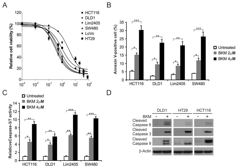
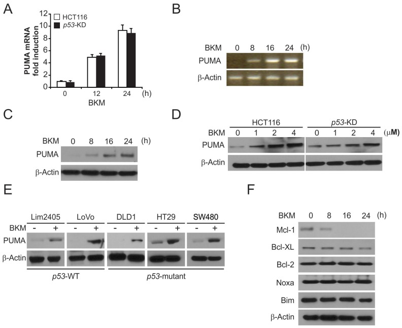
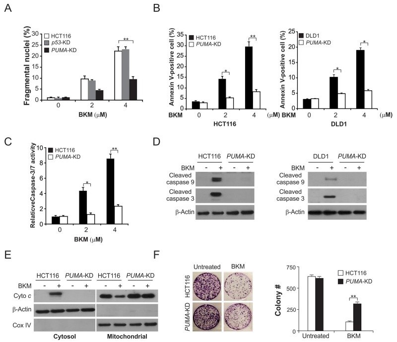
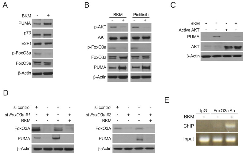
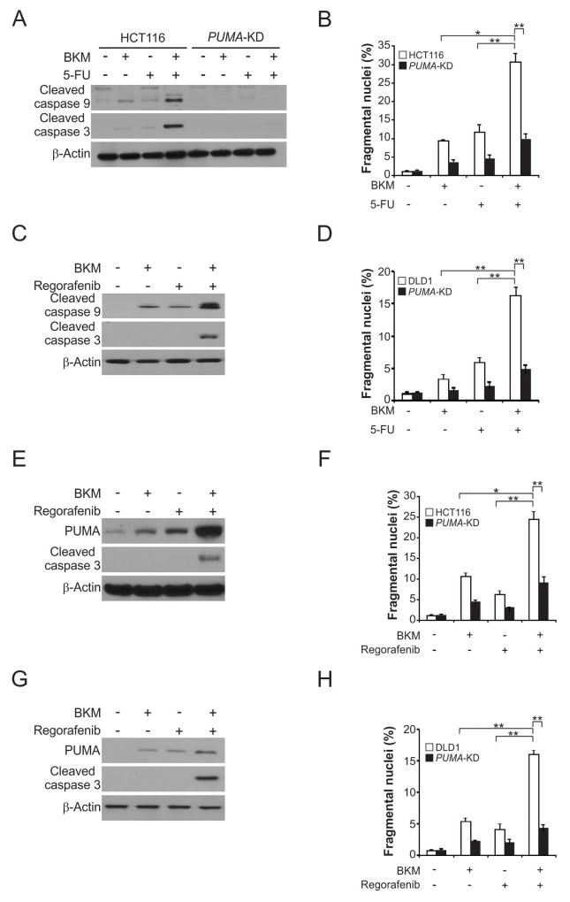
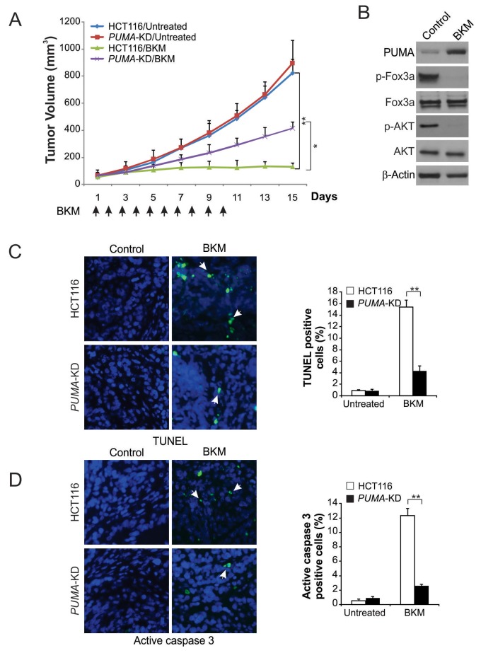
NVP-BKM120, a potent and highly selective PI3K inhibitor, is currently being investigated in phase I/II clinical trials. The mechanisms of action of NVP-BKM120 in colon cancer cells are unclear. In the present study, we investigated how NVP-BKM120 suppresses colon cancer cells growth and potentiates effects of other chemotherapeutic drugs. We found that NVP-BKM120 treatment enhance PUMA induction irrespective of p53 status through the FoxO3a pathway following AKT inhibition. Furthermore, PUMA is required for NVP-BKM120-induced apoptosis in colon cancer cells. In addition, NVP-BKM120 also synergized with 5-Fluorouracil or regorafenib to induce marked apoptosis via PUMA induction. Deficiency of PUMA suppressed apoptosis and antitumor effect of NVP-BKM120 in xenograft model. These results demonstrate a key role of PUMA in mediating the anticancer effects of NVP-BKM120 and suggest that PUMA could be used as an indicator of NVP-BKM120 sensitivity, and also have important implications for it clinical applications.

Keywords: FoxO3a; NVP-BKM120; PUMA; apoptosis; colon cancer.


Conflict of interest statement

CONFLICTS OF INTEREST The authors declare no conflicts of interest.

Figures

Figure 1
Figure 1. NVP-BKM120 induces apoptosis in CRC
(A) The indicated cell lines were treated with increasing dose of NVP-BKM120 for 72 hours. Cell proliferation was determined by MTS assay. (B) The indicated cell lines were treated with increasing NVP-BKM120 for 24 hours at indicated concentrations. Apoptosis was analyzed by Annexin V/PI staining followed by flow cytometry. (C) The indicated cell lines were treated with NVP-BKM120 for 24 hours at indicated concentrations. Caspase 3/7 activity was determined by fluorogenic analysis. (D) The indicated cell lines were treated with 4μmol/LNVP-BKM120 for 24 hours. Cleaved caspase 3, 8 and 9 were analyzed by Western blotting. Results in (A), (B) and (C) were expressed as means ± SD of 3 independent experiments. ***, P<0.001;**, P<0.01; *, P<0.05.
Figure 2
Figure 2. NVP-BKM120 induces p53-independent PUMA induction
(A) Parental and p53-KD HCT116 cells were treated with NVP-BKM120 at indicated time point. PUMA mRNA induction by NVP-BKM120 was analyzed by real-time reverse transcriptase (RT) PCR, with β-actin as a control. (B) HCT116 cells were treated with 4 μmol/L NVP-BKM120 at indicated time point. Total RNA was extracted, and PUMA mRNA expression was analyzed by semiquantitive reverse transcription PCR (RT-PCR). β-actin was used as a control. (C) HCT116 cells were treated with 4 μmol/L NVP-BKM120 at indicated time point. PUMA expression was analyzed by Western blotting. (D) Parental and p53-KD HCT116 cells were treated with NVP-BKM120 for 24 hours at indicated concentration. PUMA expression was analyzed by Western blotting. (E) Indicated colon cancer cell lines with different p53 status were treated with 4 μmol/L NVP-BKM120 for 24 hours. PUMA expression was analyzed by Western blotting. (F) HCT116 cells were treated with 4 μmol/L NVP-BKM120 at indicated time point. Indicated protein expression was analyzed by Western blotting.
Figure 3
Figure 3. PUMA is required for the apoptotic activity of NVP-BKM120
(A) Parental, p53-KD and PUMA-KD HCT116 cells were treated with NVP-BKM120 at indicated concentration for 24 hours. Apoptosis was analyzed by a nuclear fragmentation assay. (B) Parental and PUMA-KD HCT116 or DLD1 cells were treated with NVP-BKM120 at indicated concentration for 24 hours. Apoptosis was analyzed by Annexin V/PI staining followed by flow cytometry. (C) Parental and PUMA-KO HCT116 cells were treated with NVP-BKM120 for 24 hours. Caspase 3/7 activity was determined by fluorogenic analysis. (D) Parental and PUMA-KD HCT116 or DLD1 cells were treated with 4 μmol/L NVP-BKM120 for 24 hours. Cleaved caspase 3 and 9 were analyzed by Western blotting. (E) The cytoplasm and mitochondria were fractionated from parental and PUMA-KO HCT116 cells treated with 4 μmol/L NVP-BKM120 for 24 hours. The distribution of cytochrome c was analyzed by Western blotting. β-Actin and cytochrome oxidase subunit IV (Cox IV) were analyzed as the control for loading and fractionation. (F) Parental and PUMA-KD HCT116 cells were treated with 4 μmol/L NVP-BKM120 for 24 hours. Colony formation assay was done by seeding an equal number of treated cells in 12-well plates, and then staining attached cells with crystal violet 14 days later. Left, representative pictures of colonies; Right, quantification of colony numbers. Results in (A), (B), (C) and (F) were expressed as means ± SD of 3 independent experiments.**, P<0.01; *, P<0.05.
Figure 4
Figure 4. Induction of PUMA by NVP-BKM120 is mediated through the AKT inhibition
(A) HCT116 cells were treated with 4μmol/L NVP-BKM120 for 24 hours. PUMA, p73, E2F1, p-FoxO3a and FoxO3a expression was analyzed by Western blotting. (B) HCT116 cells were treated with 4μmol/L NVP-BKM120 or 4μmol/L Pictilisib for 24 hours. Indicated protein levels were analyzed by Western blotting. (C) HCT116 cells were transfected with active AKT plasmid for 6 hours, and then treated with 4μmol/L NVP-BKM120 for 24 hours. PUMA and AKT expression was analyzed by Western blotting. (D) HCT116 cells were transfected with either a control scrambled siRNA or FoxO3a siRNA for 24 hours, and then treated with 4μmol/L NVP-BKM120 for 24 hours. FoxO3a and PUMA expression was analyzed by Western blotting. siRNA FoxO3a #1 from Santa Cruz Biotechnology and siRNA FoxO3a #2 Thermo Fisher Scientific. (E) Chromatin immunoprecipitation (ChIP) was performed using anti-FoxO3a antibody on HCT116 cells following NVP-BKM120 treatment for 12 hours. ChIP with the control IgG was used as a control. PCR was carried out using primers surrounding the FoxO3a binding sites in the PUMA promoter.
Figure 5
Figure 5. NVP-BKM120 synergizes with 5-FU or regorafenib to induce apoptosis via PUMA in CRC
(A) Parental and PUMA-KD HCT116 cells were treated with 2 μmol/L NVP-BKM120, 20 mg/L 5-fluorouracil (5-FU), or their combination for 24 hours. Cleaved caspase 3 and 9 were analyzed by Western blotting. (B) Parental and PUMA-KD HCT116 cells were treated 2 μmol/L NVP-BKM120, 20 mg/L 5-FU, or their combination for 24 hours. Apoptosis was analyzed by a nuclear fragmentation assay. (C) Parental and PUMA-KD DLD1 cells were treated with 2 μmol/L NVP-BKM120, 20 mg/L 5-FU or their combination for 24 hours. Cleaved caspase 3 and 9 were analyzed by Western blotting. (D) Parental and PUMA-KD DLD1 cells were treated 2 μmol/L NVP-BKM120, 20 mg/L 5-FU, or their combination for 24 hours. Apoptosis was analyzed by a nuclear fragmentation assay. (E) HCT116 cells were treated with 2 μmol/L NVP-BKM120, μmol/L regorafenib, or their combination for 24 hours. PUMA and cleaved caspase 3 were analyzed by Western blotting. (F) Parental and PUMA-KD HCT116 cells were treated 2 μmol/L NVP-BKM120, 20 μmol/L regorafenib, or their combination for 24 hours. Apoptosis was analyzed by a nuclear fragmentation assay. (G) DLD1 cells were treated with 2 μmol/L NVP-BKM120, 20 μmol/L regorafenib, or their combination for 24 hours. PUMA and cleaved caspase 3 were analyzed by Western blotting. (H) Parental and PUMA-KD DLD1 cells were treated 2 μmol/L NVP-BKM120, 20 μmol/Lregorafenib, or their combination for 24 hours. Apoptosis was analyzed by a nuclear fragmentation assay. Results in (B), (D), (F) and (H) were expressed as means ± SD of 3 independent experiments. **, P<0.01; *, P<0.05.
Figure 6
Figure 6. PUMA mediates the antitumor effects of NVP-BKM120 in a xenograft model
(A) Nude mice were injected s.c. with 4 × 10⁶ parental and PUMA-KD HCT116 cells. After 1 week, mice were treated with 40 mg/kg NVP-BKM120 or buffer for 10 consecutive days. Tumor volume at indicated time points after treatment was calculated and plotted (n=6 in each group). Arrows indicate NVP-BKM120 injection. (B) Parental HCT116 xenograft tumors were treated with 40 mg/kg NVP-BKM120 or the control buffer as in (A) for 4 consecutive days. Indicated protein in representative tumors were analyzed by Western blotting. (C) Paraffin-embedded sections of tumor tissues from mice treated as in (B) were analyzed by TUNEL staining. Left, representative TUNEL staining pictures; Right, TUNEL-positive cells were counted and plotted. (D) Tissue sections from (C) were analyzed by active caspase 3 staining. Left, representative staining pictures; Right, active caspase 3-positive cells were counted and plotted. Results of (A), (C) and (D) were expressed as means ± SD of 3 independent experiments. **, P<0.01; *, P<0.05.

Similar articles

Cited by

References

    1. Vanhaesebroeck B, Stephens L, Hawkins P. PI3K signalling: the path to discovery and understanding. Nat Rev Mol Cell Biol. 2012;13:195–203. https://doi.org/10.1038/nrm3290. - DOI - PubMed
    1. Fruman DA, Rommel C. PI3K and cancer: lessons, challenges and opportunities. Nat Rev Drug Discov. 2014;13:140–56. https://doi.org/10.1038/nrd4204. - DOI - PMC - PubMed
    1. Engelman JA. Targeting PI3K signalling in cancer: opportunities, challenges and limitations. Nat Rev Cancer. 2009;9:550–62. https://doi.org/10.1038/nrc2664. - DOI - PubMed
    1. Bunney TD, Katan M. Phosphoinositide signalling in cancer: beyond PI3K and PTEN. Nat Rev Cancer. 2010;10:342–52. https://doi.org/10.1038/nrc2842. - DOI - PubMed
    1. Speranza MC, Nowicki MO, Behera P, Cho CF, Chiocca EA, Lawler SE. BKM-120 (Buparlisib): a phosphatidyl-inositol-3 kinase inhibitor with anti-invasive properties in glioblastoma. Sci Rep. 2016;6:20189. https://doi.org/10.1038/srep20189. - DOI - PMC - PubMed