. 2017 Sep 15;8(47):83100-83113.
doi: 10.18632/oncotarget.20912. eCollection 2017 Oct 10.

Regulation of FBXO4-mediated ICAM-1 protein stability in metastatic breast cancer

Affiliations

Regulation of FBXO4-mediated ICAM-1 protein stability in metastatic breast cancer

Jae-Hyeok Kang et al. Oncotarget. .

Abstract

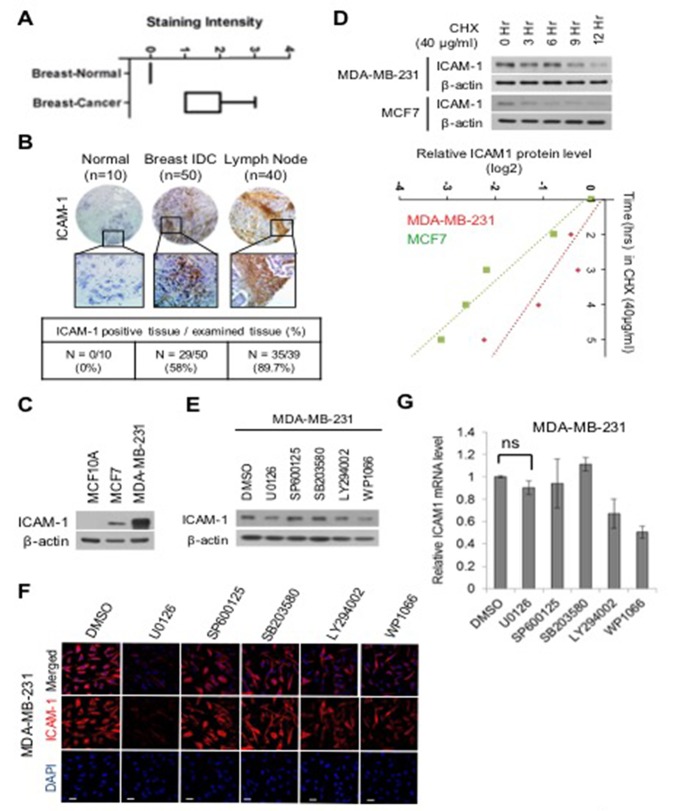
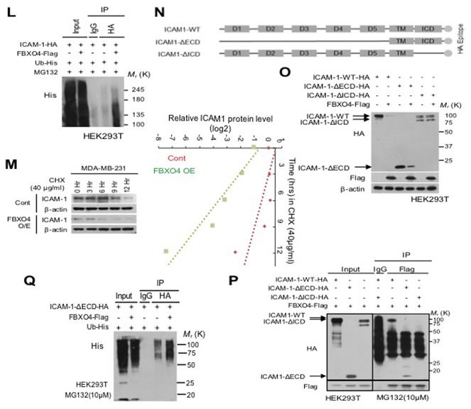
Advanced or progressive cancers share common traits such as altered transcriptional modulation, genetic modification, and abnormal post-translational regulation. These processes influence protein stability and cellular activity. Intercellular adhesion molecule-1 (ICAM-1) is involved in the malignant progression of various human cancers, including breast, liver, renal, and pancreatic cancers, but protein stability has not been deal with in metastatic breast cancer. Additionally, the relevance of the stability maintenance of ICAM-1 protein remains obscure. Here, we identified a novel interaction of E3 ligase FBXO4 that is specifically presented to ICAM-1. To understand how FBXO4 modulates ICAM-1 stability, we investigated ICAM-1-overexpressing or knockdown metastatic/non-metastatic breast cancers. ICAM-1 was found to influence tumor progression and metastasis, whereas FBXO4 regulated aggressive tumorigenic conditions. These results demonstrate that FBXO4 is a major regulator of ICAM-1 stability and that alterations in the stability of ICAM-1 can influence therapeutic outcome in metastatic cancer.

Keywords: E3 ligase; FBXO4; intercellular adhesion molecule-1; metastatic breast cancer; stability.


Conflict of interest statement

CONFLICTS OF INTEREST The authors declare that they have no conflicts of interest.

Figures

Figure 1
Figure 1. ERK regulates the stability of ICAM-1 in breast cancer
(A) Analysis of ICAM-1 data from publicly available human target protein databases (http://www.proteinatlas.org/). (B) Representative micrographs of human breast cancer tissue. Quantification of ICAM-1 intensity in tissues of normal breast cancer, advanced breast cancer, and metastatic breast cancer. (C) Endogenous ICAM-1 in normal epithelial cells, metastatic breast cancer cells, or non-metastatic breast cancer cells was detected by western blot analysis. (D) CHX pulse-chase experiments the analysis of the stability of ICAM-1 in metastatic or non-metastatic breast cancer cells that were treated with cycloheximide (CHX, 40 μg /mL). Western blot data were quantified using ImageJ software. (E-G) Western blotting (E) ICC (F), and qRT-PCR (G) were performed to detect the influence of the inhibition of various signaling pathways in metastatic breast cancer cells that were incubated for 24 hours. (H) Pulse-chase experiments for the analysis of ICAM-1 stability, which was affected by the inhibition of ERK signaling in metastatic or non-metastatic breast cancer cells that were treated with CHX. (I) Immunoprecipitation and western blotting for the demonstration of the polyubiquitination of ICAM-1 in metastatic breast cancer cells under ERK inhibition or after the treatment with DMSO and control IgG antibodies for 48 hours. The proteasome inhibitor, MG132 was added to stabilize ubiquitination. (J) Relationship of the stability of ICAM-1 with pERK signaling after EGF stimulation in non-metastatic breast cancer cells and SK-BR3 cells was analyzed by western blotting (J) and ICC (K). (L, M) Expression levels of ICAM-1 and pERK proteins and mRNAs in non-metastatic breast cancer cells after EGF stimulation and U0126 treatment were analyzed by western blotting (L) and conventional RT-PCR. (M). All western blot data were quantified using ImageJ software. The results are expressed as mean ± SD of three different experiments. (Single asterisks (*) indicate a significant difference (P < 0.05), double asterisks (**) indicate a highly significant difference (P < 0.01), triple asterisks (***) indicated the highest significant difference.)
Figure 2
Figure 2. FBXO4, which is an E3 ligase, reduces the stability of ICAM-1 in breast cancer cells
(A) qRT-PCR was performed to detect the changes in the level of F-BOX mRNA expression in metastatic breast cancer cells corresponding to U0126 treatment. (B, C) Western blotting of selected F-BOX candidates was confirmed to detect changes in the stability of ICAM-1 after U0126 treatment, which caused the knockdown of F-BOX protein in metastatic breast cancer cells (B) and analyzed by qRT-PCR under the same condition (C). (D) Western blotting for the analysis of changes in ICAM-1 expression due to the knockdown of selected F-BOX candidates in non-metastatic breast cancer cells. (E, F) Immunoprecipitation and western blot analysis demonstrated the interaction between ICAM-1 and FBXO4. (G-H) For additional confirmation, HEK293T cells that were transfected with ICAM-1-HA or FBXO4-Flag for 48 hours were examined. Cell lysate was collected after MG132 treatment for 6 hours before subjecting to immunoprecipitation and western blot analysis. (I) Comparison of the level of transcriptional or post-transcriptional expression that was confirmed in HEK293T cells using ICAM-1-HA and FBXO4-Flag was performed using western blotting and conventional PCR. (J) Immunofluorescence images of HEK293T cells with the overexpression or knockdown of ICAM-1-HA alone or together with FBXO4-Myc. Scale bar = 100 μm. (K) Binding affinity of FBXO4 and ICAM-1 was assessed by in situ PLA with HEK293T cells, which were transfected with ICAM-1-HA or FBXO4-Myc. (L) HEK293T cells were transfected as described in the panel labels. Immunoprecipitation and western blotting were performed using either an anti-HA antibody for pooling HA-tagged ICAM-1 or an anti-Flag antibody against FBXO4-Flag after MG132 treatment. (M) CHX pulse-chase assay for ICAM-1 stability degradation by FBXO4 detected by western blotting. (N) Schematic of ICAM-1 structure and the deletion mutant construct, which was generated for mapping the interaction domain of ICAM-1-FBXO4. (O) Protein expression of WT and ICAM-1-deletion construct by FBXO4, HEK293T cells, which were co-transfected with ICAM-1-WT-HA, ICAM-1-∆ECD, ICAM-1-∆ICD, and FBXO4-Flag, were analyzed by western blotting. (P) Wild-type HA-tagged ICAM-1-deletion construct (ICAM-1-∆ECD or ICAM-1-∆ICD) were co-transfected with FBXO4-Flag. Cell lysate was collected after MG132 treatment before subjecting to immunoprecipitation and western blot analysis. (Q) Ubiquitination was analyzed as follows. HEK293T cells were co-transfected with ICAM-1-∆ECD-HA and RBXO4-Flag for 48 hours. The cell lysate was treated with MG132 before subjecting to co-IP. All western blot data were quantified using ImageJ software. Also, the IP and co-IP fractions were then subjected to western blot analysis with mouse anti-HA Ab or mouse anti-Flag Ab (co-IP). (*P < 0.05, **P < 0.01 or ***P<0.001.)
Figure 3
Figure 3. FBXO4 regulates EMT and metastasis in breast cancer
(A) Expression of the FBXO4 gene between the luminal (metastatic) and basal subtypes (non-metastatic) of breast cancer was detected by conventional RT-PCR. (B, C) Levels of the EMT-related proteins, vimentin, SLUG, ICAM-1, and FBXO4 in FBXO4-overexpressing metastatic (B) or FBXO-knockdown non-metastatic breast cancer cells (C) were detected by western blot analysis. β-actin served as a loading control. (D, E) Immunofluorescence for analyzing the expression of ICAM-1, FBXO4, ZEB1, and vimentin in FBXO4-overexpressing metastatic breast cancer cells (D) or FBXO-knockdown non-metastatic breast cancer cells (E). Scale bar = 10 μm. (F, G) Invasion and migration of metastatic or non-metastatic cancer cells with the overexpression (F) or knockdown (G) of FBXO4. (H, I) Levels of protein expression in metastatic (H) or non-metastatic breast cancer cells (I) after the overexpression or knockdown of FBXO4 alone or together with ICAM-1. β-actin served as a loading control. (J-L) Transplantation of control MDA-MB-231 LM-1 or FBXO4-overexpressing LM-1 cells into fat pads of mice (n=5). The mice were sacrificed 4 weeks later. Representative images of lung metastasis, which were detected by H&E staining. (K) Number of metastatic foci in the lung that was counted for EBXO4-positive cells. (L) Bright-field images of the primary tumor tissue that was derived from an in vivo experiment, wherein it was stained with the EMT-related markers, ICAM-1 and FBXO4, and analyzed by IHC. Advanced breast cancer cells served as control. (M) Representative images of the expression of the FBXO4 and ICAM-1 proteins as determined by IHC of the clinical specimen. Normal breast tissue served as control (at 200× magnification). (N) Kaplan-Meier plot of patients who survived metastasis stratified by FBXO4 expression. Data were obtained from the breast cancer dataset from Oncomine. All data are expressed as the mean of data collected from three independent experiments.(*P < 0.05,**P < 0.01 or ***P<0.001.)
Figure 4
Figure 4. miR-340, which is induced by ERK, regulates FBXO4
(A, B) Analysis of miRNA data from publicly available databases for miRNA target prediction; miRNA.org (http://www.microrna.org/microrna/home.do). (A) Level of miRNA expression in metastatic breast cancer corresponding to U0126 treatment was analyzed by qRT-PCR. (B, C) Expression levels of FBXO4 protein and mRNA in non-metastatic breast cancer cells that were transfected with selected miRNA candidates were analyzed by western blotting and qRT-PCR. (D, E) Expression levels of FBXO4 and ICAM-1 protein and mRNA after the transfection of non-metastatic breast cancer cells with miR340 or miR410 were detected by western blotting (D) and qRT-PCR (E). (F and H) Representative immunofluorescence image of the expression levels of ICAM-1, FBXO4, and EMT-related marker proteins in response to miR340 with or without FBXO4, which was analyzed by western blotting (F) and ICC (H). (G) Levels of ICAM-1 and FBXO4 mRNA expression in non-metastatic breast cancer cells that were transfected with miR340 alone or together with FBXO4. The results of the mRNA experiments were normalized to the mRNA level of β-actin. (I) Schematic model of regulation of ICAM-1 stabilization dependent corresponding to FBXO4 in tumor cells. (*P < 0.05, **P < 0.01 or ***P<0.001.)

References

    1. Hanahan D, Weinberg RA. Hallmarks of cancer: the next generation. Cell. 2011;144:646–74. - PubMed
    1. Badve S, Dabbs DJ, Schnitt SJ, Baehner FL, Decker T, Eusebi V, Fox SB, Ichihara S, Jacquemier J, Lakhani SR, Palacios J, Rakha EA, Richardson AL, et al. Basal-like and triple-negative breast cancers: a critical review with an emphasis on the implications for pathologists and oncologists. Mod Pathol. 2011;24:157–67. - PubMed
    1. Zhang K, Rodriguez-Aznar E, Yabuta N, Owen RJ, Mingot JM, Nojima H, Nieto MA, Longmore GD. Lats2 kinase potentiates Snail1 activity by promoting nuclear retention upon phosphorylation. EMBO J. 2012;31:29–43. - PMC - PubMed
    1. Duperray A, Languino LR, Plescia J, McDowall A, Hogg N, Craig AG, Berendt AR, Altieri DC. Molecular identification of a novel fibrinogen binding site on the first domain of ICAM-1 regulating leukocyte-endothelium bridging. J Biol Chem. 1997;272:435–41. - PubMed
    1. Guo X, Ramirez A, Waddell DS, Li Z, Liu X, Wang XF. Axin and GSK3- control Smad3 protein stability and modulate TGF- signaling. Genes Dev. 2008;22:106–20. - PMC - PubMed

LinkOut - more resources