. 2017 May 12;8(48):83509-83522.
doi: 10.18632/oncotarget.17840. eCollection 2017 Oct 13.

Novel mechanisms for crotonaldehyde-induced lung edema

Affiliations

Novel mechanisms for crotonaldehyde-induced lung edema

Yue Li et al. Oncotarget. .

Abstract

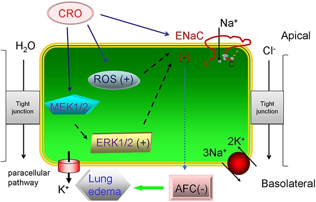
Background: Crotonaldehyde is a highly noxious α,β-unsaturated aldehyde in cigarette smoke that causes edematous acute lung injury.

Objective: To understand how crotonaldehyde impairs lung function, we examined its effects on human epithelial sodium channels (ENaC), which are major contributors to alveolar fluid clearance.

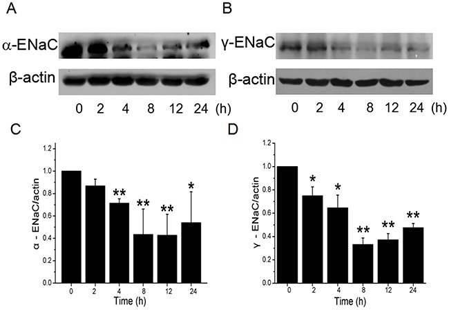
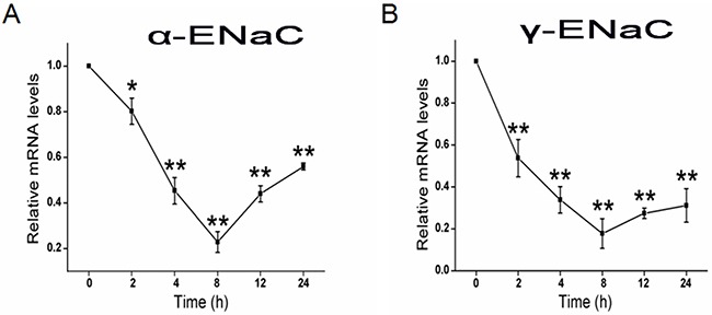
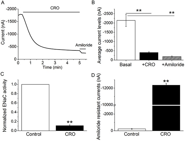
Methods: We studied alveolar fluid clearance in C57 mice and ENaC activity was examined in H441 cells. Expression of α- and γ-ENaC was measured at protein and mRNA levels by western blot and real-time PCR, respectively. Intracellular ROS levels were detected by the dichlorofluorescein assay. Heterologous αβγ-ENaC activity was observed in an oocyte model.

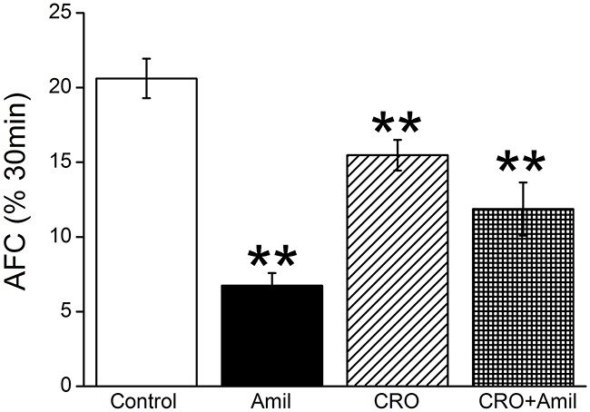
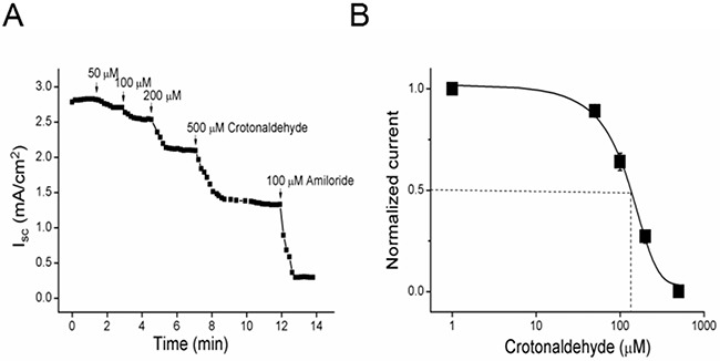
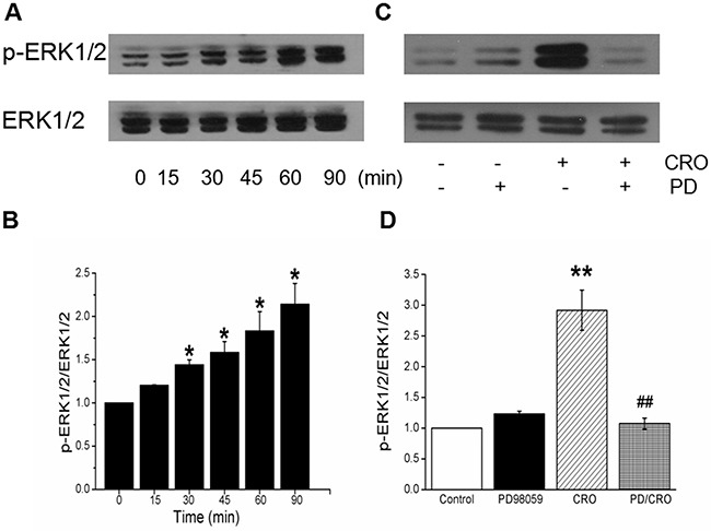
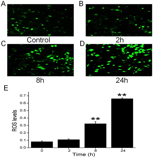
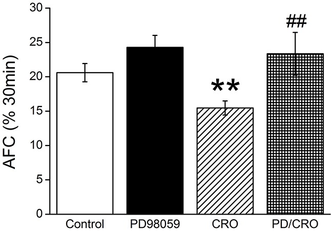
Results: Our results showed that crotonaldehyde reduced transalveolar fluid clearance in mice. Furthermore, ENaC activity in H441 cells was inhibited by crotonaldehyde dose-dependently. Expression of α- and γ-subunits of ENaC was decreased at the protein and mRNA level in H441 cells exposed to crotonaldehyde, which was probably mediated by the increase in phosphorylated extracellular signal-regulated protein kinases 1 and 2. ROS levels increased time-dependently in cells exposed to crotonaldehyde. Heterologous αβγ-ENaC activity was rapidly eliminated by crotonaldehyde.

Conclusion: Our findings suggest that crotonaldehyde causes edematous acute lung injury by eliminating ENaC activity at least partly via facilitating the phosphorylation of extracellular signal-regulated protein kinases 1 and 2 signal molecules. Long-term exposure may decrease the expression of ENaC subunits and damage the cell membrane integrity, as well as increase the levels of cellular ROS products.

Keywords: alveolar fluid clearance; crotonaldehyde; epithelial sodium channels; lung injury; reactive oxygen species.


Conflict of interest statement

CONFLICTS OF INTEREST The authors declare that they have no conflicts of interest.

Figures

Figure 1
Figure 1. CRO downregulates mouse alveolar fluid clearance in vivo
Anesthetized mice were intratracheally instilled with 5% bovine serum albumin dissolved in physiologic saline solution, administered alone for the control group (Control) or containing amiloride (Amil, 1mM), crotonaldehyde (CRO, 80 μM), or both (CRO+Amil) for exposed mice. The reabsorption rate was computed as the percentage of instilled volume after 30 min (% 30min). Average AFC values are presented as mean ± SE, one-way ANOVA. **P < 0.01, compared with control group, n = 8.
Figure 2
Figure 2. Short-circuit current level in H441 monolayers is reduced by CRO in a dose-dependent manner
(A) Representative short-circuit current (Isc) traces after treatment of monolayers with 50, 100, 200, or 500 μM CRO, and amiloride was then applied. Amiloride-sensitive currents are defined as the difference between the total current and the amiloride-resistant current, with the basal amiloride-sensitive current set as 100%. (B) Normalized amiloride-sensitive currents were plotted as a dose-dependent curve (n = 9). The raw data were calculated by fitting with the Boltzmann equation, and the IC50 value was calculated to be 108.1 μM.
Figure 3
Figure 3. Effects of CRO on the protein expression level of ENaC α- and γ-subunits in H441 cells
H441 cells were exposed to 80 μM CRO for 0 to 24 h and then proteins were extracted and analysed by western blot. (A, B) Western blots of α- and γ-ENaC protein demonstrating reductions in levels over time. Blots for β-actin were used as internal controls. (C, D) Graphical representation of data obtained from three sets of western blot assays for which bands were quantified using gray analysis (α-ENaC/β-actin and γ-ENaC/β-actin). Data are shown as the mean ± SE, *P < 0.05, **P < 0.01, compared with control.
Figure 4
Figure 4. CRO reduces the transcriptional expression of ENaC α- and γ-subunits
H441 cells were treated with CRO for 0 to 24 h and then RNA samples were isolated for real-time PCR assays. (A, B) Relative levels of mRNA were calculated as α- or γ-ENaC/GAPDH ratios. *P < 0.05, **P < 0.01, compared with control; average of 3 experiments for each type of subunit.
Figure 5
Figure 5. CRO elevates ERK1/2 phosphorylation level in H441 cells, and PD98059 inhibits CRO-induced ERK1/2 phosphorylation
(A) Western blots assays for ERK1/2 phosphorylation in H441 cells exposed to 80 μM CRO for 0 to 90 min. (B) Graphical representation of data obtained from three sets of western blot assays for which bands were quantified using gray analysis (p-ERK1/2/ERK1/2). (C) Representative western blot of phosphorylated ERK1/2 in untreated H441 cells (Control), after pretreatment with 40 μM PD98059 (PD98059), after treatment with 80 μM CRO (CRO), and after pretreatment with PD98059 for 30 min prior to the addition of CRO (PD98059/CRO). Lanes shown in this figure are from the same western blot. (D) Ratio of the band densities for p-ERK1/2 and ERK1/2 in H441 cells. Data were obtained using three western blots and are shown as the mean ± SE, *P < 0.05, **P < 0.01, compared with control, ##P < 0.01, compared with CRO.
Figure 6
Figure 6. Effects of CRO on oxidative stress in H441 cells
Production of ROS was measured by fluorescence microscopy using the fluorogenic substrate 2’,7’-dichlorofluorescein diacetate, which is oxidized to fluorescent 2’,7’-dichlorofluorescein. (A) Air-subjected control cell culture. (B-D) Cells exposed to 80 μM CRO for 2 h, 8 h, and 24 h. (E) The ROS levels were determined from the summary fluorescence intensity measured from the images of H441 cells (n = 25 in each group) that had been treated for different periods with CRO. Levels were corrected for background fluorescence. Data are shown as the mean ± SE, **P < 0.01, compared with control.
Figure 7
Figure 7. CRO alters the activity of heterologous human αβγ-ENaC channels expressed in oocytes and impairs properties of the plasma membrane
Oocytes were perfused with CRO and amiloride continuously, and whole cell currents were monitored every 10 s. (A) Representative inward current traces in the presence of CRO and amiloride. Currents were digitized at -120 mV, and the period of drug application is indicated by solid horizontal lines. (B) Average currents at -120 mV (mean ± SE). **P < 0.01 compared with basal level. (C) Normalized ENaC activity. Oocytes expressing human αβγ-ENaC were incubated with CRO for 24 h, and ENaC activity was evaluated. Amiloride-sensitive currents reflecting ENaC activity are defined as the difference between the total current and the amiloride-resistant current, with the basal amiloride-sensitive current set as 100%. (D) Average amiloride-resistant currents at -120 mV (mean ± SE), reflecting oocyte permeability. **P < 0.01 compared with control. n = 8 from four different frogs.
Figure 8
Figure 8. ERK1/2 phosphorylation involved in CRO-reduced mouse alveolar fluid clearance in vivo
Anesthetized mice were intratracheally instilled with 5% bovine serum albumin dissolved in physiologic saline solution, administered alone for the control group (Control) or containing PD98059 (40 μM), crotonaldehyde (CRO, 80 μM), or both (PD/CRO) for exposed mice. The reabsorption rate was computed as the percentage of instilled volume after 30 min (% 30min). Average AFC values are presented as mean ± SE, one-way ANOVA. **P < 0.01, compared with control, ##P < 0.01, compared with CRO, n = 6-8.
Figure 9
Figure 9. Schematic diagram summarizing the effects of CRO on alveolar fluid transport
Cell exposure to CRO results in: reductions in ENaC activity via enhanced ERK1/2 phosphorylation, decreased transcription and translation of ENaC subunits, and elevation of levels of ROS products. These activities lead to the reduction of AFC and contribute to edematous acute lung injury.

Similar articles

Cited by

References

    1. Eaton AF, Yue Q, Eaton DC, Bao HF. ENaC activity and expression is decreased in the lungs of protein kinase C-α knockout mice. Am J Physiol Lung Cell Mol Physiol. 2014;307:L374–L385. - PMC - PubMed
    1. Althaus M, Clauss WG, Fronius M. Amiloride-sensitive sodium channels and pulmonary edema. Pulm Med. 2011;2011:830320. - PMC - PubMed
    1. Downs CA, Trac DQ, Kreiner LH, Eaton AF, Johnson NM, Brown LA, Helms MN. Ethanol alters alveolar fluid balance via nadph oxidase (NOX) signaling to epithelial sodium channels (ENaC) in the lung. PLoS One. 2013;8:e54750. - PMC - PubMed
    1. Kellenberger S, Schild L. International union of basic and clinical pharmacology. XCI. Structure, function, and pharmacology of acid-sensing ion channels and the epithelial Na+ channel. Pharmacol Rev. 2015;67:1–35. - PubMed
    1. Joo NS, Jeong JH, Cho HJ, Wine JJ. Marked increases in mucociliary clearance produced by synergistic secretory agonists or inhibition of the epithelial sodium channel. Sci Rep. 2016;6:36806. - PMC - PubMed

LinkOut - more resources