. 2018 Jan 1;495(1):749-754.
doi: 10.1016/j.bbrc.2017.11.066. Epub 2017 Nov 11.

Long-range stress transmission guides endothelial gap formation

Affiliations

Long-range stress transmission guides endothelial gap formation

C Corey Hardin et al. Biochem Biophys Res Commun. .

Abstract

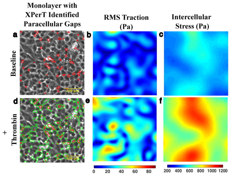
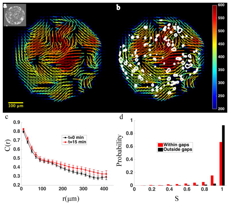
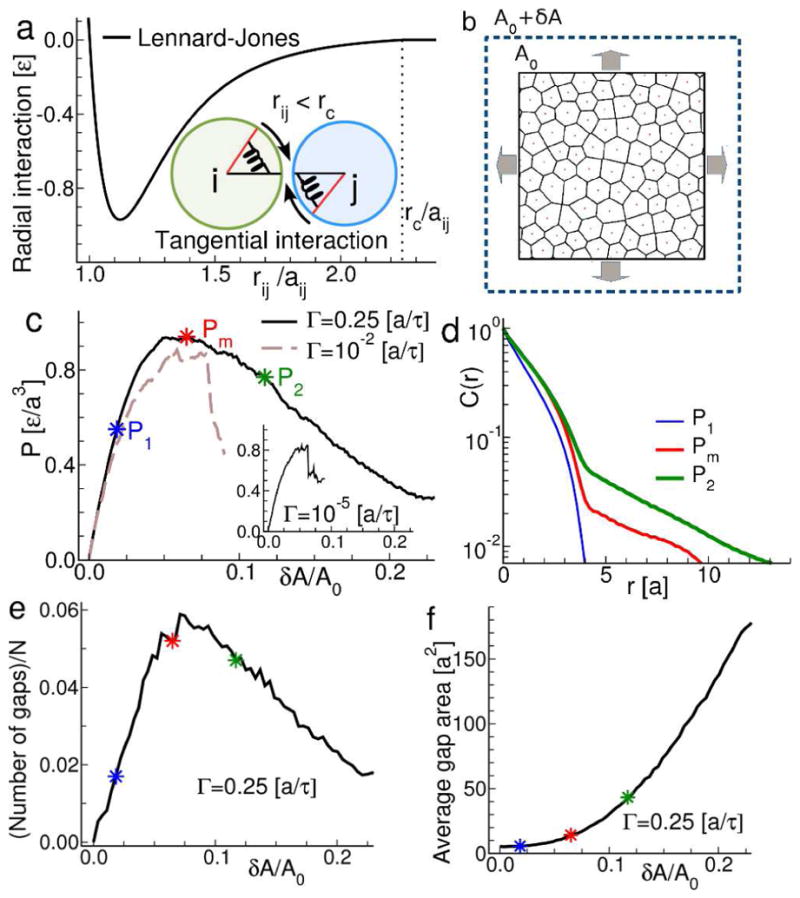
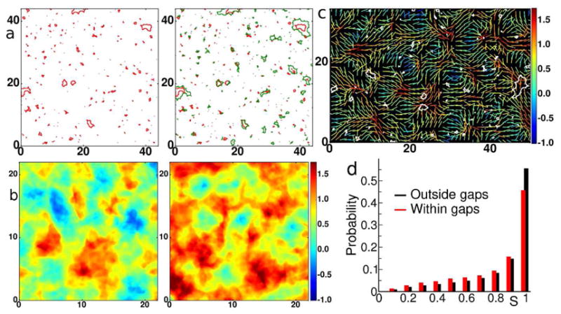
In endothelial gap formation, local tractions exerted by the cell upon its basal adhesions are thought to exceed balancing tensile stresses exerted across the cell-cell junction, thus causing the junction to rupture. To test this idea, we mapped evolving tractions, intercellular stresses, and corresponding growth of paracellular gaps in response to agonist challenge. Contrary to expectation, we found little to no relationship between local tensile stresses and gap formation. Instead, we discovered that intercellular stresses were aligned into striking multi-cellular domains punctuated by defects in stress alignment. Surprisingly, gaps emerged preferentially not at stress hotspots, as predicted, but rather at stress defects. This unexpected behavior is captured by a minimal model of the cell layer as a jammed assembly of cohesive particles undergoing plastic rearrangements under tension. Together, experiments and model suggest a new physical picture in which gap formation, and its consequent effect on endothelial permeability, is determined not by a local stress imbalance at a cell-cell junction but rather by emergence of non-local, cooperative stress reorganization across the cellular collective.

Keywords: Barrier function; Endothelium; Gaps; Stress; Traction.


Figures

Figure 1
Figure 1. Agonist induced gaps co-localize with baseline gaps
a. 10x phase contrast image of interior of micropatterned monolayer of Human Pulmonary Artery Cells adherent upon a polyacrylamide substrate. At baseline, the monolayer features an array of small XPerT identified paracellular gaps (red). b. The corresponding Fourier Transform Traction Microscopy (FTTM) map of tractions (Pa) is spatially heterogeneous. c. Monolayer Stress Microscopy (MSM) map of intercellular stress (Pa) is also heterogeneous but features larger spatial domains of similar tension. d. 15 minutes after treatment with 0.3 U/ml thrombin the monolayer permeability has increased as evidenced by the larger array of paracellular gaps (green contours). There is substantial overlap between the location of pre- and post-thrombin gaps indicating that much of the increase in permeability has occurred through gap growth rather than de novo gap formation. e. FTTM measured tractions 15 min after thrombin treatment demonstrate an increase in both traction magnitude and heterogeneity. f. MSM map of intercellular stress 15 min after thrombin treatment similarly shows an increase in tension and spatial heterogeneity.
Figure 2
Figure 2. Increases in monolayer permeability preferentially occur in vicinity of orientation defects
a. Cells are micropatterned into circular islands (inset, panel a). Within these islands, intercellular stress demonstrates regions of high orientational order. Lines representing orientation of maximal principal stress vectors tend to align in parallel over large domains. At domain boundaries, stress orientation ordering breaks down, creating regions of orientation defects. b. After treatment with thrombin (0.3 U/ml), the size and number of paracellular gaps increase. Contours of thrombin induced paracellular gaps are superimposed on map of baseline stress orientation (stress magnitude is in Pascals). Gaps (white outlines) tend to localize to areas that feature baseline orientation defects. c. The ordering of intercellular stress is reflected in a large spatial correlation length. The graph depicts the spatial autocorrelation function of intercellular stress before (black circles) and 15minutes after (red circles) thrombin treatment (n=13). Error bars represent standard error of the mean. Correlated domains of intercellular stress are 10–20 cells in diameter. d. Probability distribution of orientation order parameter, calculated for baseline monolayer, in areas which become disrupted by gaps (red) after thrombin treatment, compared with areas which do not become disrupted by gaps (black), illustrating the tendency of gaps to form in disordered regions.
Figure 3
Figure 3. Endothelial monolayer modeled as a tensed, disordered assembly of cohesive particles far from equilibrium
a. We construct a minimalist model of the monolayer as an assembly of particles, poly-disperse in size, joined by an attractive inter-particle interaction potential. The pair potential has components that act along the inter-particle contact radius and also tangential to it, to mimic the mechanics of actual cell-cell junctions. b. To mimic the action of contractile agonists such as thrombin, the model is strained by a symmetric, bi-axial expansion. The imposed external strain is balanced by a build-up of internal tensions. c. Load curve for the model monolayer subjected to biaxial strain. At slow rates of expansion, G, the stress-strain relationship is relatively linear (inset) and terminates with abrupt fracture. As G is increased the relationship changes, indicating the onset of plastic deformation and yielding rather than abrupt fracture. At the same time, the peak internal tension generated prior to failure is increased (solid and dashed lines). d. Spatial autocorrelation function of the model monolayer at different points on the load curves. The onset of plastic behavior is associated with the emergence of long-range correlations. e. Number of gaps (rescaled by the total number of particles N). f. Average gap area as a function of strain. In the yielding regime, permeability increases primarily by growth of existing gaps. Close to the maximum of the load curve, a few gaps of relatively small size are present. Further increasing the strain beyond the yielding threshold causes a rapid increase in gap area with a less dramatic increase in gap number. This is in contrast to the small and fairly constant average gap area measured in the linear regime, where the number of gaps rapidly increases.
Figure 4
Figure 4. The model monolayer mimics endothelial gap formation
a. Red contours are the gaps at the yielding transition corresponding to the point labeled Pm in Figure 3c. Green contours are gaps with further increase of strain corresponding to moving from Pm to P2 in the yield curve of Figure 3c. The further increase in permeability preferentially occurs via gap growth as evidenced by comparing the maps of inter-particle gaps at the onset of the yielding regime (left panel) and after (right panel). b. Maps of average normal stress (in units of [ εa3]) in the model monolayer corresponding to P1 (left panel) and Pm (right panel) indicate that the yielding regime is associated with pronounced spatial heterogeneities in inter-particle stress. c. Lines represent orientation of maximal principal stress vectors at P1 and the color indicates the value of the local normal stress (measured in units of [ εa3]). Superimposed are the gaps (white contours) measured at Pm. d. Probability distribution of the orientation order parameter S in areas which become disrupted by gaps at Pm(red), compared to the overall distribution at P1 (black), illustrating the tendency of large gaps to grow in disordered regions.

Similar articles

Cited by

References

    1. Hotchkiss RS, Karl IE. The Pathophysiology and Treatment of Sepsis. New England Journal of Medicine. 2003;348:138–150. - PubMed
    1. Ware L, Mathay M. The Acute Respiratory Distress Syndrome. New England Journal of Medicine. 2000;342:1334–1349. - PubMed
    1. Mehta D, Bhattacharya J, Matthay MA, Malik AB. Integrated control of lung fluid balance. American Journal of Physiology - Lung Cellular and Molecular Physiology. 2004;287:1081–1090. - PubMed
    1. Petit V, Thiery JP. Focal Adhesions: Structure and Dynamics. Biology of the Cell. 2012;92:477–494. - PubMed
    1. Prasain N, Stevens T. The actin cytoskeleton in endothelial cell phenotypes. Microvascular Research. 2009;77:53–63. - PMC - PubMed

Publication types

LinkOut - more resources