Review
. 2017 Dec 1;41(4):578-593.
doi: 10.1152/advan.00061.2017.

Autonomic regulation of the immune system in cardiovascular diseases

Affiliations
Review

Autonomic regulation of the immune system in cardiovascular diseases

François M Abboud et al. Adv Physiol Educ. .

Abstract

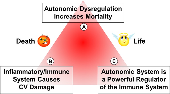
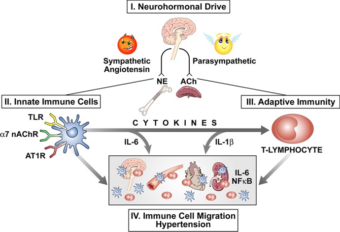
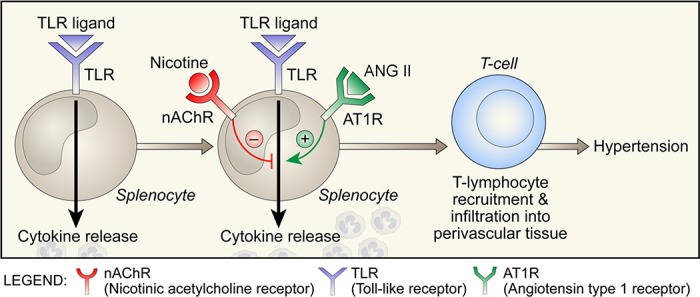
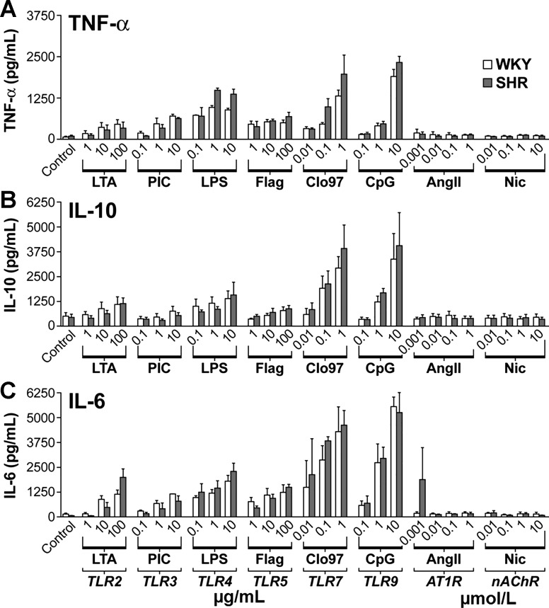
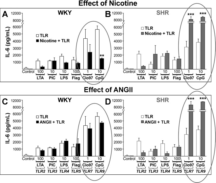
The autonomic nervous system is a powerful regulator of circulatory adjustments to acute hemodynamic stresses. Here we focus on new concepts that emphasize the chronic influence of the sympathetic and parasympathetic systems on cardiovascular pathology. The autonomic neurohumoral system can dramatically influence morbidity and mortality from cardiovascular disease through newly discovered influences on the innate and adaptive immune systems. Specifically, the end-organ damage in heart failure or hypertension may be worsened or alleviated by pro- or anti-inflammatory pathways of the immune system, respectively, that are activated through neurohumoral transmitters. These concepts provide a major new perspective on potentially life-saving therapeutic interventions in the deadliest of diseases.

Keywords: cytokines; hypertension; immune system; parasympathetic; sympathetic.


Figures

Fig. 1.
Fig. 1.
Discovery of sensory receptors and autonomic neurotransmitters. Sensory afferents from the carotid body, carotid sinus, and aortic arch relay their signals through the petrosal and nodose ganglia to the nucleus tractus solitarius. The sympathetic ganglia neurons are the soma of the sympathetic efferent innervation releasing norepinephrine (NE). The parasympathetic cholinergic (ACh) innervation of the heart and gut originates in the dorsal motor nucleus of the vagus in the medulla (red lines).
Fig. 2.
Fig. 2.
Hemodynamics of syncope: sympathoinhibition, hypotension, bradycardia, and vasodilatation (finger plethysmography). The asterisk marks the sudden loss of sympathetic activity. [From Wallin and Sundlof (81) with permission from Elsevier.]
Fig. 3.
Fig. 3.
Muscle sympathetic nerve activity (SNA) in heart failure. Left: direct recordings of muscle SNA from the peroneal nerve in patients with heart failure are shown. SNA is highest in the patient with the most severe heart failure. MAP, mean arterial pressure; HR, heart rate; bpm, beats/min; CI, cardiac index. Right: bar graphs show the marked increase in muscle SNA in congestive heart failure (CHF) compared with age-matched controls. The SNA increases progressively with the left ventricular (LV) filling pressure. Values are means ± SE; n, no. of subjects. [Left panel from Ferguson et al. (21) with permission from Elsevier; right panel from Leimbach et al. (46) with permission from Wolters Kluwer Health.]
Fig. 4.
Fig. 4.
Prolonged survival with sympathoinhibition and vagal stimulation (VS). Improved survival in heart failure in dogs with carotid sinus baroreceptor nerve stimulation (top left) and in rats with vagus nerve stimulation (top right) is shown. Bottom: the survival was dramatically prolonged and associated with significant reductions in norepinephrine (NE) levels (shaded line and bar). Values are means ± SE; nos. in parentheses, no. of animals. SS, sham stimulation. *P < 0.01. [Right panels from Li et al. (49) and left panels from Zucker et al. (88) with permission from Wolters Kluwer Health.]
Fig. 5.
Fig. 5.
The neuroimmune triangle. The schematic portrays the fatal influences of autonomic dysregulation in A, the pathological damage of the immune system in B, and the powerful modulation of the immune response by the autonomic system in C. CV, cardiovascular.
Fig. 6.
Fig. 6.
T-cell dependence of ANG II hypertension. Left: shows that vascular infiltration with CD3+ lymphocytes (purple-stained cells) and expression of T-cell receptors (TcR; red cells) are increased with ANG II infusion. Right: the pressor response to ANG II is restored in RAG1−/− mice with T-cell but not B-cell infusions. Values are means ± SE. *P < 0.01 vs. C57BL/6. [Modified from Guzik et al. (27) with permission.]
Fig. 7.
Fig. 7.
IL-6 dependence of ANG II hypertension. IL-6 knockout (KO) prevents ANG II-hypertension seen in wild type (WT; left), but constriction of afferent renal arterioles is preserved in the KO (right). [From Brand et al. (9) with permission from Wolters Kluwer Health.]
Fig. 8.
Fig. 8.
ANG II-induced splenic sympathetic overactivity enhances gene expression of cytokines. Central ANG II infusion increases splenic and renal sympathetic nerve discharge (SND; left, tracings) and activates immune system (right; bar graphs). Increased cytokine gene expression in spleen with ANG II (light shaded bars) vs. artificial cerebrospinal fluid (aCSF; solid bars) is reduced significantly with splenic denervation (dark shaded bars). Values are means ± SE. *Significantly different from aCSF-treated splenic-intact and ANG II-treated splenic-denervated, P < 0.05. †Significantly different from ANG II-treated splenic-denervated, P < 0.05. [From Ganta et al. (22).]
Fig. 9.
Fig. 9.
Myocardial infarction (MI) increases sympathetic activity to bone marrow. Schematic shows that MI accelerates atherosclerosis by an immunological process involving activation of the sympathetic nervous system (SNS) to the bone marrow (BM), releasing hematopoietic, stem, and progenitor cells, and activation of the immune system of the spleen and migration of macrophages and monocytes to the coronaries. SCF, stem cell factor. [From Dutta et al. (19) with permission from Macmillan Publishers Ltd.]
Fig. 10.
Fig. 10.
Tonic suppressive vagal influence on the immune system. Vagotomy increases NF-κB expression at 2 and 4 wk (W) after surgery (A) and enhances cytokine release from CD4+ T-cell activation (solid bars vs. open bars; B). Values are means ± SE. *P < 0.05 and **P < 0.005 (A). *P < 0.01 (B). [panel B was modified from Karimi et al. (42) with permission from Elsevier.]
Fig. 11.
Fig. 11.
Vagal nerve stimulation attenuates the LPS-induced TNF response and endotoxin shock. Left: schematic shows vagal efferent activation of the reticuloendothelial system and cholinergic (ACh) receptors on macrophage. Right, AC: show that vagal denervation (VGX) worsens the fall in mean arterial blood pressure (MABP) and enhances the serum and liver TNF responses to LPS (hatched bars). Vagal nerve stimulation (STIM) attenuates the LPS-induced TNF responses and prevents hypotension, endotoxin, and shock (shaded bars and line). Values are means ± SE. *P < 0.05 and **P < 0.005 vs. sham + LPS. #P < 0.05 vs. VGX + LPS. [Right panel was modified from Borovikova et al. (8) with permission from Macmillan Publishers Ltd.]
Fig. 12.
Fig. 12.
α7-Nicotinic receptors are protective in two-kidney-one-clip (2K1C) hypertension. Proinflammatory state (IL-6, IL-Iβ, and TNF-α), as well as cardiac hypertrophy [left ventricular weight/body weight (LVW/BW)] and glomerulosclerosis in 2K1C hypertension (WT; solid bars), are enhanced in α7-nicotinic receptors of KO mice (shaded bars). nAChR, nicotinic ACh receptor. Values are means ± SE. *P < 0.05 and **P < 0.01 compared with sham (unpaired t-test). #P < 0.05 compared with WT (unpaired t-test). [From Li et al. (48) with permission from Wolters Kluwer Health.]
Fig. 13.
Fig. 13.
Pathways of autonomic modulation of inflammation cells and their migration to end organs. Schematic portrays the proinflammatory influence of angiotensin II and increased sympathetic neurohumoral drive (NE) balanced off by an anti-inflammatory influence of parasympathetic activity (ACh). Receptors on the innate immune cells modulate cytokine release that activates the adaptive immune system and the migration of immune cells to end organs, causing hypertension. nAChR, nicotinic ACh receptor; AT1R, angiotensin II type 1 receptor.
Fig. 14.
Fig. 14.
Regulation of cytokine release from immune cells during toll-like receptor (TLR) activation. Schematic shows opposing influences of activation of α7-nAChR by nicotine and AT1R by ANG II on cytokine release.
Fig. 15.
Fig. 15.
Cytokine responses of splenocytes to activation of TLR effects of specific TLR ligands on TNF-α (A), IL-10 (B), and IL-6 (C) are dose related and equivalent in WKY (open bars) and SHR (shaded bars) splenocytes. Direct effects of nicotine (Nic) and ANG II are negligible. Values are means ± SE. LTA, lipoteichoic acid; PIC, poly I:C; Flag, flagellin; CpG, CpG2395. [From Harwani et al. (31) with permission from Wolters Kluwer Health.]
Fig. 16.
Fig. 16.
Markedly enhanced IL-6 responses to TLR by nicotine (A and B) and ANG II (C and D) in SHR splenocytes (B and D) and not in WKY (A and C). IL-6 release in response to TLR 7 (Cl097) and TLR9 (CpG) ligands ex vivo is inhibited by nicotine in WKY (solid bars) and markedly enhanced in SHR (shaded bars). ANG II also enhances the response in SHR but not in WKY. Values are means ± SE. ***P < 0.001 and **P < 0.01 indicate significantly different responses to TLR alone (open bars) vs. TLR with exposure to either nicotine or ANG II (solid and shaded bars). [From Harwani et al. (31) with permission from Wolters Kluwer Health.]
Fig. 17.
Fig. 17.
In vivo cytokine responses to Cl097. In vivo responses of serum levels of IL-6 and IL-1β to TLR7 ligand (Cl097) are increased in SHR (solid bars) and suppressed in WKY (shaded bars) by nicotine. Nos. above each bar graph, no. of animals in the particular group. Values are means ± SE. *P < 0.05. **P < 0.01. [From Harwani et al. (31) with permission from Wolters Kluwer Health.]
Fig. 18.
Fig. 18.
Cell sorting of activated macrophages in SHR vs. WKY. Activated macrophages (CD161+, CD8+) are increased in SHR vs. WKY and increase further with nicotine in SHR. [From Harwani et al. (31) with permission from Wolters Kluwer Health.]
Fig. 19.
Fig. 19.
Pronounced increases of CD161+ splenocytes in SHR over WKY is age related and antecedes hypertension. A: age-related increases in CD161+ cells in splenocyte population are associated with increases in arterial pressure in SHR (shaded bars) vs. WKY (black bars). B: the increases of CD161+ cells in SHR over WKY are not caused by an increase in the splenocyte population and spleen size (C) nor on CD4+ (D) or CD8+ cells (E). Values are means ± SE. [From Singh et al. (71) with permission from Elsevier.]
Fig. 20.
Fig. 20.
End-organ overexpression of CD161+ cells in SHR. Overexpression of CD161+ cell population in kidney and aorta of SHRs vs. WKYs is shown. FSC, forward scatter. [From Singh et al. (71) with permission from Elsevier.]
Fig. 21.
Fig. 21.
Importance of RORγt in Th17 programming. Activated innate immune cells induce Th17 programming of naive CD4+ T cells that includes overexpression of RORγt. MHC, myosin heavy chain; TCR, T-cell receptor. [From Singh et al. (71) with permission from Elsevier.]
Fig. 22.
Fig. 22.
Pronounced gene expression of RORγt and Il-17f in SHR vs. WKY. Proinflammatory cytokine gene induction of splenocytes into Th17 lineage by T-cell receptor activation includes a marked overexpression of RORγt and an enhanced response of Il-17f, but not Il-17a, in SHR compared with WKY. Values are means ± SE. Untr, untreated. *Statistically significant difference (P < 0.05). [From Singh et al. (71) with permission from Elsevier.]
Fig. 23.
Fig. 23.
Effects of IL-17A (C and D) and IL-17F (A and B) on endothelial dysfunction. Selective loss of cholinergic (ACh) endothelial-dependent vasorelaxation is seen with IL-17F (A) but not IL-17A (C). Neither 17F (B) nor 17A (D) reduced nonendothelial-mediated relaxation with sodium nitroprusside (SNP). Values are means ± SE. *Statistically significant difference (P < 0.05). [From Singh et al. (71) with permission from Elsevier.]

Similar articles

Cited by

References

    1. Abboud F, Thames MD, Mark AL. Role of cardiac afferent nerves in regulation of circulation during coronary occlusion and heart failure. In: Disturbances in Neurogenic Control of the Circulation, edited by Abboud FM, Fozzard HA, Gilmore JP, Reid DJ. Bethesda, MD: American Physiological Society, 1981, p. 65–86.
    1. Abboud FM. Ventricular syncope: is the heart a sensory organ? N Engl J Med 320: 390–392, 1989. doi:10.1056/NEJM198902093200609. - DOI - PubMed
    1. Abboud FM, Harwani SC, Chapleau MW. Autonomic neural regulation of the immune system: implications for hypertension and cardiovascular disease. Hypertension 59: 755–762, 2012. doi:10.1161/HYPERTENSIONAHA.111.186833. - DOI - PMC - PubMed
    1. Adamik J, Henkel M, Ray A, Auron PE, Duerr R, Barrie A. The IL17A and IL17F loci have divergent histone modifications and are differentially regulated by prostaglandin E2 in Th17 cells. Cytokine 64: 404–412, 2013. doi:10.1016/j.cyto.2013.05.010. - DOI - PMC - PubMed
    1. Anderson C, McKinley M, Martelli D, and McAllen R.. Letter to the editor: Parasympathetic innervation of the rodent spleen? Am J Physiol Heart Circ Physiol 309: H2158, 2015. doi:10.1152/ajpheart.00766.2015. - DOI - PubMed

MeSH terms

LinkOut - more resources