. 2018 Jan 3;38(1):108-119.
doi: 10.1523/JNEUROSCI.1550-17.2017. Epub 2017 Nov 14.

A Conserved Cytoskeletal Signaling Cascade Mediates Neurotoxicity of FTDP-17 Tau Mutations In Vivo

Affiliations

A Conserved Cytoskeletal Signaling Cascade Mediates Neurotoxicity of FTDP-17 Tau Mutations In Vivo

Farah H Bardai et al. J Neurosci. .

Abstract

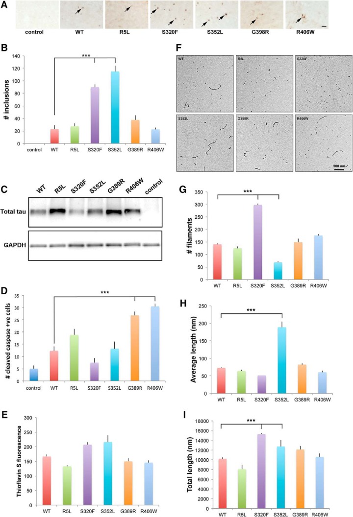
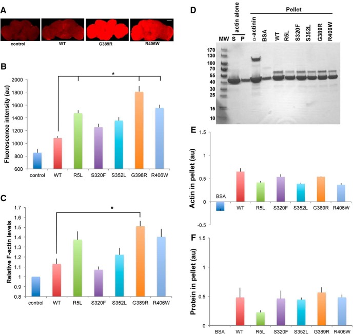
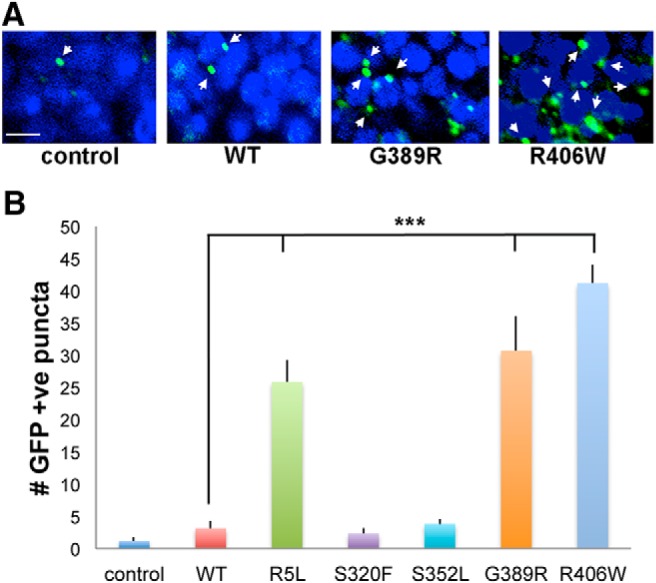
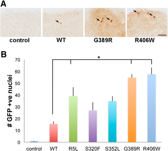
The microtubule binding protein tau is strongly implicated in multiple neurodegenerative disorders, including frontotemporal dementia and parkinsonism linked to chromosome 17 (FTDP-17), which is caused by mutations in tau. In vitro, FTDP-17 mutant versions of tau can reduce microtubule binding and increase the aggregation of tau, but the mechanism by which these mutations promote disease in vivo is not clear. Here we take a combined biochemical and in vivo modeling approach to define functional properties of tau driving neurotoxicity in vivo We express wild-type human tau and five FTDP-17 mutant forms of tau in Drosophila using a site-directed insertion strategy to ensure equivalent levels of expression. We then analyze multiple markers of neurodegeneration and neurotoxicity in transgenic animals, including analysis of both males and females. We find that FTDP-17 mutations act to enhance phosphorylation of tau and thus promote neurotoxicity in an in vivo setting. Further, we demonstrate that phosphorylation-dependent excess stabilization of the actin cytoskeleton is a key phosphorylation-dependent mediator of the toxicity of wild-type tau and of all the FTDP-17 mutants tested. Finally, we show that important downstream pathways, including autophagy and the unfolded protein response, are coregulated with neurotoxicity and actin cytoskeletal stabilization in brains of flies expressing wild-type human and various FTDP-17 tau mutants, supporting a conserved mechanism of neurotoxicity of wild-type tau and FTDP-17 mutant tau in disease pathogenesis.SIGNIFICANCE STATEMENT The microtubule protein tau aggregates and forms insoluble inclusion bodies known as neurofibrillary tangles in the brain tissue of patients with a variety of neurodegenerative disorders, including Alzheimer's disease. The tau protein is thus widely felt to play a key role in promoting neurodegeneration. However, precisely how tau becomes toxic is unclear. Here we capitalize on an "experiment of nature" in which rare missense mutations in tau cause familial neurodegeneration and neurofibrillary tangle formation. By comparing the biochemical activities of different tau mutations with their in vivo toxicity in a well controlled Drosophila model system, we find that all mutations tested increase phosphorylation of tau and trigger a cascade of neurotoxicity critically impinging on the integrity of the actin cytoskeleton.

Keywords: Alzheimer's; Drosophila; tau.


Figures

Figure 1.
Figure 1.
Expression of tau in transgenic animals. Quantitative real-time PCR reveals equivalent levels of tau transcripts in flies expressing 0N4R wild-type and FTDP-17-linked forms of tau under the control of the pan-neuronal elav-GAL4 driver. Values are normalized to wild type. Experiments were run in triplicate and the experiment was repeated at least three times. There were no significant differences in relative transcript levels (p = 0.5, ANOVA). Flies are 1 d old. Full genotypes are provided in Figure 1-1.
Figure 2.
Figure 2.
Phosphorylation and turnover of tau in transgenic animals. A, Western blots showing the phosphorylation of tau at proline-directed sites in flies expressing tau using the pan-neuronal elav-GAL4 driver. Tau-1 recognizes nonphosphorylated tau. The blot was reprobed with a phosphorylation-independent antibody (total tau) to assess total (phosphorylated and unphosphorylated) tau and with an antibody to GAPDH to illustrate equivalent protein loading. All blots were repeated a minimum of three times. B, Quantitative analysis of tau phosphorylation at proline-directed sites reveals significant increases in phosphorylation of tau at AT270 and PHF1 in flies expressing R5L, G389R, or R406W mutant tau compared with flies expressing wild-type tau (p < 0.05, Tukey's HSD). Control is elav-GAL4/+. Full genotypes are provided in Figure 2-1. Flies are 1 d old. C, Tau turnover following conditional expression of tau variants in adult neurons using the elav-GeneSwitch driver. Full genotypes are provided in Figure 2-1. The blot was reprobed with an antibody to GAPDH to illustrate equivalent protein loading. All blots were repeated four times. D, Quantitative analysis of tau turnover reveals that WT, S320F, and S352L are less stable than R406W and E14 (p < 0.05, Tukey's HSD). Full statistical analysis is presented in Figure 2-1.
Figure 3.
Figure 3.
Caspase cleavage in tau transgenic animals. A, Neurons with activated caspase as monitored by cleavage of the transgenic reporter UAS-CD8-PARP (40 aa at caspase cleavage site)-Venus in flies expressing tau using the pan-neuronal elav-GAL4 driver. Arrows indicate cells with caspase activation. Scale bar, 3 μm. B, Quantitative analysis of the number of neurons with activated caspase in the entire brains of control animals or in flies expressing human wild-type or FTDP-17 mutant forms of tau. Six brains were analyzed per genotype. Control is elav-GAL4/+; UAS-CD8-PARP-Venus/+. Full genotypes are provided in Figure 3-1. Flies are 30 d old. **p < 0.01, *p < 0.05, ANOVA with Tukey's HSD. Full statistical analysis is presented in Figure 3-1.
Figure 4.
Figure 4.
Stress pathway activation in tau transgenic animals. A, Neurons with activated JNK signaling as monitored by the puc-lacZ reporter and immunostaining for β-galactosidase, which is directed to the nucleus (Bier et al., 1989), in flies expressing tau using the pan-neuronal elav-GAL4 driver. Arrows indicate positive nuclei. Scale bar, 10 μm. B, Quantitative analysis of the number of neurons with activated JNK in the entire brains of control animals, or in flies expressing human wild-type or FTDP-17 mutant forms of tau. Six brains were analyzed per genotype. Control is elav-GAL4/+; puc-lacZ/+. Full genotypes are provided in Figure 4-1. Flies are 30 d old. **p < 0.01, ANOVA with Tukey's HSD. Full statistical analysis is presented in Figure 4-1.
Figure 5.
Figure 5.
In vivo and in vitro aggregation of tau. A, Inclusions as identified by AT8 immunostaining in tissue sections from flies expressing tau using the pan-glial repo-GAL4 driver. Arrows indicate inclusions. Scale bar, 10 μm. B, Quantitative analysis of the number of inclusions in sections from the midportion of the medulla neuropil in control animals or in flies expressing human wild-type or FTDP-17 mutant forms of tau. Six brains were analyzed per genotype. C, Western blot analysis showing the expression of tau expressed in glia. The blot is reprobed for GAPDH to illustrate equivalent protein loading. Control is repo-GAL4/+ in AC. D, Quantitative analysis of the number of neurons with activated caspase in the entire brains of control animals or in flies expressing human wild-type or FTDP-17 mutant forms of tau. Six brains were analyzed per genotype. Control is UAS-CD8-PARP-Venus/repo-GAL4. Full genotypes are provided in Figure 5-1. Flies are 30 d old in AD. E, Polymerization of tau measured by thioflavin S staining. F, Electron micrographs of polymerization reaction mixtures. GI, Quantification of polymerized tau protein in electron micrographs. The graphs display the total number of tau filaments per image (G), the mean length of filaments (in nanometers; H), and the total length of filaments (I). Images in the electron micrographs (F) were quantified using Image-Pro Plus version 6.0, as described in the Materials and Methods. ***p < 0.001, ANOVA with Tukey's HSD. Full statistical analysis is presented in Figure 5-1.
Figure 6.
Figure 6.
Interaction of tau with actin in vivo and in vitro. A, F-actin staining with fluorescent phalloidin in whole fly brains from flies expressing tau using the pan-neuronal elav-GAL4 driver. Scale bar, 50 μm. B, Quantitative analysis of phalloidin fluorescence in the brains of control animals or flies expressing human wild-type or FTDP-17 mutant forms of tau. Three brains were analyzed per genotype. C, F-actin ELISA in homogenates of brains from control animals or flies expressing human wild-type or FTDP-17 mutant forms of tau. Each experiment was performed with two technical replicates. The experiments were repeated three times. *p < 0.05, ANOVA with Tukey's HSD. Full statistical analysis is presented in Figure 6-1. Control is elav-GAL4/+. Full genotypes are provided in Figure 6-1. Flies are 30 d old. DF, Increased amounts of F-actin are recovered in the pellet following incubation with α-act as a positive control or with human wild-type or FTDP-17 mutant forms of tau (D), as assessed by quantitative analysis of the amount of pelleted actin (E) or protein bound in the pellet (F). BSA is used as a negative control. The experiment was repeated twice.
Figure 7.
Figure 7.
Autophagy in tau transgenic animals. A, Activation of autophagy in flies expressing tau using the pan-neuronal elav-GAL4 driver as monitored by the accumulation of the transgenic reporter UAS-Atg8a-GFP and immunostaining for GFP. Arrows indicate GFP-positive puncta. Scale bar, 3 μm. B, Quantitative analysis of the number of puncta in a section through the calyx of the mushroom body in the brains of control animals, or flies expressing human wild-type or FTDP-17 mutant forms of tau. Six brains were analyzed per genotype. Control is elav-GAL4/+; UAS-Atg8a-GFP/+. Full genotypes are provided in Figure 7-1. Flies are 30 d old. ***p < 0.001, ANOVA with Tukey's HSD. Full statistical analysis is presented in Figure 7-1.
Figure 8.
Figure 8.
Unfolded protein response in tau transgenic animals. A, Neurons with activation of the unfolded protein response in flies expressing tau using the pan-neuronal elav-GAL4 driver as monitored by the transgenic reporter UAS-Xbp1-EGFP and immunostaining for GFP. GFP is directed to the nucleus via the Xbp1 nuclear localization sequence (Ryoo et al., 2007). Arrows indicate GFP-positive nuclei. Scale bar, 10 μm. B, Quantitative analysis of the number of neurons with activation of the unfolded protein response in the entire brains of control animals or flies expressing human wild-type or FTDP-17 mutant forms of tau. Six brains were analyzed per genotype. Control is elav-GAL4/+; UAS-Xbp1-EGFP/+. Full genotypes are provided in Figure 8-1. Flies are 30 d old. *p < 0.05, ANOVA with Tukey's HSD. Full statistical analysis is presented in Figure 8-1.

Similar articles

Cited by

References

    1. Alonso Adel C, Mederlyova A, Novak M, Grundke-Iqbal I, Iqbal K (2004) Promotion of hyperphosphorylation by frontotemporal dementia tau mutations. J Biol Chem 279:34873–34881. 10.1074/jbc.M405131200 - DOI - PubMed
    1. Bateman JR, Lee AM, Wu CT (2006) Site-specific transformation of Drosophila via phiC31 integrase-mediated cassette exchange. Genetics 173:769–777. 10.1534/genetics.106.056945 - DOI - PMC - PubMed
    1. Bier E, Vaessin H, Shepherd S, Lee K, McCall K, Barbel S, Ackerman L, Carretto R, Uemura T, Grell E (1989) Searching for pattern and mutation in the Drosophila genome with a P-lacZ vector. Genes Dev 3:1273–1287. 10.1101/gad.3.9.1273 - DOI - PubMed
    1. Bischof J, Maeda RK, Hediger M, Karch F, Basler K (2007) An optimized transgenesis system for Drosophila using germ-line-specific phiC31 integrases. Proc Natl Acad Sci U S A 104:3312–3317. 10.1073/pnas.0611511104 - DOI - PMC - PubMed
    1. Bodea LG, Eckert A, Ittner LM, Piguet O, Götz J (2016) Tau physiology and pathomechanisms in frontotemporal lobar degeneration. J Neurochem 138:71–94. 10.1111/jnc.13600 - DOI - PMC - PubMed

Publication types

LinkOut - more resources