Review
. 2017 Nov 15;144(22):4078-4090.
doi: 10.1242/dev.120402.

Developing a sense of touch

Affiliations
Review

Developing a sense of touch

Blair A Jenkins et al. Development. .

Abstract

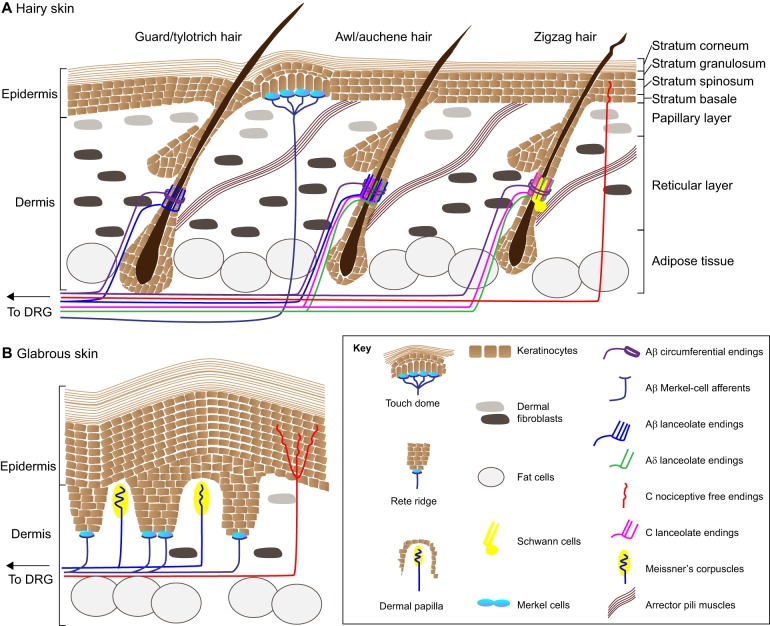
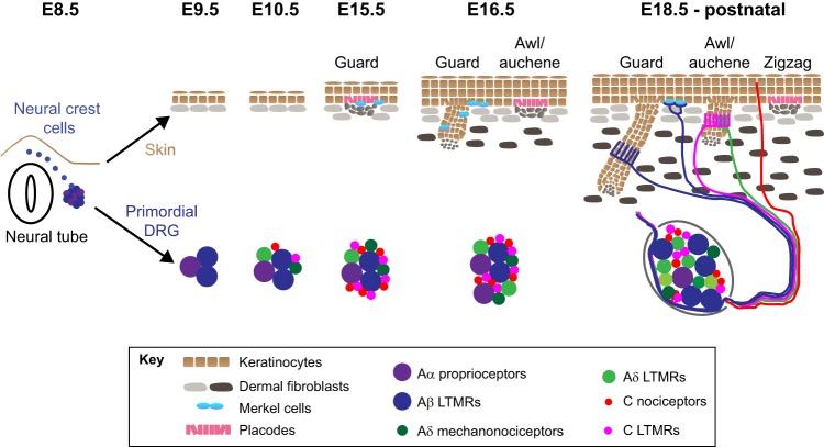
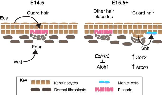
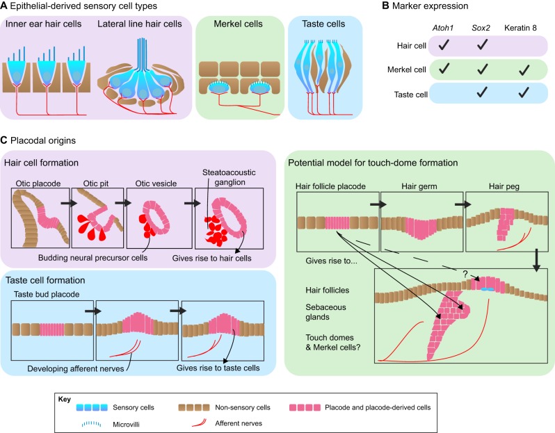
The sensation of touch is mediated by mechanosensory neurons that are embedded in skin and relay signals from the periphery to the central nervous system. During embryogenesis, axons elongate from these neurons to make contact with the developing skin. Concurrently, the epithelium of skin transforms from a homogeneous tissue into a heterogeneous organ that is made up of distinct layers and microdomains. Throughout this process, each neuronal terminal must form connections with an appropriate skin region to serve its function. This Review presents current knowledge of the development of the sensory microdomains in mammalian skin and the mechanosensory neurons that innervate them.

Keywords: Axon guidance; Hair follicle; Merkel cell; Placode; Skin; Touch.


Conflict of interest statement

Competing interestsThe authors declare no competing or financial interests.

Figures

Fig. 1.
Fig. 1.
Mechanosensory end organs in skin. The touch receptors of hairy and glabrous skin are highly diverse. (A) Hairy skin is decorated with distinct types of hair follicles. Guard/tylotrich hairs are the most rare hair type, and also the longest. Awl/auchene hairs and zigzag hairs make up the bulk of the hairs in the mouse coat. Each hair type is associated with a unique complement of sensory neurons. Lanceolate and circumferential endings wrap around the bulge region of hair follicles, which stretches between the sebaceous gland and the connection site of arrector pili muscles. Note that all lanceolate endings intercalate with the protrusions of terminal Schwann cells, one of which is shown in the schematic (yellow). Other neurons innervate touch domes, which are discrete, raised zones of the skin adjacent to guard hairs. These neurons innervate epidermal Merkel cells (teal). (B) Instead of hairs, hallmarks of glabrous skin include invaginations of the epidermis called rete ridges. Dermal zones between rete ridges are called dermal papillae. Merkel cells and their associated afferents are found at the base of rete ridges, whereas Meissner's corpuscles protrude into dermal papillae.
Fig. 2.
Fig. 2.
Timelines of skin and sensory neuron development. The skin originates from the ectoderm (brown) and mesoderm (gray), which give rise to epidermis and dermis, respectively. Guard hairs are specified as epidermal placodes (pink) with dermal clusters or condensates of fibroblasts beneath them (gray); Merkel cells (teal) appear within and adjacent to developing guard hairs by E15.5. At roughly E16.5, a new crop of placodes appears that will become awl/auchene hairs. From E18.5 onward, additional waves of folliculogenesis generate zigzag hairs. All hair types elongate down into the dermis until P21, when folliculogenesis is complete. Development of dorsal root ganglia (DRG) occurs in parallel with skin development. At E8.5, neural crest cells delaminate from the neural tube and coalesce to form DRG. Large diameter mechanosensitive neurons are born in the first wave of specification at E9.5. Small diameter neurons are specified beginning one day later at E10.5. DRG neuron precursors continue to proliferate and generate large and small diameter neurons until E13.5. From E13.5 onward, somatosensory afferents elongate from DRG to innervate the skin. A subset of the tactile endings that form in the skin from E18.5 onward are illustrated.
Fig. 3.
Fig. 3.
Factors involved in Merkel-cell formation. Guard, or tylotrich, hairs are specified at E14.5 in the trunk skin of mice. This process is driven by epidermal expression of Eda and dermal expression of Edar, which is induced by Wnt signaling in the dermis. The birth of nascent hair follicles is marked by the presence of epidermal placodes (pink) and the formation of dermal condensates (gray). One day later, Merkel cells (teal) appear with budding guard hairs. Their induction is postulated to result from Shh expression in the hair follicle. This leads to the expression of Atoh1 and Sox2, which encode transcription factors that work in concert to initiate and maintain Merkel cell specification. Ezh1/2 expression surrounding other hair follicle types represses Atoh1, preventing ectopic Merkel cell formation.
Fig. 4.
Fig. 4.
Mechanoreceptor development: insights from other sensory systems. Epidermal Merkel cells share many similarities with other sensory cell types, such as hair cells of the inner ear and taste cells. (A) All of these cell types possess apical protrusions of their cell membranes and are innervated by sensory afferents. (B) All of these cell types express similar complements of genes during their development. (C) Hair cells and taste cells develop from placodes. Based on this, we postulate that Merkel cells also arise from placodes; a potential model of placode-based Merkel cell formation is depicted.
Fig. 5.
Fig. 5.
Planar cell polarity genes organize sensory structures across species. Frizzled genes in Drosophila and mice serve as a good example of how homologous planar cell polarity genes might influence tissue patterning. Bristles on the D. melanogaster wing are disrupted in clones with aberrant Frizzled signaling. Similarly, the apical microvilli in hair cells of the mammalian cochlea, which are usually highly organized, splay out in all directions in frizzled mutant cochleae. Strikingly, frizzled genes are also required for the patterning of hair follicles and Merkel cells in mouse (Mus musculus) dorsal skin; in frizzled mutants, hairs are oriented randomly rather than in a rostral-to-caudal direction, and Merkel cells form a ring around the entire follicle rather than forming a crescent on the caudal side. KO, knockout; WT, wild type.

References

    1. Abraira V. E. and Ginty D. D. (2013). The sensory neurons of touch. Neuron 79, 618-639. 10.1016/j.neuron.2013.07.051 - DOI - PMC - PubMed
    1. Adrian E. D. and Zotterman Y. (1926a). The impulses produced by sensory nerve endings: part 3. Impulses set up by Touch and Pressure. J. Physiol. 61, 465-483. 10.1113/jphysiol.1926.sp002308 - DOI - PMC - PubMed
    1. Adrian E. D. and Zotterman Y. (1926b). The impulses produced by sensory nerve-endings: part II. The response of a Single End-Organ. J. Physiol. 61, 151-171. 10.1113/jphysiol.1926.sp002281 - DOI - PMC - PubMed
    1. Agarwala S., Duquesne S., Liu K., Boehm A., Grimm L., Link S., König S., Eimer S., Ronneberger O. and Lecaudey V. (2015). Amotl2a interacts with the Hippo effector Yap1 and the Wnt/β-catenin effector Lef1 to control tissue size in zebrafish. Elife 4, e08201 10.7554/eLife.08201 - DOI - PMC - PubMed
    1. Agelli M. and Clegg L. X. (2003). Epidemiology of primary Merkel cell carcinoma in the United States. J. Am. Acad. Dermatol. 49, 832-841. 10.1016/S0190-9622(03)02108-X - DOI - PubMed

Publication types

LinkOut - more resources