. 2017 Nov 28;114(48):E10438-E10445.
doi: 10.1073/pnas.1715812114. Epub 2017 Nov 14.

Structural basis for antibody recognition of the NANP repeats in Plasmodium falciparum circumsporozoite protein

Affiliations

Structural basis for antibody recognition of the NANP repeats in Plasmodium falciparum circumsporozoite protein

David Oyen et al. Proc Natl Acad Sci U S A. .

Erratum in

Abstract

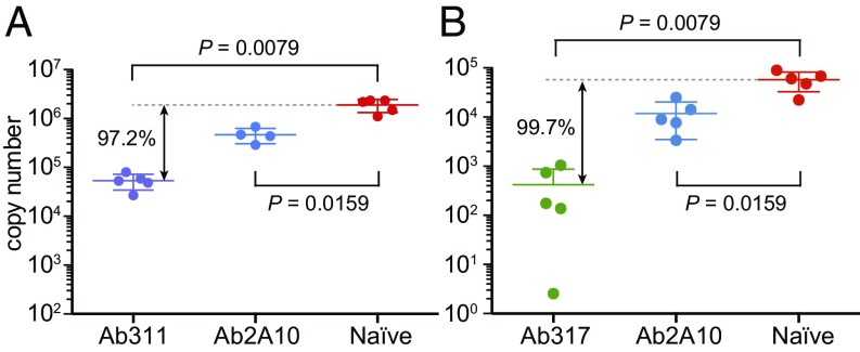
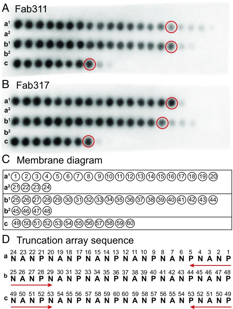
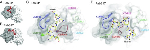
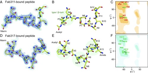
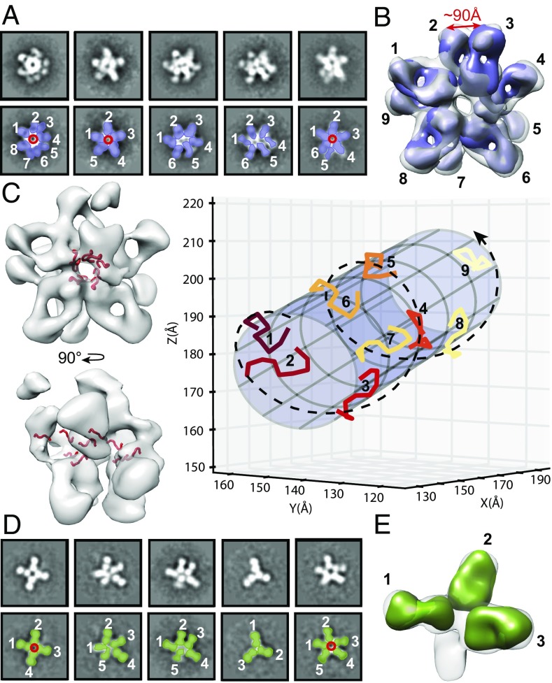
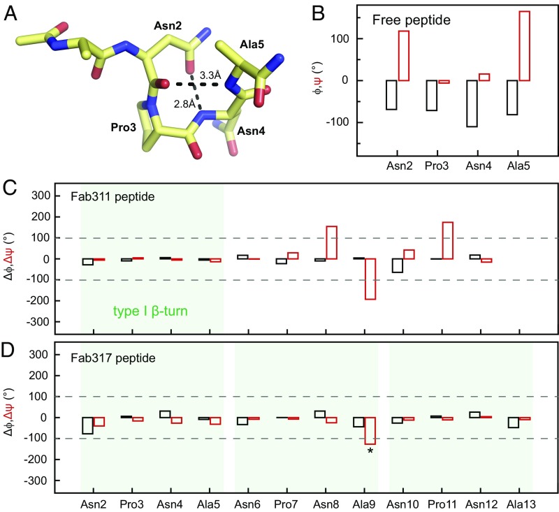
Acquired resistance against antimalarial drugs has further increased the need for an effective malaria vaccine. The current leading candidate, RTS,S, is a recombinant circumsporozoite protein (CSP)-based vaccine against Plasmodium falciparum that contains 19 NANP repeats followed by a thrombospondin repeat domain. Although RTS,S has undergone extensive clinical testing and has progressed through phase III clinical trials, continued efforts are underway to enhance its efficacy and duration of protection. Here, we determined that two monoclonal antibodies (mAbs 311 and 317), isolated from a recent controlled human malaria infection trial exploring a delayed fractional dose, inhibit parasite development in vivo by at least 97%. Crystal structures of antibody fragments (Fabs) 311 and 317 with an (NPNA)3 peptide illustrate their different binding modes. Notwithstanding, one and three of the three NPNA repeats adopt similar well-defined type I β-turns with Fab311 and Fab317, respectively. Furthermore, to explore antibody binding in the context of P. falciparum CSP, we used negative-stain electron microscopy on a recombinant shortened CSP (rsCSP) construct saturated with Fabs. Both complexes display a compact rsCSP with multiple Fabs bound, with the rsCSP-Fab311 complex forming a highly organized helical structure. Together, these structural insights may aid in the design of a next-generation malaria vaccine.

Trial registration: ClinicalTrials.gov NCT01857869.

Keywords: EM; X-ray crystallography; antibodies; circumsporozoite protein; malaria.


Conflict of interest statement

Conflict of interest statement: W.V. and D.E. are employees of and own equity in Atreca, Inc.

Figures

Fig. 1.
Fig. 1.
Antibody inhibition of malaria infection in mice. Parasite liver load 40 h post challenge with a chimeric P. berghei strain as assessed by qPCR for P. berghei-specific 18S rRNA after administration of Ab311 (100 μg) (A) and Ab317 (300 μg) (B). Significant protection is observed compared with naive mice, with 97.2% and 99.7% inhibition of parasite development for Ab311 and Ab317, respectively, while a previously reported antibody, 2A10 (300 μg dose) (24) showed only 75–82% inhibition of the parasite liver load. The P values were determined using the Mann–Whitney U test. Only four data points are available for 2A10 in A because one mouse died.
Fig. 2.
Fig. 2.
Epitope mapping using truncation peptide arrays. (A and B) The PepSpot membrane is shown for Fab311 (A) and Fab317 (B) and consists of five rows of the spotted peptides (a1, a2, b1, b2, and c). Dark spots indicate strong Fab binding. (C and D) Schematic of the location of the peptide spots on the membrane. The numbers within the circles refer to the numbers in the truncation array sequence (D). Rows a1 and a2 correspond to a truncation array starting from the C terminus of the (NANP)6 peptide, rows b1 and b2 are truncations from the N terminus, and row c represents truncations from both the N terminus and C terminus simultaneously. The peptides that appear to have the minimal number of repeats for strong Fab binding are circled in red in A and B.
Fig. 3.
Fig. 3.
Crystal structures of (NPNA)3 peptides in complex with Fab311 and Fab317. (A and B) Surface representation of the variable domains of Fab311 (A) and Fab317 (B) with the (NPNA)3 peptide represented by a red tube. The heavy- and light-chain variable domains are colored dark and light gray, respectively. (C and D) Paratope representation for Fab311 (C) and Fab317 (D) with a transparent dark gray surface for the heavy chain and a transparent light gray surface for the light chain. The underlying CDR loops are shown in cartoon representation and are colored green (H1), blue (H2), red (H3), light green (L1), light blue (L2), and pink (L3). The (NPNA)3 peptide is shown in stick representation (yellow carbons). The N terminus of each peptide is indicated (Nterm).
Fig. 4.
Fig. 4.
Structural analysis of antibody-bound peptides. (A and D) 2Fo-Fc electron density maps contoured at 2.0σ (blue) and 0.8σ (cyan) for peptide bound to Fab311 (A) and Fab317 (D). The peptide is shown in stick representation (yellow carbons). (B and E) Type I β-turns are highlighted by transparent green circles for peptide bound to Fab311 (B) and Fab317 (E). Intrapeptide hydrogen bonds that emulate a pseudo 310 turn between the first Asn sidechain and amide backbone of the third residue in the turn are shown as black dashed lines. (C and F) Ramachandran plots for the dihedral angles of Fab311-bound peptide (C) and Fab317-bound peptide (F). Residues that have typical dihedral angles indicative of canonical NPNA type I β-turns are colored green; otherwise they are colored red. The β-sheet region is in the dark shaded region of the plot in the upper left quadrant, and the α-helical region is in the central region on the left around ψ of −30°. The Fab311-bound and Fab317-bound peptides have one and three canonical type I β-turns, respectively.
Fig. 5.
Fig. 5.
nsEM for rsCSP bound to Fab311 and Fab317. (A and D) Five selected representative class averages for the rsCSP–Fab311 (A) and rsCSP–Fab317 (D) complexes, false colored to show the location of rsCSP (red), Fab311 (purple), and Fab317 (green). Fabs are labeled by number in white. (B) The refined 3D model confirmed the presence of multiple densities for Fab311, for which a total count of nine Fab311s could be observed at the low threshold level. The distance between the heavy-chain C termini of Fab311 nos. 2 and 3 was measured at 90 Å, which could be accommodated in an IgG. (C) The (NPNA)3 peptide (red) from the Fab311 crystal structure was docked into the 3D model (Left) and revealed a helical shape looking into the central hole of the rsCSP complex and along its length (Right). The docked peptides in the nsEM map for the rsCSP–Fab311 complex were fitted to a cylinder with a radius of 15.2 Å. The peptides are colored using a color progression, and the helical organization is shown by the dashed spiral line. (D and E) Reaching convergence for the rsCSP–Fab317 complex (E) was difficult due to the various stoichiometries seen in the 2D class averages (D). The unmodeled blob may be a remnant of a fourth Fab fragment that is present in some of the complexes.
Fig. 6.
Fig. 6.
Comparison of dihedral angles shows similarities between the bound and free peptides. (A) X-ray structure of the free ANPNA peptide shows a type I β-turn in which the Asn2 (residue i) OD1 also hydrogen bonds to the backbone amide of Asn4 (i + 2) (27). (B) Plot of the dihedral angles for the NPNA unit in the ANPNA X-ray structure; ϕ and ψ are shown in black and red, respectively. (C and D) Plots of the dihedral angle differences between each of the NPNA units for the peptide bound to Fab311 (C) or Fab317 (D) and the NPNA unit of the free peptide; Δϕ and Δψ are shown in black and red, respectively. Type I β-turns are highlighted by transparent green boxes. The dihedral angle differences are relatively small within each NPNA type I β-turn, except for Ala9 Δψ in the Fab317 peptide (asterisk). This deviation from the NPNA type I β-turn in solution reflects a change in direction at the end of the NPNA repeat rather than a disruption of the canonical type I β-turn.

References

    1. World Health Organization . World Malaria Report 2016. WHO; Geneva: 2016.
    1. WHO Malaria Vaccine Funders Group . Malaria Vaccine Technology Roadmap. WHO; Geneva: 2013.
    1. Agnandji ST, et al. RTS,S Clinical Trials Partnership First results of phase 3 trial of RTS,S/AS01 malaria vaccine in African children. N Engl J Med. 2011;365:1863–1875. - PubMed
    1. RTS,S Clinical Trials Partnership Efficacy and safety of the RTS,S/AS01 malaria vaccine during 18 months after vaccination: A phase 3 randomized, controlled trial in children and young infants at 11 African sites. PLoS Med. 2014;11:e1001685. - PMC - PubMed
    1. RTS,S Clinical Trials Partnership Efficacy and safety of RTS,S/AS01 malaria vaccine with or without a booster dose in infants and children in Africa: Final results of a phase 3, individually randomised, controlled trial. Lancet. 2015;386:31–45. - PMC - PubMed

Publication types

MeSH terms

Associated data