

Facilitation of information processing in the primary somatosensory area in the ball rotation task
- PMID: 29138504
- PMCID: PMC5686197
- DOI: 10.1038/s41598-017-15775-x
Facilitation of information processing in the primary somatosensory area in the ball rotation task
Abstract
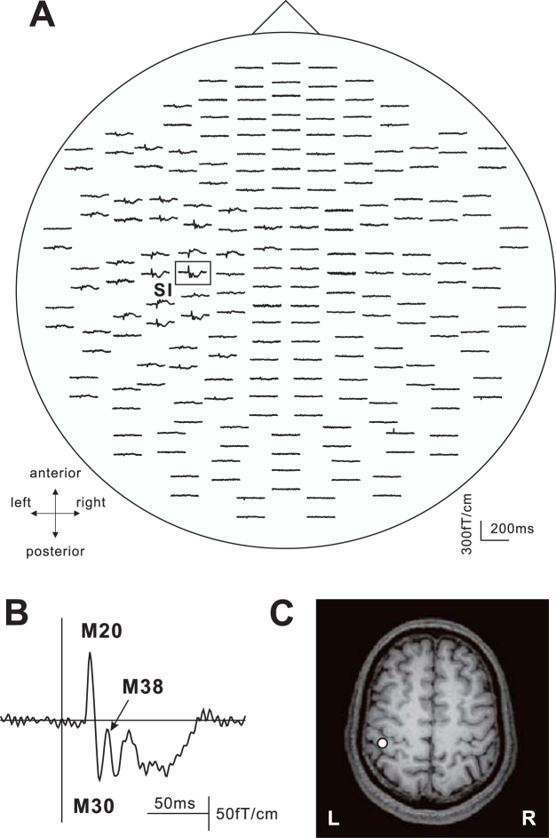
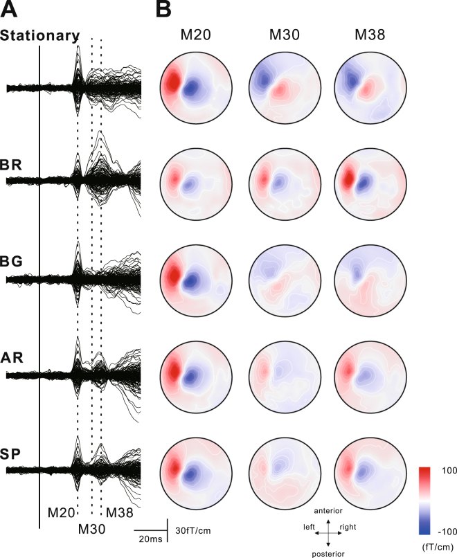
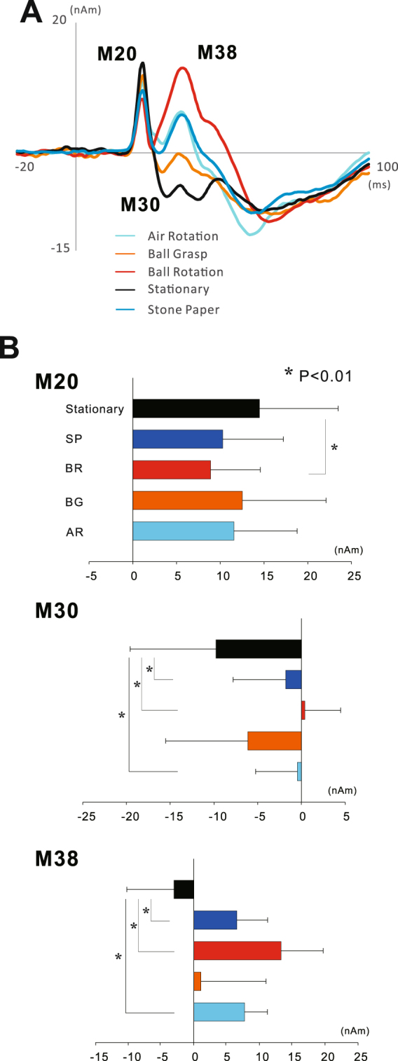
Somatosensory input to the brain is known to be modulated during voluntary movement. It has been demonstrated that the response in the primary somatosensory cortex (SI) is generally gated during simple movement of the corresponding body part. This study investigated sensorimotor integration in the SI during manual movement using a motor task combining movement complexity and object manipulation. While the amplitude of M20 and M30 generated in the SI showed a significant reduction during manual movement, the subsequent component (M38) was significantly higher in the motor task than in the stationary condition. Especially, that in the ball rotation task showed a significant enhancement compared with those in the ball grasping and stone and paper tasks. Although sensorimotor integration in the SI generally has an inhibitory effect on information processing, here we found facilitation. Since the ball rotation task seems to be increasing the demand for somatosensory information to control the complex movements and operate two balls in the palm, it may have resulted in an enhancement of M38 generated in the SI.
Conflict of interest statement
The authors declare that they have no competing interests.
Figures
Similar articles
-
Dexterous manual movement facilitates information processing in the primary somatosensory cortex: A magnetoencephalographic study.Eur J Neurosci. 2021 Jul;54(2):4638-4648. doi: 10.1111/ejn.15310. Epub 2021 Jun 4. Eur J Neurosci. 2021. PMID: 33987876 Free PMC article.
-
Centrifugal regulation of human cortical responses to a task-relevant somatosensory signal triggering voluntary movement.Neuroimage. 2006 Sep;32(3):1355-64. doi: 10.1016/j.neuroimage.2006.05.015. Epub 2006 Jun 27. Neuroimage. 2006. PMID: 16806987
-
Intracortical modulation of somatosensory evoked fields during movement: evidence for selective suppression of postsynaptic inhibition.Brain Res. 2012 Jun 12;1459:43-51. doi: 10.1016/j.brainres.2012.04.023. Epub 2012 Apr 20. Brain Res. 2012. PMID: 22564923
-
The premotor cortex and nonstandard sensorimotor mapping.Can J Physiol Pharmacol. 1996 Apr;74(4):469-82. doi: 10.1139/cjpp-74-4-469. Can J Physiol Pharmacol. 1996. PMID: 8828893 Review.
-
Functional somatotopy in sensorimotor cortex.Neuroreport. 2005 Mar 15;16(4):313-6. doi: 10.1097/00001756-200503150-00001. Neuroreport. 2005. PMID: 15729128 Review.
Cited by
-
Dexterous manual movement facilitates information processing in the primary somatosensory cortex: A magnetoencephalographic study.Eur J Neurosci. 2021 Jul;54(2):4638-4648. doi: 10.1111/ejn.15310. Epub 2021 Jun 4. Eur J Neurosci. 2021. PMID: 33987876 Free PMC article.
-
Frequency dependence of cortical somatosensory evoked response to peripheral nerve stimulation with controlled afferent excitation.J Neural Eng. 2025 Mar 28;22(2):026035. doi: 10.1088/1741-2552/adc204. J Neural Eng. 2025. PMID: 40101361 Free PMC article.
-
Effects of repetitive practice of motor tasks on somatosensory gating.Front Hum Neurosci. 2023 Mar 29;17:1131986. doi: 10.3389/fnhum.2023.1131986. eCollection 2023. Front Hum Neurosci. 2023. PMID: 37063102 Free PMC article.
-
Effective Connectivity for Decoding Electroencephalographic Motor Imagery Using a Probabilistic Neural Network.Sensors (Basel). 2021 Sep 30;21(19):6570. doi: 10.3390/s21196570. Sensors (Basel). 2021. PMID: 34640888 Free PMC article.
-
Effects of Complex Movement on the Excitability of the Ipsilateral Primary Motor Cortex and Spinal Motoneurons Contralateral to the Movement: A Comparison of Ball Rotation and Grasping Tasks with Equivalent Muscle Activity.Brain Sci. 2025 Feb 10;15(2):171. doi: 10.3390/brainsci15020171. Brain Sci. 2025. PMID: 40002504 Free PMC article.
References
-
- Craggs MD, Rothwell JC, Rushton DN. Gating of somatosensory evoked potentials by active and passive movements in man [proceedings] J Physiol. 1979;295:96P–97P. - PubMed
MeSH terms
LinkOut - more resources
Full Text Sources
Other Literature Sources
