. 2017:2017:6742427.
doi: 10.1155/2017/6742427. Epub 2017 Aug 23.

Anti-Inflammatory Strategy for M2 Microglial Polarization Using Retinoic Acid-Loaded Nanoparticles

Affiliations

Anti-Inflammatory Strategy for M2 Microglial Polarization Using Retinoic Acid-Loaded Nanoparticles

Marta Machado-Pereira et al. Mediators Inflamm. 2017.

Abstract

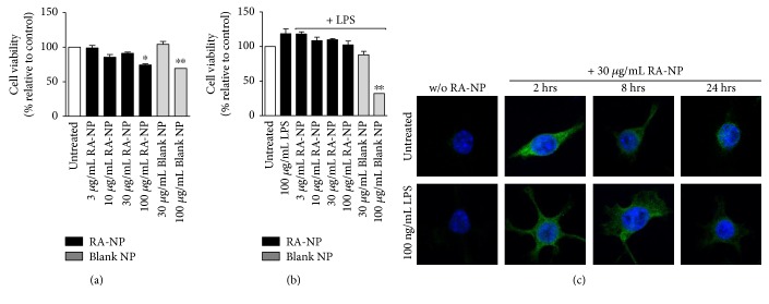
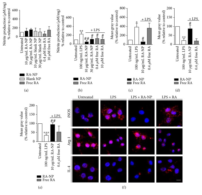
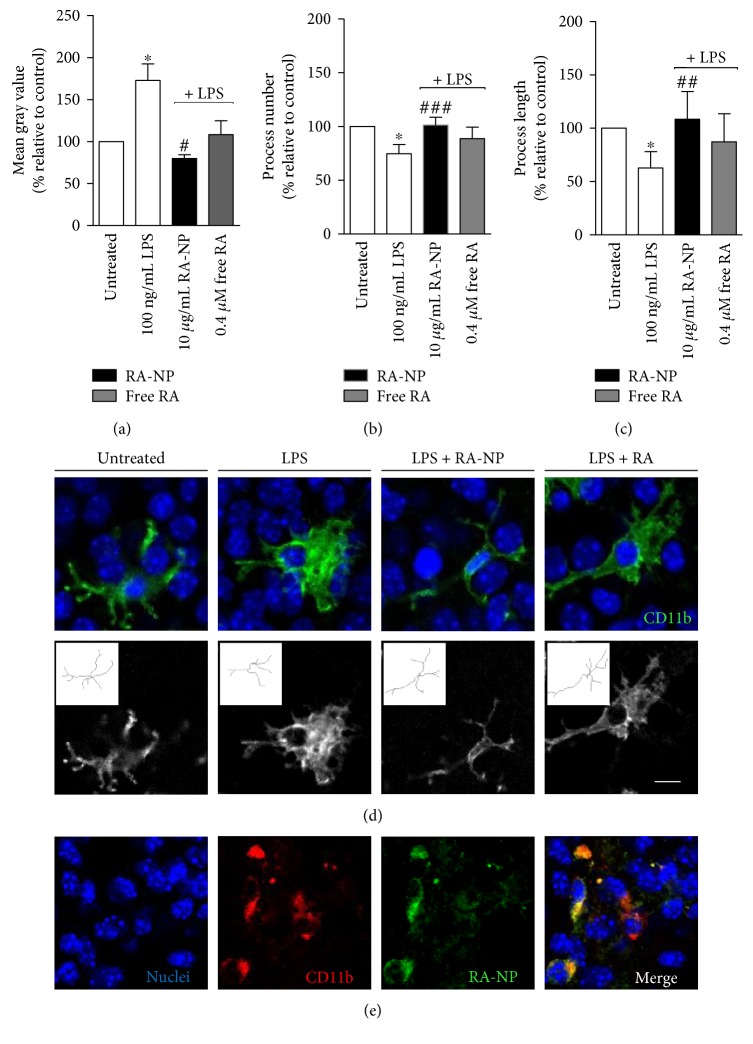
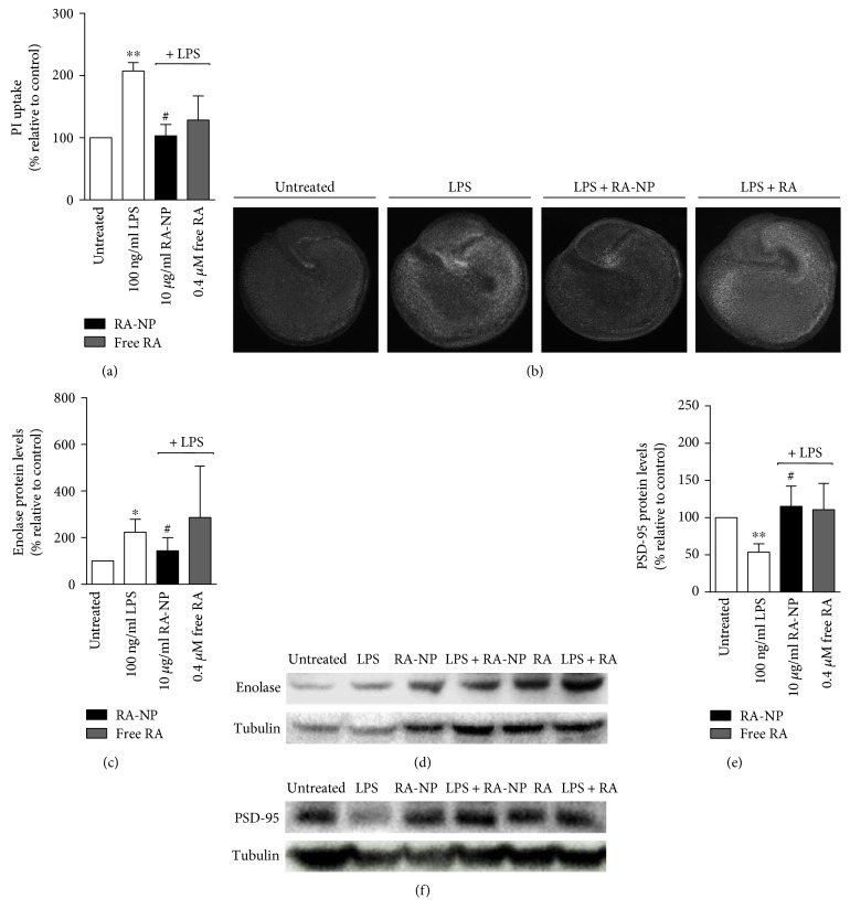
Inflammatory mechanisms triggered by microglial cells are involved in the pathophysiology of several brain disorders, hindering repair. Herein, we propose the use of retinoic acid-loaded polymeric nanoparticles (RA-NP) as a means to modulate microglia response towards an anti-inflammatory and neuroprotective phenotype (M2). RA-NP were first confirmed to be internalized by N9 microglial cells; nanoparticles did not affect cell survival at concentrations below 100 μg/mL. Then, immunocytochemical studies were performed to assess the expression of pro- and anti-inflammatory mediators. Our results show that RA-NP inhibited LPS-induced release of nitric oxide and the expression of inducible nitric oxide synthase and promoted arginase-1 and interleukin-4 production. Additionally, RA-NP induced a ramified microglia morphology (indicative of M2 state), promoting tissue viability, particularly neuronal survival, and restored the expression of postsynaptic protein-95 in organotypic hippocampal slice cultures exposed to an inflammatory challenge. RA-NP also proved to be more efficient than the free equivalent RA concentration. Altogether, our data indicate that RA-NP may be envisioned as a promising therapeutic agent for brain inflammatory diseases.


Figures

Figure 1
Figure 1
RA-NP did not compromise microglia cell viability. (a) Microglial cells were treated with RA-NP and blank NP (24 hours) to assess toxicity, using MTT assay. Cytotoxicity was induced at concentrations starting at 100 μg/mL (n = 3; p < 0.05 and ∗∗p < 0.01 compared to untreated cells). (b) LPS (100 ng/mL) did not potentiate cytotoxicity; only blank nanoparticles (100 μg/mL) in the presence of LPS significantly compromised viability (n = 3; ∗∗p < 0.01 compared to untreated cells). (c) RA-NP (30 μg/mL) internalization by microglial cells was observed by confocal microscopy over the course of 24 hours, in the absence (top row) or presence of LPS (bottom row). Scale bar 10 μm.
Figure 2
Figure 2
RA-NP induced an M2 microglial phenotype under inflammatory challenge. (a) None of the cell treatments (RA-NP, blank NP or free RA) affected NO levels in the absence of a stimulus. (b) RA-NP (10 μg/mL) and free RA (0.4 and 10 μM) inhibited NO production in the presence of 100 ng/mL LPS (24 hours) (n = 4; ∗∗p < 0.01 compared to untreated cells; #p < 0.05, ##p < 0.01 compared to LPS). (c) RA-NP (10 μg/mL) decreased LPS-induced iNOS expression while free RA had no effect (n = 3–6; p < 0.05 compared to untreated cells, #p < 0.05 compared to LPS). (d) RA-NP (10 μg/mL) increased LPS-inhibited Arg-1 expression. Free RA (0.4 μM) had no effect (n = 3–6; ∗∗p < 0.01 compared to untreated cells, #p < 0.05 compared to LPS). (e) RA-NP (10 μg/mL) increased IL-4 expression while free RA had no effect (n = 3–6; ∗∗∗p < 0.001 compared to untreated cells, ##p < 0.01 compared to LPS). (f) Representative confocal images depicting expression of iNOS, Arg-1, and IL-4 after cell treatments (in red; top, middle, and bottom panels, resp.). Nuclear staining in blue. Scale bar 10 μm.
Figure 3
Figure 3
RA-NP modulated microglia activation and morphology in LPS-treated hippocampal slice cultures. Murine organotypic hippocampal slice cultures (P7) were treated with RA-NP (10 μg/mL) or free RA (0.4 μM), and their effect on cell morphology was quantified in an inflammatory context (100 ng/mL LPS, 24 hours). (a) RA-NP treatment significantly reduced cell bodies; free RA (0.40 μM) had no effect (n = 4; p < 0.05 compared to untreated cells; #p < 0.05 compared to LPS). (b) RA-NP treatment (10 μg/mL) significantly promoted a higher number of microglial processes while free RA (0.40 μM) had no effect (n = 4; p < 0.05 compared to untreated cells, ###p < 0.001 compared to LPS). (c) RA-NP treatment (10 μg/mL) significantly promoted an increase in length of microglial processes. Free RA had no effect (n = 4; p < 0.05 compared to untreated cells, ##p < 0.01 compared to LPS). (d) Representative brain slices were stained for CD11b (green; top panel), and skeletonized microglial cells are shown in the bottom panel. Nuclear staining in blue. (e) Microglial cells (in red) internalized RA-NP (in green). Colocalization is highlighted in the merged image. Nuclear staining in blue. Scale bar 10 μm.
Figure 4
Figure 4
RA-NP promoted tissue viability and enhanced neuronal protection after an inflammatory challenge. (a) RA-NP (10 μg/mL) protected from LPS-induced toxicity while free RA (0.40 μM) had no effect (n = 3; ∗∗p < 0.01 compared to untreated cells, #p < 0.05 compared to LPS). (b) Representative images depicting cell death on organotypic hippocampal slice cultures. Slices were counterstained with propidium iodide (PI). (c) RA-NP (10 μg/mL) significantly counteracted the LPS effect by decreasing enolase levels. (n = 3; p < 0.05 compared to untreated cells, #p < 0.05 compared to LPS). (d) RA-NP (10 μg/mL) significantly inhibited the LPS effect by increasing PSD-95 levels (n = 3; ∗∗p < 0.01 compared to untreated cells, #p < 0.05 compared to LPS). Free RA (0.40 μM) had no effect on both markers. Representative images depicting the effect of RA-NP treatment on neuronal damage (e) and synaptic function (f). Additional controls with RA-NP alone and free RA are also shown.

References

    1. Szalay G., Martinecz B., Lénárt N., et al. Microglia protect against brain injury and their selective elimination dysregulates neuronal network activity after stroke. Nature Communications. 2016;7, article 11499 doi: 10.1038/ncomms11499. - DOI - PMC - PubMed
    1. Miyamoto A., Wake H., Ishikawa A. W., et al. Microglia contact induces synapse formation in developing somatosensory cortex. Nature Communications. 2016;7, article 12540 doi: 10.1038/ncomms12540. - DOI - PMC - PubMed
    1. Thameem Dheen S., Kaur C., Ling E.-A. Microglial activation and its implications in the brain diseases. Current Medicinal Chemistry. 2007;14:1189–1197. - PubMed
    1. Vinet J., van Weering H. R., Heinrich A., et al. Neuroprotective function for ramified microglia in hippocampal excitotoxicity. Journal of Neuroinflammation. 2012;9:p. 27. doi: 10.1186/1742-2094-9-27. - DOI - PMC - PubMed
    1. Mantovani A. Reflections on immunological nomenclature: in praise of imperfection. Nature Immunology. 2016;17:215–216. doi: 10.1038/ni.3354. - DOI - PubMed

LinkOut - more resources