. 2018 Jan;41(1):364-372.
doi: 10.3892/ijmm.2017.3249. Epub 2017 Nov 9.

Vitamin D receptor activation influences the ERK pathway and protects against neurological deficits and neuronal death

Affiliations

Vitamin D receptor activation influences the ERK pathway and protects against neurological deficits and neuronal death

Jie Yuan et al. Int J Mol Med. 2018 Jan.

Abstract

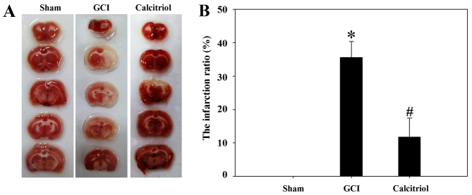
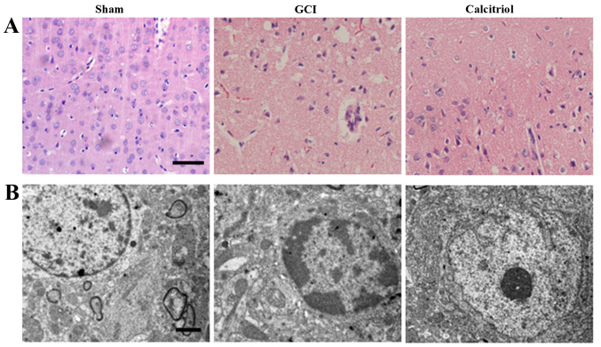
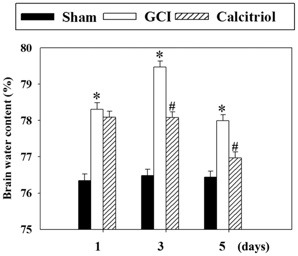
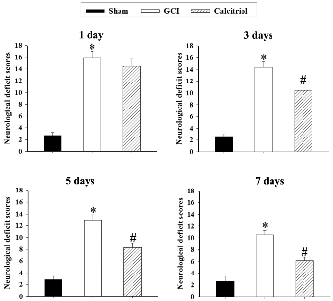
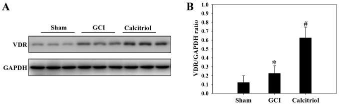
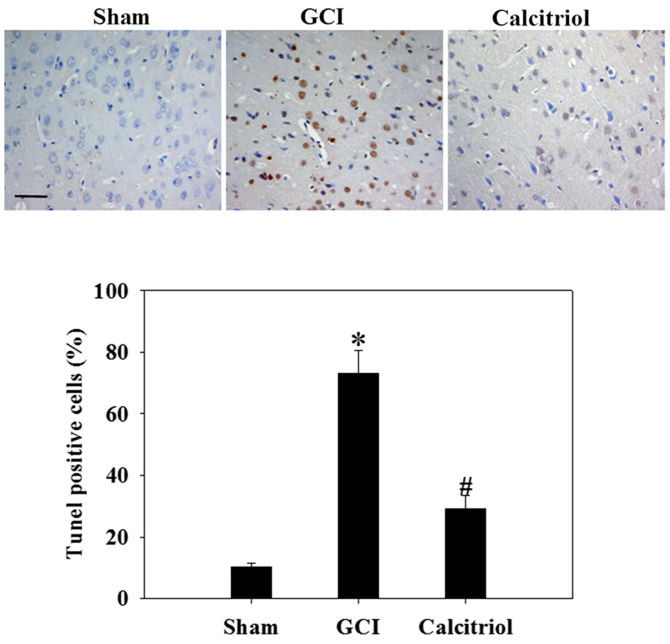
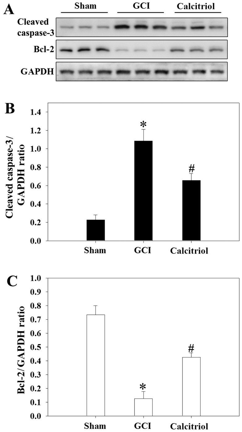
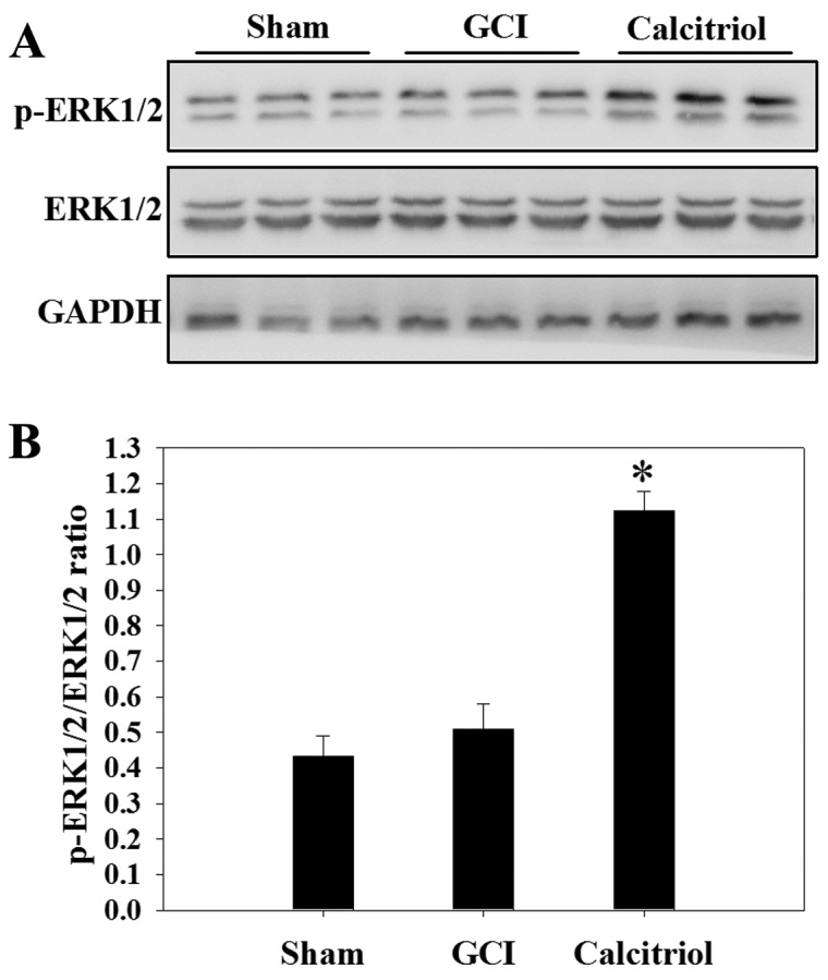
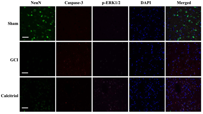
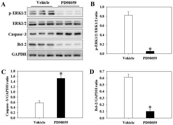
Previous studies have demonstrated that global cerebral ischemia (GCI) causes neurological deficits and neuronal cell apoptosis. Calcitriol, a biologically active metabolite of vitamin D, exerts its endocrinological influence via nuclear vitamin D receptor. It is being assessed as an emerging therapeutic strategy in models of various medical conditions, including acute brain injury. The purpose of the present study was to investigate the neuroprotective effects of calcitriol on GCI and further refine the potential underlying mechanisms. A total of 145 male rats were assigned to 5 groups as follows: Sham group, GCI group, calcitriol treatment group, PD98059 treatment group and vehicle-treated group. Brain water content and neurologic severity score were assessed to evaluate the brain edema and neurological deficits of rats. Histopathological changes and ultrastructures of cells were observed via hematoxylin and eosin stain and transmission electron microscopy, respectively. Immunofluorescent staining and western blot analysis were used to assess the expression of proteins and their co-localization at the molecular level. The results demonstrated that post-GCI administration of calcitriol attenuated brain edema and improved neurological function in rats. Calcitriol also caused marked extracellular signal-regulated kinase 1/2 pathway activation, and thereby attenuated neuronal apoptosis. The present study provided novel clues for understanding the mechanisms by which calcitriol exerts its neuroprotective activity in a rat model of GCI.


Figures

Figure 1
Figure 1
The effects of calcitriol on the brain infarct volume after GCI. (A) Representative images of 2,3,5-triphenyltetrazolium chloride-stained coronal sections were obtained at 7 days after GCI insult. A notable cerebral infarction region was observed in the GCI group. (B) Quantified relative region of cerebral infarction. Values are expressed as the mean ± standard error (n=5 for each group). After GCI, the infarct volume was significantly increased and the infarct ratio was obviously lower after administration of calcitriol. *P<0.001 vs. Sham group; #P<0.005 vs. GCI group. GCI, global cerebral ischemia.
Figure 2
Figure 2
Morphological detection results in the cortex region of the brain after GCI. (A) Assessment of histopathologic changes in the experimental groups (hematoxylin and eosin staining; scale bar, 50 μm). The nuclei of normal neurons were round and stained pale, whereas nuclei of dying neurons were pyknotic and darkly stained after GCI. The pathological changes of cortex neurons in the calcitriol-treated group were improved compared with those in the GCI group. (B) Ultrastructures of neurons were observed via transmission electron microscopy in the experimental groups (scale bar, 2 μm). In the Sham group, the neurons in the cortex had large round nuclei, the double nuclear membranes were clear and complete, and the outer membrane enclosed the periphery of the mitochondria. In the GCI group, neurons were irregular and exhibited chromatin condensation, cytoplasm dissolution and vacuole formation. The nuclear membranes and cell organelles were dissolved or absent. In the calcitriol group, the damage to the neurons was alleviated. GCI, global cerebral ischemia.
Figure 3
Figure 3
Effect of calcitriol treatment on brain edema. The brain water content was determined at 1, 3 and 5 days following GCI. Values are expressed as the mean ± standard error (n=5 for each time-point). The cerebral water content was significantly increased at 1, 3 and 5 days after GCI, while it was attenuated by treatment with calcitriol at 3 and 5 days. *P<0.001 vs. Sham group; #P<0.001 vs. GCI group. GCI, global cerebral ischemia; d, days.
Figure 4
Figure 4
Effect of calcitriol treatment on GCI-induced neurological deficits. The temporal change of neurological deficits was determined at 1, 3, 5 and 7 days following GCI and calculated as the NSS. Values are expressed as the mean ± standard error (n=5 for each group). Rats exhibited significant neurological deficits after GCI, while calcitriol significantly improved the NSS at 3, 5 and 7 days. *P<0.001 vs. Sham group; #P<0.005 vs. GCI group. GCI, global cerebral ischemia; NSS, neurologic severity score.
Figure 5
Figure 5
Effect of calcitriol treatment on VDR expression. (A) Western blot analysis was used to assess the levels of VDR in the cortex of rats at 3 days after GCI. (B) The quantitative expression levels were determined by densitometry relative to the GAPDH bands. Values are expressed as the mean ± standard error (n=5 for each group). The results indicated that the protein levels of VDR increased in the GCI group and treatment with calcitriol dramatically increased the protein expression of VDR. *P<0.05 vs. Sham group; #P<0.05 vs. GCI group. GCI, global cerebral ischemia; VDR, vitamin D receptor.
Figure 6
Figure 6
Effect of calcitriol on the apoptosis of neurons. The cortex region from the Sham, GCI and calcitriol groups was subjected to a TUNEL assay at 3 days after GCI (scale bar, 50 μm). Percentages of TUNEL-positive cells were determined. Values are expressed as the mean ± standard error (n=5 for each group). *P<0.001 vs. Sham group; #P<0.001 vs. GCI group. GCI, global cerebral ischemia; TUNEL, terminal deoxynucletidyl transferase deoxyuridine triphosphate nick end-labelling.
Figure 7
Figure 7
Effect of calcitriol on cleaved caspase-3 and Bcl-2 expression after GCI. (A) Western blot analysis indicating the levels of cleaved caspase-3 and Bcl-2 in the cortex of rats at 3 days. (B and C) The quantitative results of cleaved caspase-3 and Bcl-2 were determined as the densitometric ratio vs. the GAPDH bands. Values are expressed as the mean ± standard error (n=5 for each group). *P<0.001 vs. Sham group; #P=0.006 vs. GCI group (cleaved caspase-3) and #P=0.002 vs. GCI group (Bcl-2). GCI, global cerebral ischemia; Bcl-2, B-cell lymphoma-2.
Figure 8
Figure 8
Effect of calcitriol on the ERK1/2 pathway. (A) Western blot analysis indicated the levels of ERK1/2 and p-ERK1/2 in the cortex of rats at 3 days after GCI. (B) The quantitative results for p-ERK1/2 were expressed as the ratio to ERK1/2 as determined by densitometry of the western blot bands. Values are expressed as the mean ± standard error (n=5 for each group). *P=0.003 vs. GCI group. GCI, global cerebral ischemia; p-ERK, phosphorylated extracellular signal-regulated kinase.
Figure 9
Figure 9
Fluorescence staining for NeuN, caspase-3 and p-ERK1/2 in rat cortex, and the merged images. Immunofluorescence staining was performed at 3 days in Sham, GCI and calcitriol groups (n=5 for each group; scale bar, 50 μm; green, NeuN; red, caspase-3; purple, p-ERK1/2; blue, DAPI). GCI, global cerebral ischemia; p-ERK, phosphorylated extracellular signal-regulated kinase.
Figure 10
Figure 10
Effects of PD98059 on calcitriol-treated rats. (A) Western blot analysis was used to detect the expression levels of p-ERK1/2, caspase-3 and Bcl-2 in the cortex of rats at 3 days. (B) The quantitative results for p-ERK1/2 were expressed as the ratio to ERK1/2 as determined by densitometry of the western blot bands. The quantitative results of (C) caspase-3 and (D) Bcl-2 were expressed as the densitometric ratio vs. the GAPDH bands. Values are expressed as the mean ± standard error (n=5 for each group). *P<0.001 vs. vehicle group in (B and D); and *P=0.014 vs. vehicle group in (C). GCI, global cerebral ischemia; p-ERK, phosphorylated extracellular signal-regulated kinase; Bcl-2, B-cell lymphoma-2.

References

    1. Zhao P, Zhou R, Zhu XY, Hao YJ, Li N, Wang J, Niu Y, Sun T, Li YX, Yu JQ. Matrine attenuates focal cerebral ischemic injury by improving antioxidant activity and inhibiting apoptosis in mice. Int J Mol Med. 2015;36:633–644. doi: 10.3892/ijmm.2015.2260. - DOI - PMC - PubMed
    1. Han J, Sun L, Xu Y, Liang H, Cheng Y. Activation of PPARγ by 12/15-lipoxygenase during cerebral ischemia-reperfusion injury. Int J Mol Med. 2015;35:195–201. doi: 10.3892/ijmm.2014.1998. - DOI - PubMed
    1. Bao C, Wang Y, Min H, Zhang M, Du X, Han R, Liu X. Combination of ginsenoside Rg1 and bone marrow mesenchymal stem cell transplantation in the treatment of cerebral ischemia reperfusion injury in rats. Cell Physiol Biochem. 2015;37:901–910. doi: 10.1159/000430217. - DOI - PubMed
    1. Hellman P, Liu W, Westin G, Törmä H, Akerström G. Vitamin D and retinoids in parathyroid glands (Review) Int J Mol Med. 1999;3:355–361. - PubMed
    1. Hwang JH, Wang T, Lee KS, Joo JK, Lee HG. Vitamin D binding protein plays an important role in the progression of endometriosis. Int J Mol Med. 2013;32:1394–1400. doi: 10.3892/ijmm.2013.1506. - DOI - PubMed

MeSH terms