. 2018 Jan;41(1):353-363.
doi: 10.3892/ijmm.2017.3253. Epub 2017 Nov 13.

In vitro investigation of the mechanism underlying the effect of ginsenoside on the proliferation and differentiation of neural stem cells subjected to oxygen-glucose deprivation/reperfusion

Affiliations

In vitro investigation of the mechanism underlying the effect of ginsenoside on the proliferation and differentiation of neural stem cells subjected to oxygen-glucose deprivation/reperfusion

Jian Gao et al. Int J Mol Med. 2018 Jan.

Abstract

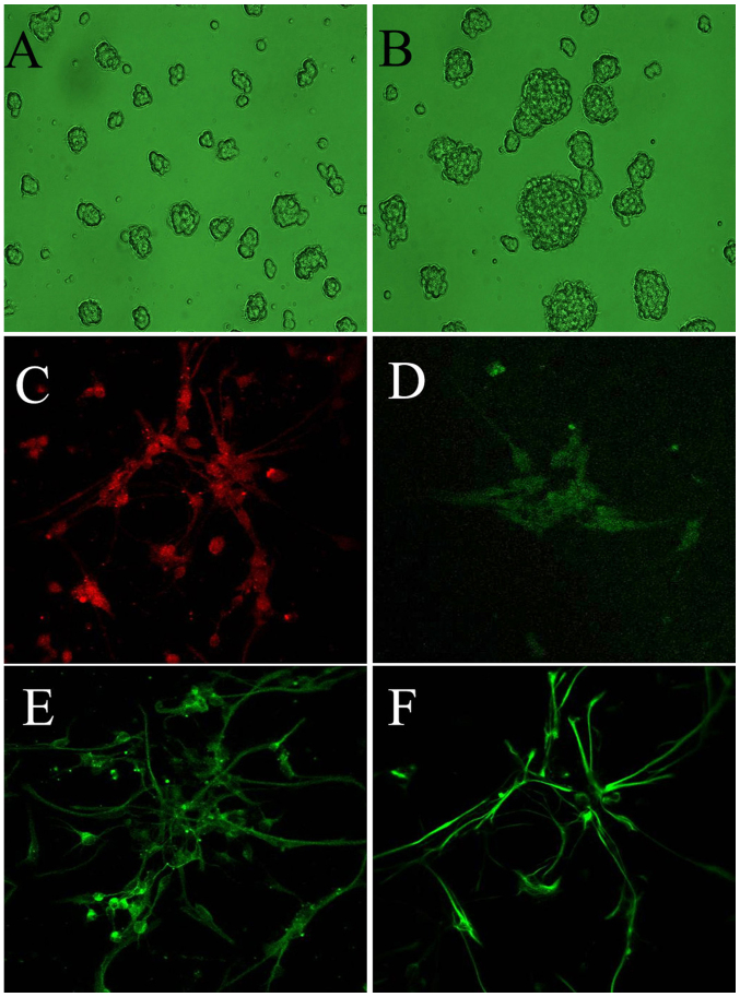
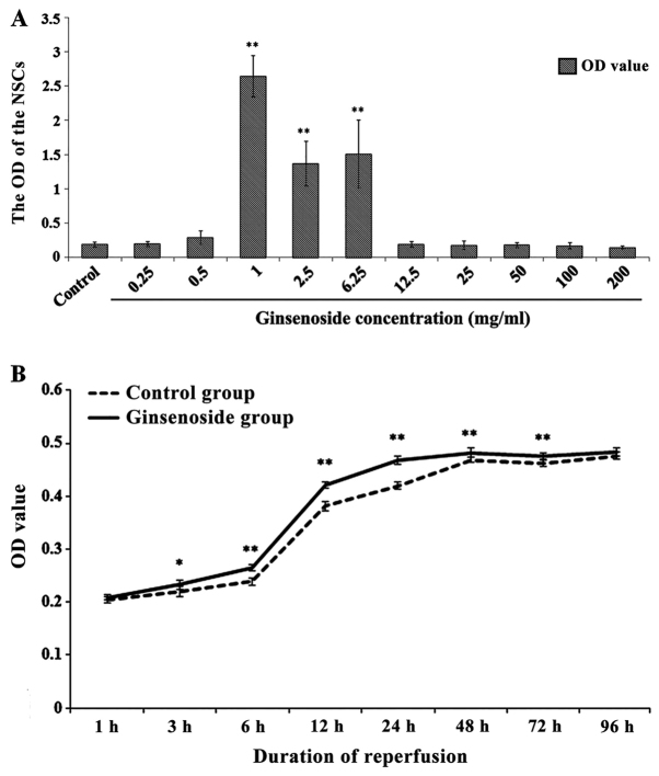
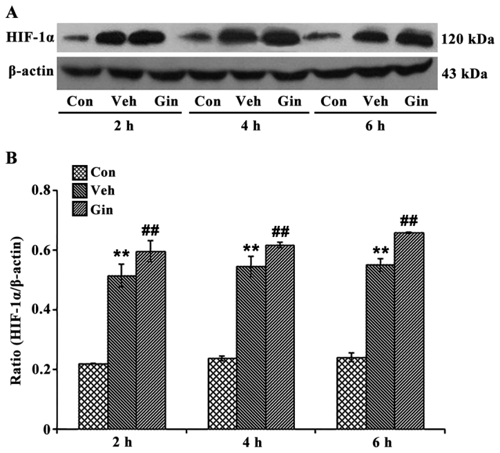
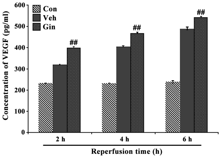
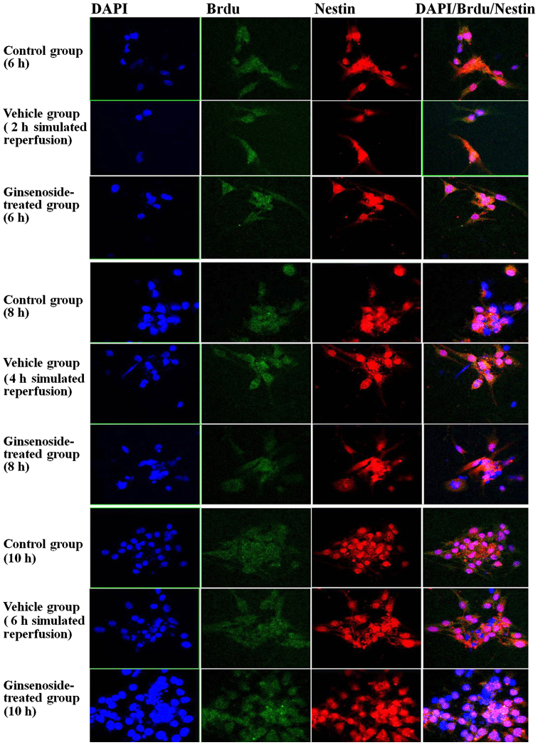
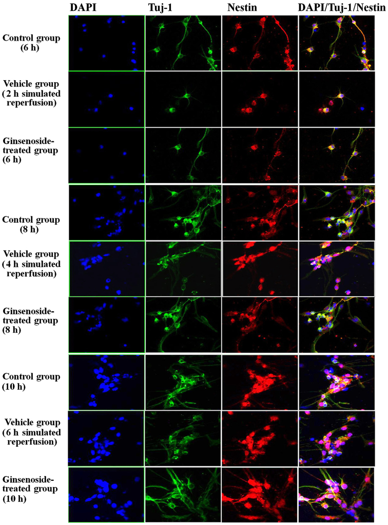
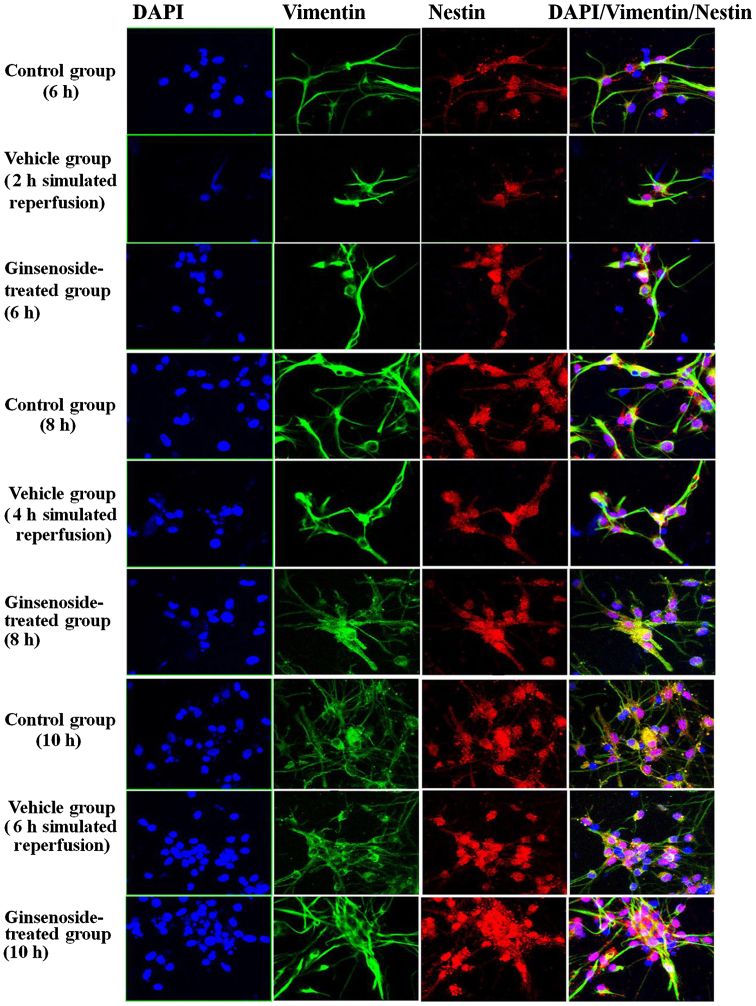
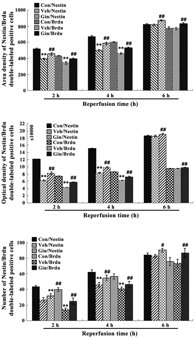
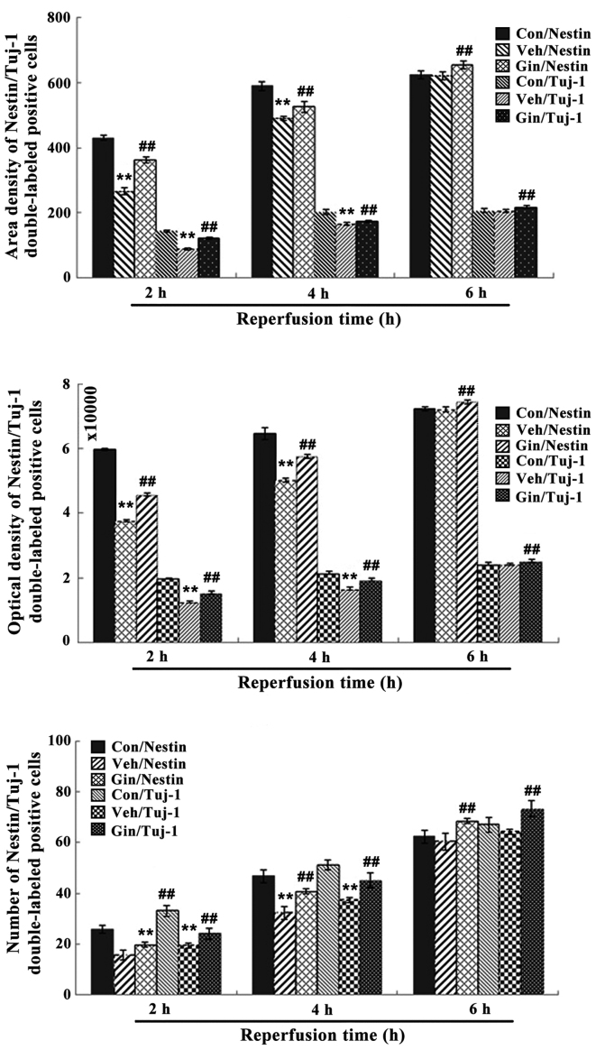
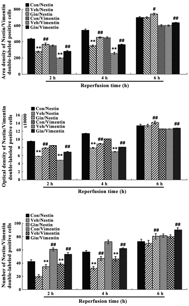
The present study comprised a series of experiments to investigate the mechanism underlying the effect of ginsenoside on the self-renewal, proliferation and differentiation of neural stem cells (NSCs) undergoing oxygen-glucose deprivation/reperfusion (OGD/R) in vitro. The NSCs, which were isolated from the hippocampus of embryonic day 17 embryo rats, were subjected to OGD/R to establish an in vitro model of brain ischemia-reperfusion, following which different doses of ginsenoside were administered to the model. The proliferation of the NSCs was determined using MTT colorimetry and nestin/bromodeoxyuridine (BrdU) immunofluorescent double-labeling. The NSCs were identified by measuring the expression of nestin, and the differentiation of NSCs was assessed through the immunofluorescent double-labeling of nestin/vimentin and nestin/neuron-specific class III β-tubulin (tuj-1). The protein levels of vascular endothelial growth factor (VEGF) and hypoxia-inducible factor-1α (HIF-1α) were detected to investigate the function and mechanism of ginsenoside on ischemic stroke using an enzyme-linked immunosorbent assay. Marked increases in the optical density, area density and numbers of nestin/BrdU-, nestin/vimentin- and nestin/tuj-1-positive cells were found in the ginsenoside-treated group. Compared with the control group, enhanced expression levels of BrdU, tuj-1 and vimentin were found in the ginsenoside-treated group, suggesting that ginsenoside may significantly promote the proliferation and differentiation of NSCs. The results of the present study also showed that ginsenoside significantly increased the protein level of HIF-1α (P<0.05) in the NSCs exposed to OGD/R. These results indicated that ginsenoside may maintain NSC replication, promote NSC proliferation and promote NSC differentiation into neurons and astrocytes. Ginsenoside may initiate the expression of downstream VEGF, which is involved in promoting the survival, self-renewal and differentiation of NSCs.


Figures

Figure 1
Figure 1
Identification of NSCs and its differentiated cells. (A) Primary culture of cell clone spheres. (B) Passage culture of cell clone spheres. Immunofluorescence images of the NSCs to show (C) nestin, (D) BrdU, (E) tuj-1 and (F) vimentin (magnification, ×200). Primary neurospheres had a regular morphology without irregularly sized processes. Volume increased with the culture time, as with the passaged cell clone spheres. The immunofluorescence staining showed that the cells were nestin-positive, with red fluorescent in the cytoplasm. The cells shown to be BrdU-positive following 4 days with proliferation condition medium, exhibited green fluorescence in the cytoplasm. NSCs, neural stem cells; BrdU; bromodeoxyuridine; tuj1, neuron-specific class III β-tubulin.
Figure 2
Figure 2
Effects of ginsenoside on proliferation and differentiation NSCs. Effects of different (A) concentrations of ginsenoside and (B) durations of reperfusion. The values were analyzed using a t-test and expressed as the mean ± standard deviation (n=8). *P<0.05 and **P<0.01 vs. control groups. NSCs, neural stem cells; OD, optical density.
Figure 3
Figure 3
Expression of HIF-1α in the neural stem cells. (A) Representative image of the protein expression of HIF-1α. (B) Bar graph quantifying the protein levels of HIF-1α. The results are expressed as the mean ± standard deviation. β-actin was used as the internal control. There were three time points of reperfusion (2, 4 and 6 h) for the groups: 4 h of OGD and simulated reperfusion for 2 h; 4 h of OGD and simulated reperfusion for 4 h; 4 h of OGD and simulated reperfusion for 6 h. **P<0.01 vs. Con; ##P<0.01 vs. Veh. Con, control group; Veh, vehicle group; Gin, ginsenoside-treated group; HIF-1α, of hypoxia-inducible factor-1α.
Figure 4
Figure 4
Concentrations of VEGF. Concentrations of VEGF (pg/ml) were determined in the three groups. ##P<0.05 vs. Veh. VEGF, vascular endothelial growth factor; Con, control group; Veh, vehicle group; Gin, ginsenoside-treated group.
Figure 5
Figure 5
Morphological observations of the number, optical density and area density of nestin/BrdU double-labeled positive cells following 2, 4 and 6 h of reperfusion. From left to right (×200): DAPI (blue), BrdU (green), Nestin (red), Merge. DAPI, 2-(4-amidinophenyl)-6-indolecarbamidine; BrdU, bromodeoxyuridine.
Figure 6
Figure 6
Morphological observation of the number, optical density and density of the nestin/tuj-1 double-labeled positive cells for 2, 4 and 6 h of reperfusion. From left to right (×200): DAPI (blue), tuj-1 (green), nestin (red), merge. DAPI, 2-(4-amidinophenyl)-6-indolecarbamidine; BrdU, bromodeoxyuridine; tuj-1, neuron-specific class III β-tubulin.
Figure 7
Figure 7
Morphological observation of the number, optical density and density of the nestin/vimentin double-labeled positive cells for 2, 4 and 6 h of reperfusion. From left to right (×200): DAPI (blue), vimentin (green), nestin (red), Merge. DAPI, 2-(4-amidinophenyl)-6-indolecarbamidine; BrdU, bromodeoxyuridine.
Figure 8
Figure 8
Nestin/BrdU double-labeled immunochemical staining results following 2, 4 and 6 h of reperfusion. Results for area density, optical density and positive number are shown. **P<0.01 vs. Con; #P<0.05 and ##P<0.01 vs. Veh. Compared with those in the control group, the number of positive cells, optical density and area density were decreased significantly between 2 and 4 h of reoxygenation (P<0.01) in the vehicle group and were significantly increased (P<0.01) in the ginsenoside-treated group, compared with those in the vehicle group. The statistical results showed that the number of positive cells, optical density and area density were decreased following 6 h of reoxygenation (P>0.05) in the vehicle group, but were significantly increased (P<0.01) in the ginsenoside-treated group, compared with those in the vehicle groups. Veh. Con, control group; Veh, vehicle group; Gin, ginsenoside-treated group; BrdU, bromodeoxyuridine.
Figure 9
Figure 9
Nestin/tuj-1 double-labeled immunochemical staining results of the control group, vehicle group and ginsenoside-treated group following 2, 4 and 6 h. of reperfusion. **P<0.01 vs. Con; ##P<0.01 vs. Veh. Compared with those in the control group, the number of positive cells, optical density and area density were significantly decreased between 2 and 4 h of reoxygenation (P<0.01) in the vehicle group, but were increased significantly (P<0.01) in the ginsenoside-treated group, compared with those in the vehicle group. The number of positive cells, optical density and area density were decreased following 6 h of reoxygenation (P>0.05) in the vehicle group, but were increased significantly (P<0.01) in the ginsenoside-treated group, compared with those in the vehicle group. Veh. Con, control group; Veh, vehicle group; Gin, ginsenoside-treated group; BrdU, bromodeoxyuridine; tuj-1, neuron-specific class III β-tubulin.
Figure 10
Figure 10
Nestin/vimentin double-labeled immunochemical staining results of the control group, vehicle group, and ginsenoside-treated group following 2, 4 and 6 h of reperfusion. **P<0.01 vs. Con; #P<0.05; ##P<0.01 vs. Veh. Compared with those in the control group, the number of positive cells, optical density and area density were decreased significantly between 2 and 4 h of reoxygenation (P<0.01) in the vehicle group, but were increased significantly (P<0.01) in the ginsenoside-treated group, compared with those in the vehicle group. The number of positive cells, optical density and area density were decreased following 6 h of reoxygenation (P>0.05) in the vehicle group, but were increased significantly (P<0.01) in the ginsenoside-treated group, compared with those in the vehicle group. Veh. Con, control group; Veh, vehicle group; Gin, ginsenoside-treated group; BrdU, bromodeoxyuridine.

Similar articles

Cited by

References

    1. Shi Q, Zhang P, Zhang J, Chen X, Lu H, Tian Y, Parker TL, Liu Y. Adenovirus-mediated brain-derived neurotrophic factor expression regulated by hypoxia response element protects brain from injury of transient middle cerebral artery occlusion in mice. Neurosci Lett. 2009;465:220–225. doi: 10.1016/j.neulet.2009.08.049. - DOI - PubMed
    1. Mehta SL, Manhas N, Raghubir R. Molecular targets in cerebral ischemia for developing novel therapeutics. Brain Res Brain Res Rev. 2007;54:34–66. doi: 10.1016/j.brainresrev.2006.11.003. - DOI - PubMed
    1. Peruzzotti-Jametti L, Donegá M, Giusto E, Mallucci G, Marchetti B, Pluchino S. The role of the immune system in central nervous system plasticity after acute injury. Neuroscience. 2014;283:210–221. doi: 10.1016/j.neuroscience.2014.04.036. - DOI - PMC - PubMed
    1. Kalluri HS, Eickstaedt J, Dempsey RJ. Oxygen glucose deprivation inhibits the growth and ERK phosphorylation of neural progenitor cells in vitro. Neurosci Lett. 2007;426:145–148. doi: 10.1016/j.neulet.2007.08.047. - DOI - PMC - PubMed
    1. Lipton P. Ischemic cell death in brain neurons. Physiol Rev. 1999;79:1431–1568. doi: 10.1152/physrev.1999.79.4.1431. - DOI - PubMed

MeSH terms